2551
Microgravity-induced changes in pituitary morphology, brain volumetry, and cerebral spinal fluid hydrodynamics: relationship to spaceflight associated neuro-ocular syndromeLarry A. Kramer1, Khader M. Hasan1, Michael B. Stenger2, Ashot Sargsyan2, and Brandon R. Macias2
1Diagnostic Imaging, UTSHC-Houston, Houston, TX, United States, 2NASA, Houston, TX, United States
Synopsis
A longitudinal study of astronauts with long duration exposure to microgravity showed intracranial volumetric expansion which did not return to baseline after a 1 year of post-flight recovery. These findings were associated with increased cerebral spinal fluid pulsatility through the cerebral aqueduct suggesting diminished intracranial compliance. Additionally, there was development of pituitary gland deformity similar to that seen in idiopathic intracranial hypertension implicating the presence of elevated intracranial pressure during spaceflight.
BACKGROUND
Spaceflight Associated Neuro-ocular Syndrome (SANS) affects ~40% of astronauts who participate in long-duration spaceflights, and these astronauts present with one or more of the following: optic disc edema, hyperopia, globe flattening, cotton-wool spots, or choroidal folds1. Intracranial pressure (ICP) that is chronically elevated above values typically observed in the upright posture on Earth is hypothesized to be a contributing factor to SANS. The purpose of this study was to use morphologic and quantitative magnetic resonance imaging (qMRI) to determine whether spaceflight alters brain tissue volume or cerebrospinal flow dynamics, and to identify factors that may predispose astronauts to develop SANS.METHODS
We performed a prospective longitudinal qMRI study at 3Tesla of 12 long-duration spaceflight astronauts (mean age 46±5 years), with measures conducted before flight and 1, 30, 90, 180 and 360 days after landing. Intracranial volume (ICV), cerebral spinal fluid (CSF) peak-to-peak velocity amplitude (CSF2Û), and volumetric CSF flow (CSFQ) were determined on each study day. The change in CSFQ (∆CSFQ) from preflight to postflight-day-1 was compared to preflight qMRI measurements. We also assessed qualitative changes in pituitary morphology from preflight to postflight-day-1.RESULTS
Significant increases in average intracranial volume (ICV) (P< .001) and CSF2Û (P = .02) were detected when comparing results from preflight and postflight-day-1. Eight (67%) astronauts developed pituitary dome flattening or concavity, and 3 individuals (25%) had posterior stalk displacement. A negative logarithmic correlation (R2=0.67) was detected for ∆CSFQ and preflight CSFQ.CONCLUSION
ICV expansion within a semi-rigid cranial compartment could decrease intracranial compliance and may contribute to the increase in CSF pulsatility. These fluid pressure impulses to the pituitary stalk hiatus may have caused the pituitary deformity we detected in the present study. Acquired pituitary deformity in astronauts is similar to that found in patients with idiopathic intracranial hypertension supporting the hypothesis that astronauts have chronic elevations in ICP during spaceflight. Preflight CSFQ shows promise in predicting the magnitude of ∆CSFQ.Acknowledgements
Supported by the NASA-JSC Human Research ProgramReferences
1. Mader TH, Gibson CR, Pass AF, et al. Optic disc edema, globe flattening, choroidal folds, and hyperopic shifts observed in astronauts after long-duration space flight. Ophthalmology2011; 118(10): 2058-69.Figures
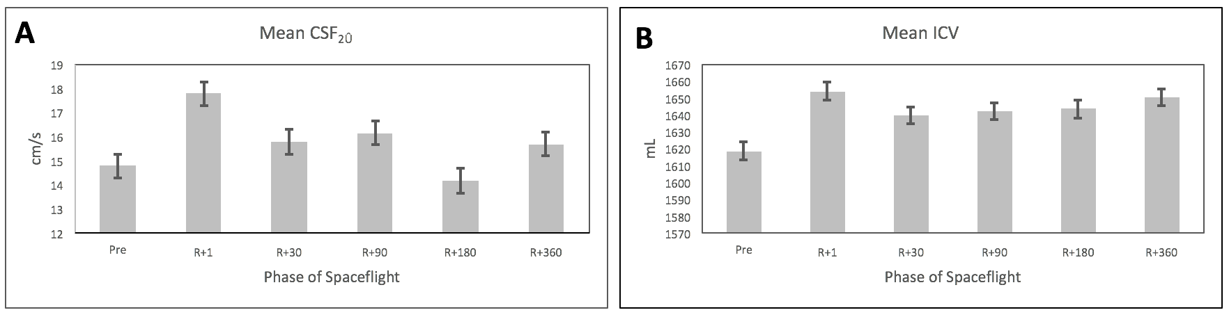
CSF hydrodynamics and intracranial volumetry. A. Longitudinal plot of mean CSF2Û pulsatility relative to phase of spaceflight. B. Longitudinal plot of mean ICV relative to phase of spaceflight.