2546
Longitudinal cerebral metabolic changes in delayed neurologic sequelae after carbon monoxide intoxication using 1H MR spectroscopy.1Dept. of Computer Science and Engineering, National Sun Yat-sen University, Kaohsiung, Taiwan, 2Dept. of Radiology, Veterans General Hospital-Kaohsiung, Kaohsiung, Taiwan, 3School of Medicine, National Yang-Ming University, Taipei, Taiwan, 4Dept. of Neurology, E-Da Hospital, Kaohsiung, Taiwan, 5School of Medicine, I-Shou University, Kaohsiung, Taiwan
Synopsis
In this study, we explored the metabolic changes in WM and GM of patients with and without DNS using 1H MRS longitudinally at onset, 1, 3, and 9 months after CO intoxication. Decreased tNAA/Cr and increased Cho/Cr were observed in WM of patients with DNS as literatures have reported. The longitudinal change of Glx/Cr and Ins/Cr in WM and GM of patients with DNS implies themselves that may provide valuable information for monitoring DNS development.
Introduction
Delayed neurologic sequelae (DNS) is commonly developed in patients with CO intoxication, which exhibits recurrent neuropsychiatric symptoms after an interval of apparent normality1,2. A neural marker for DNS is thus desirable to facilitate prediction and monitoring of neuropathology for appropriate diagnosis and treatment2,3. 1H magnetic resonance spectroscopy (MRS) has been recently used to detect metabolism changes in white matter (WM) of patients with and without DNS after CO intoxication1. In this study, we explored the metabolic changes in WM and gray matter (GM) of patients with and without DNS using 1H MRS longitudinally after CO intoxication.Methods
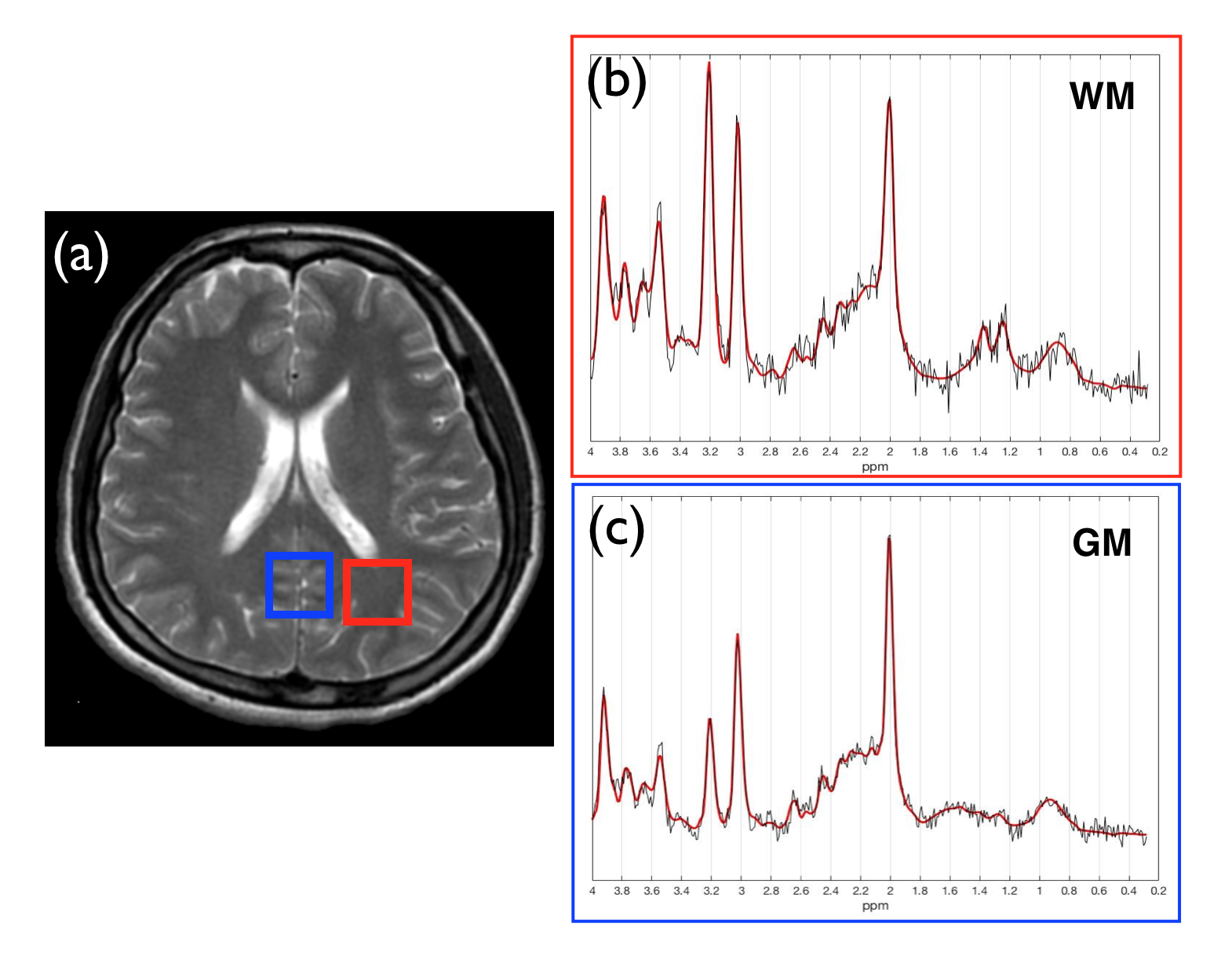
A total of 23 patients who experienced CO intoxication were participated in this study with IRB approval. Eleven patients who exhibited white matter lesion (WML) within follow-up period were categorized as WML+ and others as WML-. In WML+ group, 5 patients were diagnosed with the presence of DNS (DNS+), and others as DNS-. All MR studies were conducted on a 1.5T system (General Electric) using 8-ch head coil with single voxel spectroscopy protocol (PRESS, TR/TE = 1600/35 ms, Ave =128, voxel size = 2x2x2 cm3). MRS scans were acquired within 1 week, 1, 3, and 9 months after CO poisoning. By referencing to axial T2 images, MRS voxel were selected on the WM where WM abnormalities of DNS frequently located and nearby GM respectively (Fig.1a). Acquired spectra were analyzed by LCModel to compare the metabolic levels of NAA+NAAG/Creatine (tNAA/Cr), Choline/Creatine (Cho/Cr), Glutamate+Glutamine/Creatine (Glx/Cr), and Myo-Inositol/Creatine (Ins/Cr). Cramér-Rao lower bounds (SD%) of values larger than 15% for tNAA, Cr, Cho, and 20% for Glx were excluded in the statistical analysis.Results & Discussions
Figure 1a illustrates the MRS voxel placed at WM (red square) where WM abnormalities of DNS frequently located and the nearby gray GM (blue square). LCModel-fitted spectra of a DNS+ patient acquired at 1 month after CO intoxication demonstrate significant decreased NAA at 2.02 ppm and increased Cho at 3.2 ppm in WM (Fig.1b) whereas the spectrum in GM is considered normal (Fig.1c).
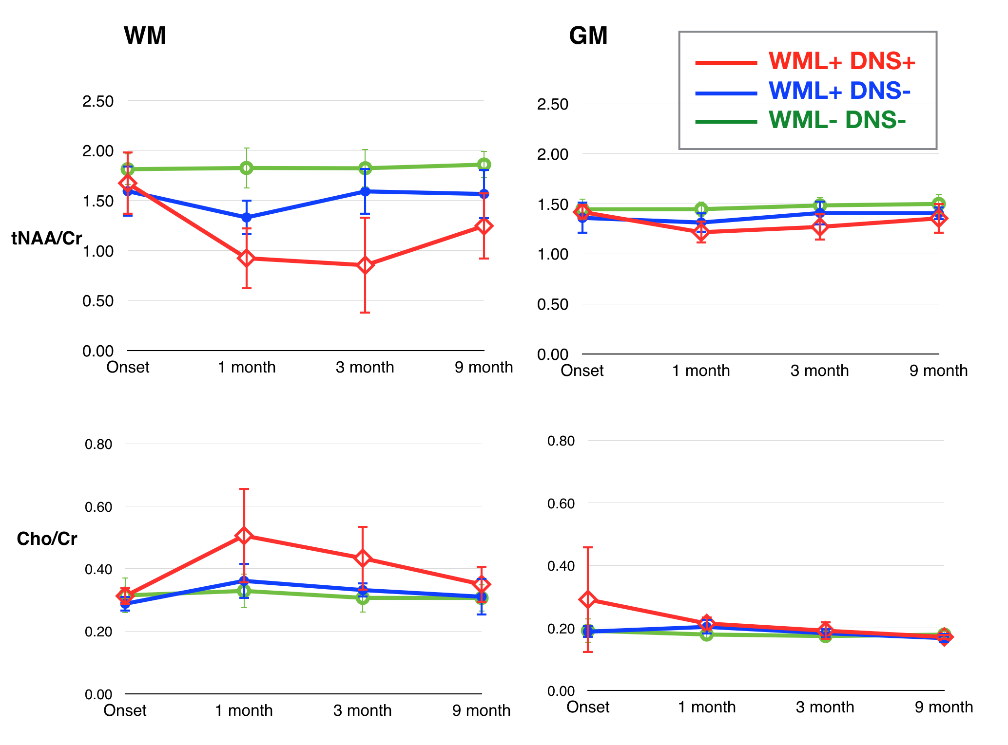
Quantitative results by LCModel of tNAA/Cr, Cho/Cr, Glx/Cr and Ins/Cr are displayed in Fig.2 and Fig.3 with their averages and standard deviations. In WM, tNAA/Cr ratios of WML+, DNS+ patients are dramatically decreasing from 1.67±0.32 (onset) to 0.92±0.31 in 1 month, 0.85±0.48 in 3 months, and slightly regain to 1.25±0.34 in 9 months. Statistic analysis implies that tNAA/Cr in WM of DNS+ (WML+) group has significant difference from DNS- (WML+,WML-) group at 1 month (p<0.0001), 3 months (p<0.001) and 9 months (p<0.001), although it is not significantly different at onset (p>0.05). In GM, tNAA/Cr also decreases slightly in DNS+ patients and shows significant difference from DNS- group at 1 (p<0.05), 3 (p<0.05), and 9 months. The Cho/Cr ratios in WM of DNS+(WML+) patients are significantly increasing in 1 and 3 months (p<0.001). In GM, only the Cho/Cr ratio at onset and 1 month has significant difference between DNS+ and DNS- groups (p<0.05). However, the change in GM is relatively minor compared with that in WM.
As the Glx/Cr ratio in WM of WML+, DNS+ patients decreases at 3 months (p<0.05) and regains in 9 months, the increased Glx/Cr in GM of WML+, DNS+ group and decreased level in WML+, DNS- at onset implies Glx/Cr may be used as a predictive indicator of DNS in WML+ patients. Although an elevation of Ins/Cr ratio may result from the presence of WML, higher Ins/Cr in DNS+ patients can suggest its potential connection with DNS.
Our results demonstrated the decreased tNAA/Cr ratio and increased Cho/Cr in WM at onset and at 1 month after CO intoxication can be observed in patients who has developed or will develop DNS. Several 1H MRS studies1 of CO intoxication with long TE has reported similar results with ours in tNAA/Cr and Cho/Cr ratios. Furthermore, the use of short TE in our study enables the investigation of WML and DNS development by means of Glx/Cr and Ins/Cr changes. Ins has been proposed as a glial marker and increased Ins has been found in patients of Parkinson’s disease4. Change in Glx has been implicated in studies of neuropsychiatric and neurological disorders5. Since DNS may develop parkinsonism with a median latency of 4 weeks and mild cognitive impairment2, Glx and Ins may provide valuable information for monitoring DNS development.
Acknowledgements
No acknowledgement found.References
- Kuroda H, Fujihara K, Mugikura S, Takahashi S, Kushimoto S, Aoki M. Altered white matter metabolism in delayed neurologic sequelae after carbon monoxide poisoning: A proton magnetic resonance spectroscopic study. J Neurol Sci 2016;360:161-169.
- Bleecker ML. Carbon monoxide intoxication. Handb Clin Neurol 2015;131:191-203.
- Beppu T, Nishimoto H, Ishigaki D, Fujiwara S, Yoshida T, Oikawa H, Kamada K, Sasaki M, Ogasawara K. Assessment of damage to cerebral white matter fiber in the subacute phase after carbon monoxide poisoning using fractional anisotropy in diffusion tensor imaging. Neuroradiology 2010;52(8):735-743.
- Firbank MJ, Harrison RM, JT OB. A comprehensive review of proton magnetic resonance spectroscopy studies in dementia and Parkinson's disease. Dement Geriatr Cogn Disord 2002;14(2):64-76.
- Sanaei Nezhad F, Anton A, Michou E, Jung J, Parkes LM, Williams SR. Quantification of GABA, glutamate and glutamine in a single measurement at 3 T using GABA-edited MEGA-PRESS. NMR Biomed 2018;31(1):e3847.
Figures