2538
Altered biochemical profiles in fetuses with congenital heart disease1Diagnostic Imaging and Radiology, Children's National Health System, Washington, DC, United States, 2Pediatrics, George Washington Univeristy, Bethesda, MD, United States, 3Developing Brain Research Laboratory, Children's National Health System, Washington, DC, United States, 4Fetal Heart Program, Children's National Health System, Washington, MD, United States
Synopsis
Brain injury is a major complication in infants with complex congenital heart disease (CHD). There is growing evidence that impaired brain development has its origins in the fetal period. We prospectively characterized in vivo fetal brain metabolic profiles in 307 fetuses (210 health fetuses and 97 with CHD). Findings from measurements of metabolite concentrations of NAA, Cr, and Cho increased with advancing GA in healthy and CHD fetuses. In CHD fetuses, tNAA/tCh ratios were significantly lower while lactate concentrations were significantly higher compared to healthy fetuses, suggesting early-life disturbances in fetal brain biochemistry.
Introduction
Congenital heart disease (CHD) is one of the most common congenital birth defects, affecting ~1% of all live births. The origins of neurodevelopmental dysfunction in infants with congenital heart disease (CHD) are increasingly finding their footprints in fetal life. Previous studies have reported disturbances in fetal brain metabolism in CHD (1,2); however, these studies have been cross-sectional in nature with modest sample sizes. The aim of this study was to prospectively compare fetal biochemical profiles in a large cohort of health and CHD fetuses using proton magnetic resonance spectroscopy (1H-MRS).Methods
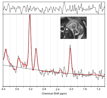
We prospectively enrolled 307 pregnant women of which, 210 had healthy fetuses and 97 had fetuses diagnosed with CHD. Pregnant women were scanned during the second and third trimester of pregnancy ranging from 18 to 39 weeks gestational age (GA). Mean GAs for healthy fetuses was 31.16 ± 5.09 weeks and 31.56 ± 4.49 weeks for CHD fetuses. SSFSE images acquired in all three planes were used as a guide to place the voxels in the central brain of the fetal brain for spectroscopic measurements. Spectra were acquired using PRESS localization sequence (TE/TR: 144/1500 ms). 192 averages of water suppressed spectra were acquired along with 16 averages of water unsuppressed spectra from a 30x30x30 mm3 voxel placed in the central brain. Phase and frequency correction were performed using programs written in Matlab and spectra were quantified in the ‘LCModel’ (3) program using water as an internal reference. Data with CRLB >20% for total Choline (tCh) were excluded from further analysis. For all other metabolites, exclusion criteria included CRLB>100%. NAA and Cre concentrations are lower in early GA, hence, exclusion criteria of CRLB>100% is used in this study which is higher than 20% that has traditionally been used to avoid biasing the metabolite concentrations to higher values (4). Statistical analysis included linear regression to assess metabolic trajectory as a function of GA and diagnostic status.Results/Discussion
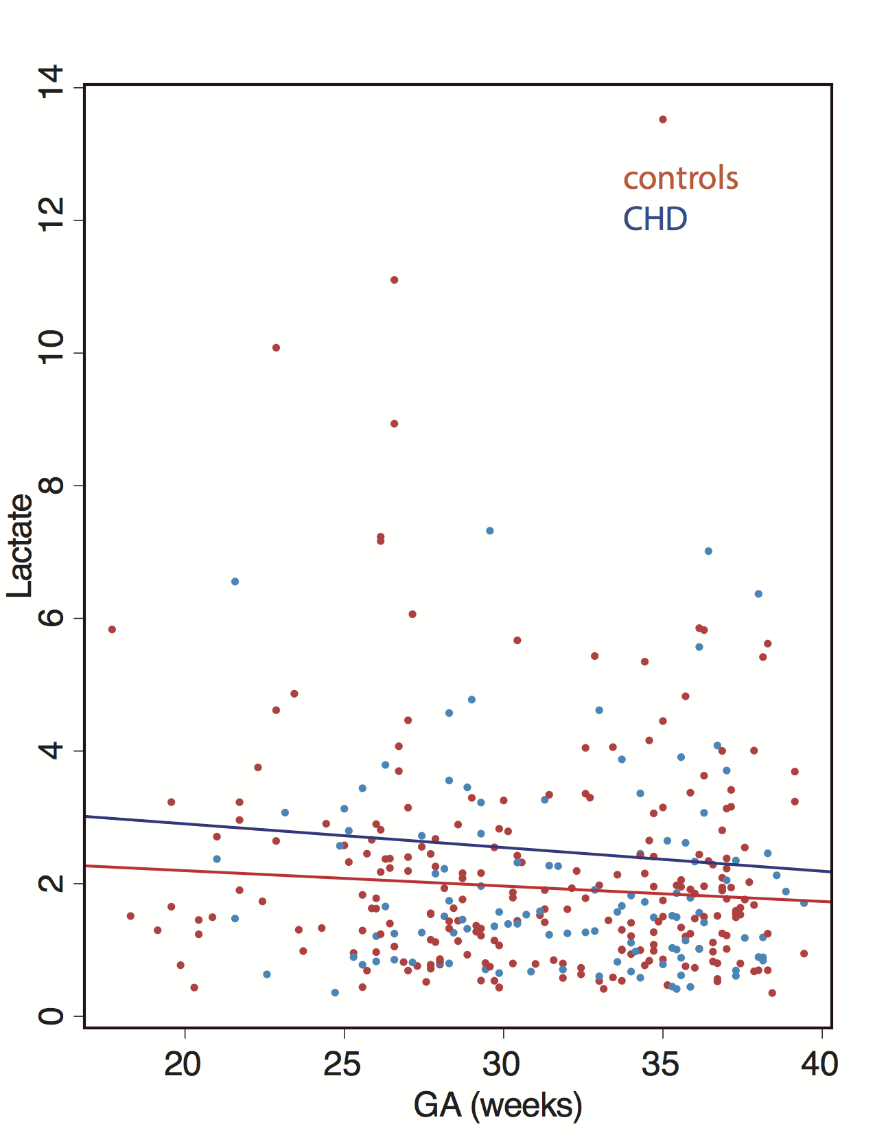
Figure 1 shows voxel placement and LCModel output of a typical spectrum acquired from the fetal brain. Figure 2 shows Lac concentrations in controls and CHD fetuses across increasing GA. Linear regression analyses with diagnostic status and GA as variables showed significant increase in tNAA, tCh, tCr with advancing GA (p<0.001). Fetuses with CHD has significantly lower tNAA/tCh ratios compared to control fetuses (p= 0.046). Our results also showed higher levels of Lac in CHD fetuses compared to healthy fetuses across all GAs (p=0.004). All conventional MRI studies for control and CHD fetuses showed no structural injury. Higher levels of brain lactate in CHD fetuses suggests the presence of anaerobic metabolism, while lower NAA/Ch levels point to neuronal injury in the absence of demonstrable injury on conventional MRI.Conclusion
In a large prospective observational study, we report decreased tNAA/tCh concentrations in fetuses diagnosed with CHD compared to control fetuses, suggesting neuronal injury and anaerobic metabolism. These data demonstrate that metabolic alterations in CHD fetuses are prevalent and may be an important early biomarker for subsequent risk of brain injury in this high-risk population.Acknowledgements
This work was supported by NIH R01HL116585-01.References
1. Limperopoulos
C, Tworetzky W, McElhinney DB, Newburger JW, Brown DW, Robertson RL, Guizard N,
McGrath E, Geva J, Annese D, Dunbar-Masterson C, Trainor B, Laussen PC, du
Plessis AJ. Brain Volume and Metabolism in Fetuses with Congenital Heart
Disease: Evaluation with Quantitative Magnetic Resonance Imaging and
Spectroscopy. Circulation 2010;121(1):26.
2. Gertsvolf
N, Votava-Smith JK, Ceschin R, Del Castillo S, Lee V, Lai HA, Bluml S, Paquette
L, Panigrahy A. Association between Subcortical Morphology and Cerebral White
Matter Energy Metabolism in Neonates with Congenital Heart Disease. Scientific
reports 2018;8(1):14057-14057.
3. Provencher
SW. Estimation of metabolite concentrations from localized in vivo proton NMR
spectra. Magnetic Resonance in Medicine 1993;30(6):672-679.
4. Kreis
R. The trouble with quality filtering based on relative Cramér-Rao lower
bounds. Magnetic Resonance in Medicine 2016;75(1):15-18.
Figures