2537
MR Spectroscopic Changes in Infants Exposed to Prenatal Opioids: A Pilot Study1Neuroscience Research Australia, Randwick, Australia, 2UNSW, Kensington, Australia, 3University of Wollongong, Wollongong, Australia, 4ANU, Canbera, Australia, 5ANU, Canberra, Australia
Synopsis
Proton spectra were obtained from the left caudate, left hippocampus and subventricular zone of the brains of one week old babies born to mothers who were opioid users. The study suggests that decreased brain volumes after prenatal opioid-exposure are associated with hippocampal spectral abnormalities with increased severity related to multiple opioid use.
Introduction
There is a rising trend in reports of neonatal abstinence syndrome (NAS), a drug-withdrawal syndrome most commonly occurring after exposure in utero to opioids [1]. The resulting postnatal withdrawal can cause failure to thrive, seizures and death if left untreated. Despite toxic cerebral effects of opioids demonstrated in animal models there are few studies of the brain in human infants and even fewer studies where the effect of post-natal environmental stressors is controlled. Here, infants were studied within one week of birth and before discharge from hospital.Methods
Of 23 infants scanned for structural MRI (1.3D T1-weighted (T1W) spoiled gradient recalled (1.2mm coronal slices; flip angle 45; repetition time 35 ms; echo time 9 ms; field of view 21 × 15 cm2; matrix 256 × 192)), 9 were scanned at a single site that included MRS in the protocol. Median gestation age, birth weight and gestation at scanning were 39 weeks, 3170g, 41 weeks respectively. Mothers used methadone (6), buprenorphine (3) and heroin (2).
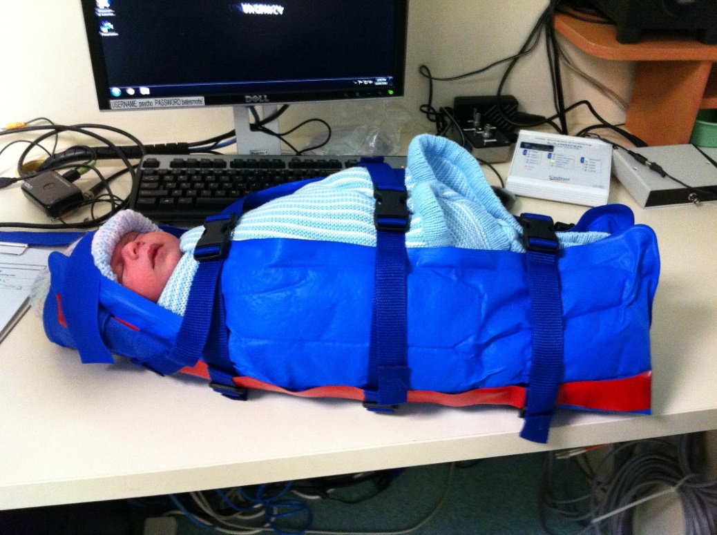
All infants were unsedated, fed and settled in an immobilizing vacubag (Fig. 1).
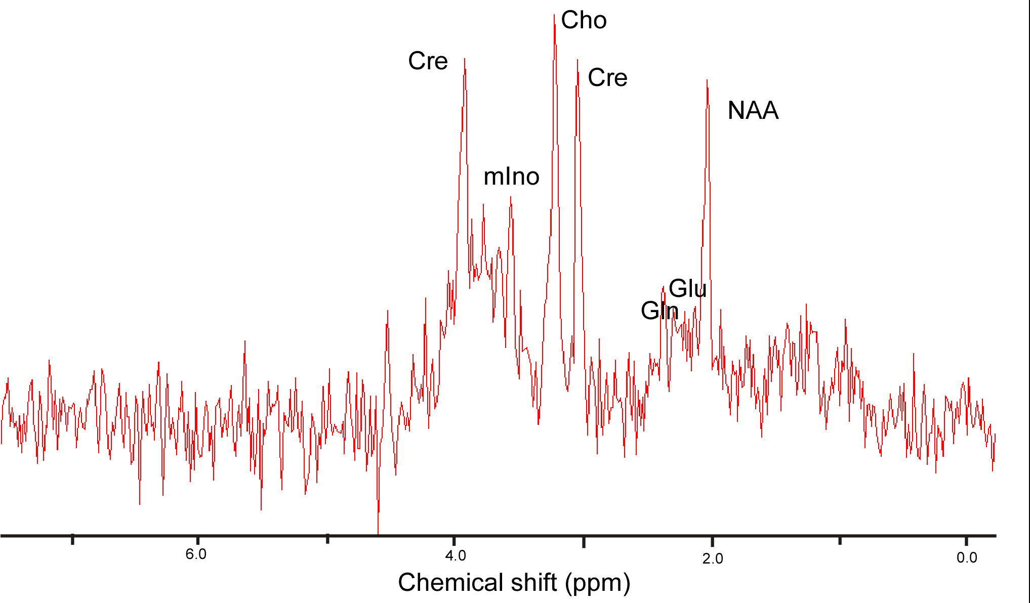
Spectra were obtained from left hippocampus, left caudate and subventricular zone region using a 3T Philips Achieva TX MRI with a 32 channel head coil (8 cm3, PRESS; TE = 38 ms, TR = 2s) as four separate 32 scan acquisitions.
Results
Mothers used methadone (6), buprenorphine (3) and heroin (2). Head circumferences were within normal ranges. Five infants required withdrawal treatment. All brains were structurally normal.
Total brain volume (TBV) correlated with head circumference (r=0.74, p=0.06). Spectral linewidths were excellent (4 – 5 Hz average) but were generally of lower signal to noise than the adult equivalents (Fig. 2).
Spectra of similar quality were obtained from all three areas. Hippocampal Gln/NAA (r=0.9,p=0.005) ratios correlated with, while NAA/Cho (r=-0.8,p=0.031) and NAA/Cr (r=-0.85,p=0.02) ratios inversely correlated with TBV (r=0.9, p= 0.005). Infants of single opioid-users had lower hippocampal NAA/Cho ratios than those of multiple opioid users (0.62 v 0.91,OR 95% CI: 0.19 (0.1-0.28), p=0.02).
Conclusions
We have previously shown decreased total brain volumes in opioid over-exposed babies along with decreased basal ganglia volumes and increased ventricles [2]. This pilot study suggests that decreased brain volumes after prenatal opioid-exposure are associated with hippocampal spectral abnormalities with increased severity related to multiple opioid use. Obtaining spectra from babies at this age is feasible.Acknowledgements
This study was funded by the Cornucopia Foundation of the Royal Hospital for Women and the Langton Centre, South East Sydney Local Health District, New South Wales, Australia. The authors acknowledge the National Imaging Facility and the staff of NeuRA Imaging.References
1. Tolia VN, Patrick SW, Bennett MM, Murthy K, Sousa J, Smith PB, Clark RH,Spitzer AR Increasing incidence of the neonatal abstinence syndrome in U.S. neonatal ICUs. N. Engl. J. Med., 2015. 372: 2118-26. 2. Yuan Q, Rubic M, Seah J, Rae C, Wright IMR, Kaltenbach K, Feller JM, Abdel-Latif ME, Chu C, Oei JL,Bob Collaborative G Do maternal opioids reduce neonatal regional brain volumes? A pilot study. J. Perinatol., 2014.Figures