2533
Comparison of MEGA-PRESS and Short Echo Time PRESS on Classification of IDH Mutation Using Machine Learning at 3T1Institute of Biomedical Engineering, Bogazici University, Istanbul, Turkey, 2Department of Molecular Biology and Genetics, Acibadem Mehmet Ali Aydinlar University, Istanbul, Turkey, 3Neuroradiology Research Center, Acibadem Mehmet Ali Aydinlar University, Istanbul, Turkey, 4Department of Neurosurgery, Acibadem Mehmet Ali Aydinlar University, Istanbul, Turkey, 5Department of Radiology, Acibadem Mehmet Ali Aydinlar University, Istanbul, Turkey
Synopsis
Isocitrate dehydrogenase (IDH) mutation is common in grade II and grade III gliomas, and results in better patient prognosis IDH mutant (IDH-mut) gliomas. Magnetic resonance spectroscopy (MRS) studies indicated an increase in 2-hydroxyglutarate (2HG) and decrease in glutamate (Glu) and glutathione (GSH) as a result of IDH mutation. The goal of this study is to compare IDH mutation classification performances of short echo-time (TE) PRESS and MEGA-PRESS by using machine learning in 60 glioma patients. Highest average classification accuracy was 75% with coarse decision trees for short TE PRESS, and 74% with ensemble of bagged of trees for MEGA-PRESS.
Introduction
Recent WHO criteria has included genetic mutations in glioma classification 1. Isocitrate dehydrogenase (IDH) mutation which is related to metabolism of the cancer tissue, is common in grade II and grade III gliomas. IDH mutant (IDH-mut) gliomas have better prognosis than IDH wild-type (IDH-wt) 2. In IDH-mut gliomas, 2-hydroxyglutarate (2HG) accumulates at tumor site as a result of the mutation 3, 4. Additionally, lower glutamate (Glu) 5, 6 and glutathione (GSH) 6, 7 values have been reported in IDH-mut gliomas. 2HG protons give rise to peaks located at 4.02, 2.25 and 1.9 ppm and all peaks of 2HG overlap with other brain metabolite peaks, such as N-acetylaspartate (NAA), creatine (Cr), glutamine (Gln), Glu, and γ-aminobutyric acid (GABA). These overlaps make 2HG detection challenging. Mescher-Garwood (MEGA) is a frequency selective refocusing technique that has been combined with Point Resolved Spectroscopy (PRESS) sequence. The aim of this study is to detect IDH mutation using machine learning algorithms by comparing the performance of a MEGA-PRESS spectral editing technique with a short echo time (TE) PRESS.Materials and Methods
Seventy-five glioma patients, whose IDH mutation status was assessed by immunohistochemistry, were included in this study. PRESS data was acquired from the solid tumor region excluding gross hemorrhage, edema and necrosis (TR=2000ms, TE=30ms, 1024 Points, BW=2000 Hz), and MEGA-PRESS data was acquired from the same region (TR=1500ms, TE=68ms, 512 Points, BW=1000 Hz) using a Siemens Tim Trio- 3T whole body scanner. At 68 ms echo time, middle peak of the 2HG triplet at 4.02 ppm is inverted, since J-coupling constants with neighboring protons at 1.90 ppm are 7 Hz (leading to an inversion at TE=142 ms) and 4.1 Hz (inversion at TE=243 ms). At difference spectra, outside peaks of the triplet at 4.02 ppm are magnified while the one in the middle is almost suppressed. Fifteen patients were excluded from the study, because of poor signal-to-noise ratio (SNR) and high full width at half maximum values. In total, 60 patients (IDH-mut:24, IDH-wt:36) were included in the analysis.
LCModel spectral fitting program was used for quantification of metabolites 8. The basis set for MEGA-PRESS sequence was simulated for TE=68ms using General Approach to Magnetic Resonance Mathematical Analysis (GAMMA) simulation library of Versatile Simulation, Pulses and Analysis (VESPA) with the prior knowledge of metabolite chemical shifts and coupling constants 9. The features for classification were selected based on previous studies 5-7 . Glycine (Glyc), GSH, 2HG, myo-inositol (Ins), lactate (Lac), total choline (Cho), total NAA and Glx (Glu+Gln) relative concentrations to Cr have been used in machine learning for both PRESS and MEGA-PRESS sequences. Classification Learner app in MATLAB R2018a (The MathWorks Inc., Natick, MA) was used to construct machine learning (ML) models, such as decision trees, support vector machine (SVM), k nearest neighbor (kNN) and ensemble of bagged trees to classify IDH-mut and IDH-wt gliomas. 10-fold cross validation was used to evaluate classifier performance. Models giving the highest accuracy was executed hundred times and average accuracy, sensitivity and specificity values were reported.
Results
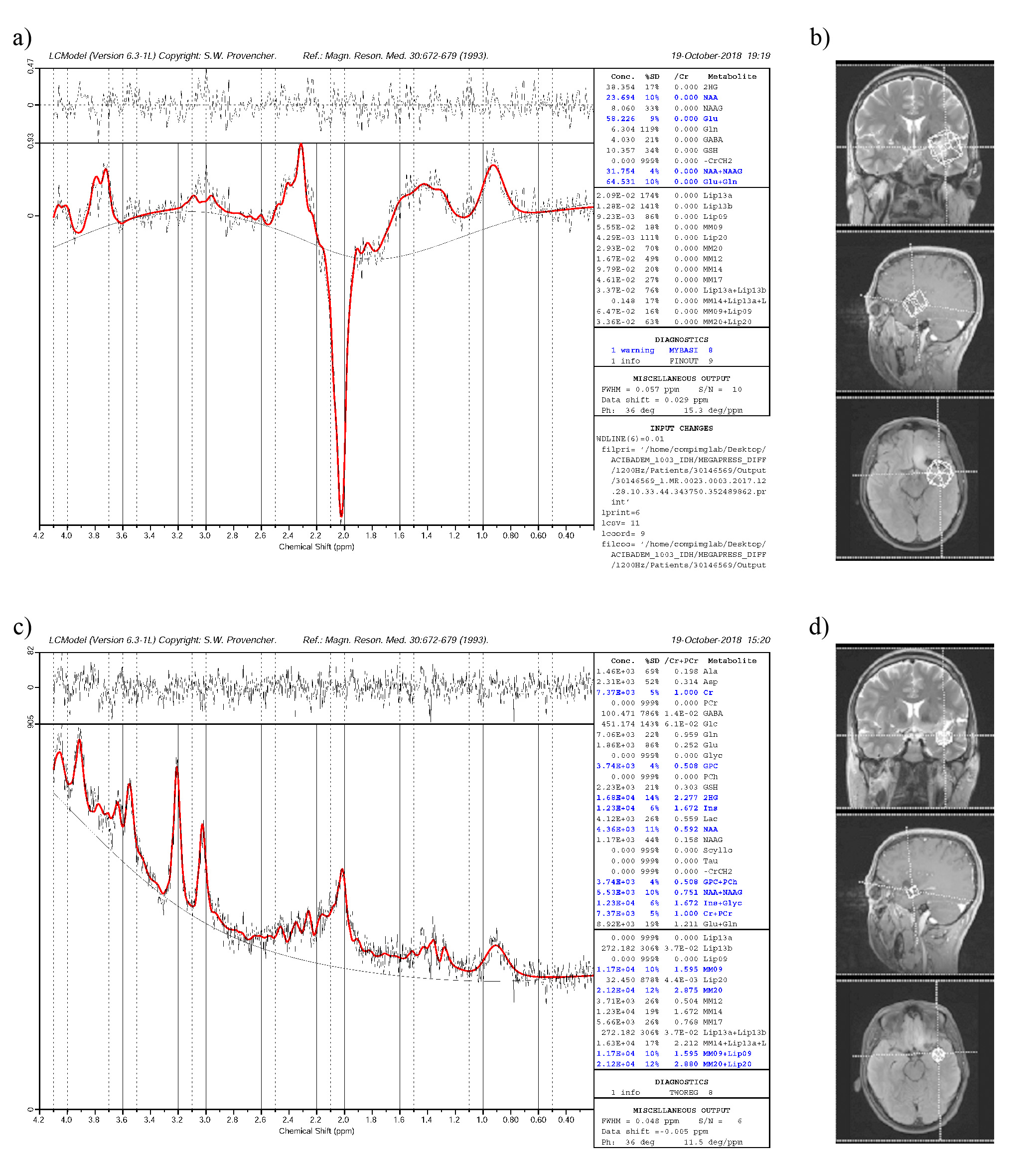
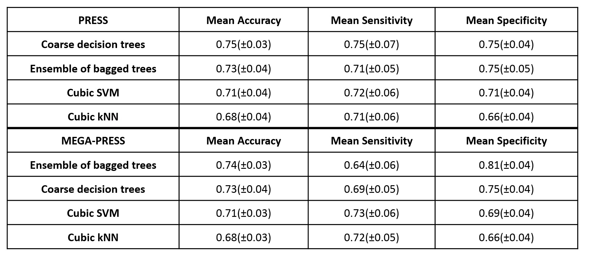
Figure 1 shows LCModel analysis results for short TE PRESS and difference spectrum of MEGA-PRESS sequence for an example IDH-mut glioma. Highest average classification accuracy obtained was 75% with coarse decision trees for short TE PRESS, and 74% with ensemble of bagged of trees for MEGA-PRESS (Table 1). Also, the highest sensitivity of 73% was achieved with cubic SVM model, and highest specificity of 81% was obtained with ensemble of bagged trees for MEGA-PRESS. Similarly, highest sensitivity and specificity values were 75% for short-TE PRESS when coarse decision trees was used.Discussion and Conclusion
This study investigated the differences of short-TE PRESS and MEGA-PRESS in terms of IDH mutation detection in gliomas. Our results indicated that IDH mutation classification results based on MR spectral profiles obtained from the two spectral data acquisition techniques were similar. The classifier results could be improved with a larger cohort size. Future studies will explore the detection efficiency of MRS for other genetic mutations of gliomas, and further optimization of machine learning models and choice of features.Acknowledgements
This project has been funded by TUBİTAK 1003 grant 216S432 and Bogazici Univesity BAP grant 10844SUP.References
1. Hoshide, R. and R. Jandial, 2016 World Health Organization Classification of Central Nervous System Tumors: An Era of Molecular Biology. World Neurosurg, 2016. 94: p. 561-562.
2. Weller, M., et al., Glioma. Nat Rev Dis Primers, 2015. 1: p. 15017.
3. Choi, C., et al., 2-hydroxyglutarate detection by magnetic resonance spectroscopy in IDH-mutated patients with gliomas. Nature medicine, 2012. 18(4): p. 624-629.
4. Andronesi, O.C., et al., Detection of 2-hydroxyglutarate in IDH-mutated glioma patients by in vivo spectral-editing and 2D correlation magnetic resonance spectroscopy. Sci Transl Med, 2012. 4(116): p. 116ra4.
5. Nagashima, H., et al., Diagnostic value of glutamate with 2-hydroxyglutarate in magnetic resonance spectroscopy for IDH1 mutant glioma. Neuro-Oncology 2016. 18(11): p. 1559-1568.
6. Ozturk-Isik, E., et al. Magnetic Resonance Spectroscopic Differences of Diffuse Glioma Groups Classified by IDH and TERT Promoter Mutations at 3T. in Conference Proceedings of ISMRM-ESMRMB . 2018. Paris, France: ISMRM. p. 955.
7. Shi, J., et al., An IDH1 mutation inhibits growth of glioma cells via GSH depletion and ROS generation. Neurological Sciences, 2014. 35(6): p. 839-45.
8. Provencher, S.W., Estimation of metabolite concentrations from localized in vivo proton NMR spectra. Magnetic Resonance in Medicine, 1993. 30(6): p. 672-679.
9. Govindaraju, V.Y., K. ; Maudsley, A.A, Proton NMR chemical shifts and coupling constants for brain metabolites. NMR Biomed, 2001. 13: p. 129-153.
Figures