2531
Neurochemical profile of the human hippocampus at 3T after traumatic brain injury1Medicine, Imperial College London, London, United Kingdom
Synopsis
The aim of this work was to investigate the metabolic profile of the hippocampus in a clinical population (moderate/severe traumatic brain injury patients in the acute phase), using an MR Spectroscopy LASER sequence at 3T. With ongoing data acquisition, preliminary results show reduced levels of total choline (tCho), metabolite reflecting membrane turnover.
INTRODUCTION:
Traumatic brain injury (TBI) is the leading cause of death and disability for people under 45 years old. TBI is a heterogeneous disorder, with damage resulting from various pathophysiological mechanisms, such as haemorrhage, edema, diffuse axonal injury and neuroinflammation1. Current molecular biomarkers for neuroinflammation, such as positron emission tomography (PET) are sensitive but are limited by a reliance on radioactive isotopes. MR Spectroscopy (MRS) is non-invasive, easily scalable and widely available in clinical settings. MRS can measure in vivo concentrations of relevant metabolites such as myo-inositol (proposed as a marker for microglial activation) and glutamate (related to astrocyte activation and excitotoxicity). The hippocampus is particularly sensitive to inflammation and neurodegeneration2 but applying MRS in this region is challenging, due to its geometry and heterogeneity. LASER MRS has proven successful in acquiring a robust metabolic profile in the human hippocampus3, due to improved voxel selection and immunity to B1 artifacts.AIM:
To investigate the metabolic profile of the hippocampus in a clinical population (moderate/severe traumatic brain injury patients in the acute phase), using an MRS LASER sequence at 3T.METHODS:
Subjects N=28 patients with moderate-severe TBI in the acute phase (0-6 weeks after injury) have been included, together with N=33 healthy controls.
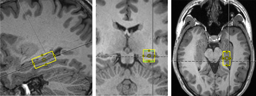
Imaging MRI was performed on a Siemens Verio 3.0T (32-channel head coil). Each subject had T1-MPRAGE (TE=2.98s, TR=2.3s, 1mm isotropic, 5min scanning time), which was then reconstructed in parallel and perpendicular to the hippocampus to guide the positioning of the voxel. After an initial fieldmap-based shim, two iterations of FASTESTMAP 4 were performed in the chosen voxel (left hippocampus 26x13x12mm), followed by MRS LASER (TE=72ms, TR=3s, NA=96, 2048 points, BW=12kHz, VAPOR water suppression, 5min scanning time). A spectrum without water suppression was also acquired from the same voxel as reference and for subsequent eddy correction (NA=4).
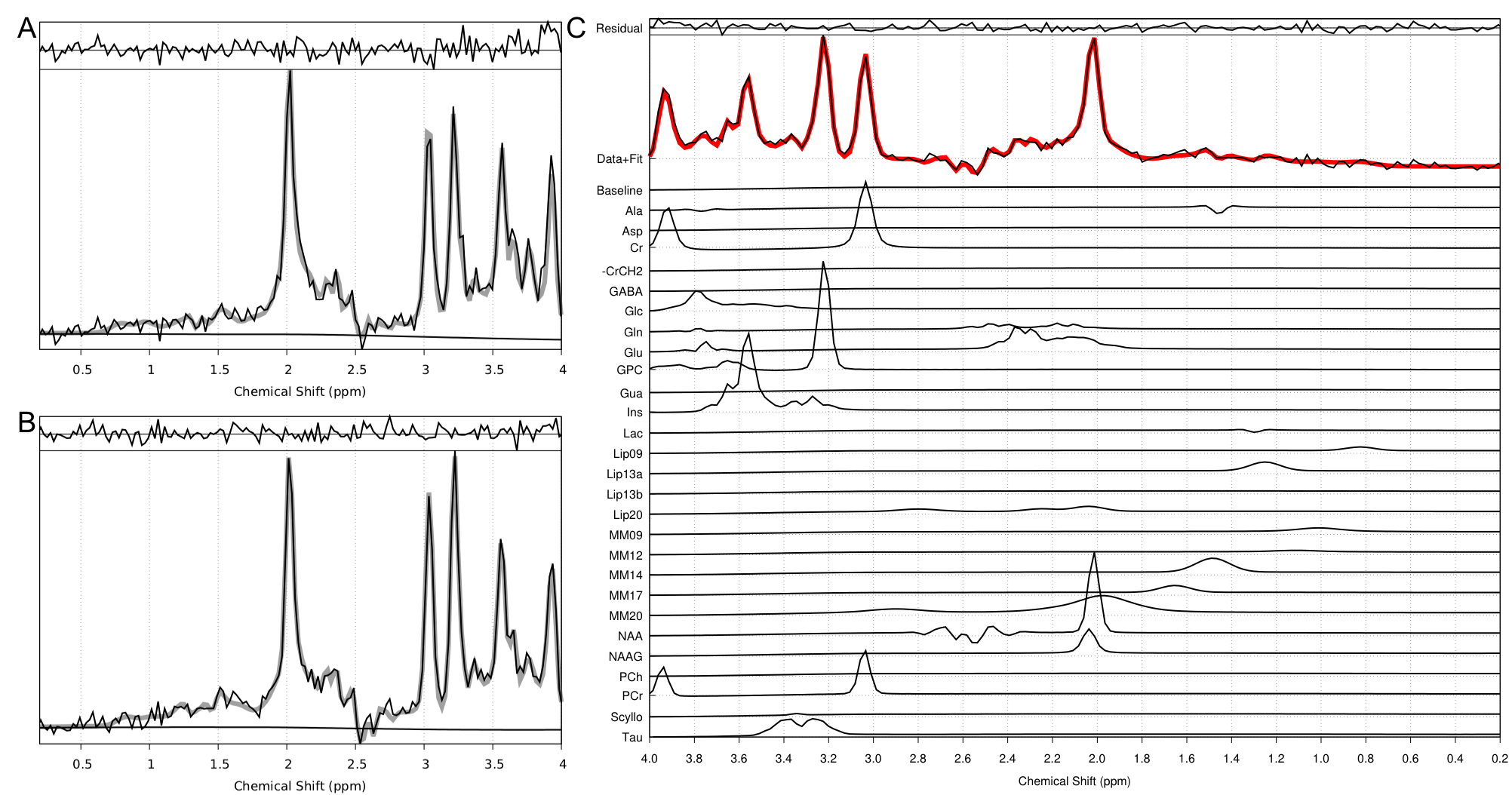
Analysis LASER MRS data was fitted in the time domain using TARQUIN5, with a simulated basis set and eddy correction performed using the unsuppressed water reference spectrum. N=7 controls and N=9 patient datasets were excluded from further analysis due to bad quality of the spectra (noisy without defined peaks, or big fitting residuals). Final numbers were N=19 patients, N=26 controls. Metabolites with consistently low SD (<30%) were included in the statistics (phosphocreatine PCr, glycerophosphocholine GPC, glutamate Glu, glutamate + glutamine Glx, myo-inositol Ins, N-acetyl-aspartate NAA and total choline tCho).
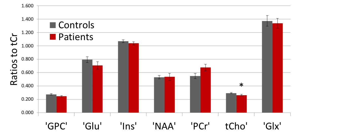
Statistics T-tests were performed to investigate differences between patients and controls, and results are expressed as average metabolic ratios to total creatine (tCr) ± standard error of the mean.
DISCUSSION:
Choline-containing compounds are involved in phospholipid synthesis and degradation, therefore reflecting membrane turnover. Several human studies have reported an increase in tCho after TBI, with animal studies reporting an early decrease after injury6. However, the interpretation of changes in the total choline resonance is not straightforward due to the multiple contributions to the signal, mostly glycerophosphocholine and phosphocholine, which could have differentiating responses or time-courses in the pathological state.
Metabolic levels are expressed as ratios to total creatine (tCr), which is conventionally used as reference in MRS studies, since it is assumed to be stable. This might not be the case in many neurodegenerative and psychiatric diseases and it is a possible confounding factor in our results.
CONCLUSION:
Our preliminary results show a decrease in total choline levels in the human hippocampus at 3T in the acute phase following traumatic brain injury.Acknowledgements
The author wants to thank Dr Małgorzata Marjańska from the University of Minnesota for helpful discussions and for facilitating the C2P agreement to use the CMRR Spectroscopy Package.References
1. Werner C, Engelhard K. Pathophysiology of traumatic brain injury. J Br J Anaesth. 2007;99(1):4‐9.
2. Mancini A, Gaetani L, Di Gregorio M et al. Hippocampal neuroplasticity and inflammation: relevance for multiple sclerosis. MS and Demyelinating Disorders. 2017; 2:2.
3. Allaili N, Valabregue R, Auerbach EJ et al. Single voxel 1H spectroscopy in the human hippocampus at 3T: characterisation of neurochemical profile and reproducibility. NMR in BioMed. 2015; 28(10):1209-1217.
4. Gruetter R and Ivan Tkac. Field mapping without reference scan using asymmetric echo-planar techniques. MRM. 2000; 43:319-323
5. Wilson M, Reynolds G, Kauppinen RA et al. A constrained least-squares approach to the automated quantitation of in vivo 1H magnetic resonance spectroscopy data. MRM. 2011; 65(1):1-12
6. Harris J, Hung-Wen Y, In-Young C et al. Altered neurochemical profile after traumatic brain injury: 1H-MRS biomarkers of pathological mechanisms. J Cereb Blood Flow Metab. 2012; 32(12):2122-2134
Figures