2500
Fast Quantification of Creatine Kinase Reaction Rate in Mouse Skeletal Muscle Using 31P Magnetic Resonance Fingerprinting1Biomedical Engineering, Case Western Reserve University, cleveland, OH, United States, 2Biomedical Engineering, Case Western Reserve University, Cleveland, OH, United States, 3Electrical and Computer Engineering, University of Illinois at Urbana-Champaign, Urbana, IL, United States, 4Electrical and Computer Engineering, University of Illinois at Urbana-Champaign, Urbana, OH, United States
Synopsis
We evaluated the accuracy and efficiency of a 31P magnetic resonance fingerprinting (MRF) method for fast measurement of the creatine kinase (CK) rate constant in mouse skeletal muscle. Our results showed consistent measurement of CK rate constant with less than 10% variations with only 6 signal averages, corresponding to 2 min acquisition.
Introduction
Phosphocreatine (PCr) is considered an important energy reservoir for maintaining a constant ATP level during heavy exercise and severe ischemia. The phosphoryl transfer between PCr and ATP reaction is catalyzed by Creatine Kinase (CK). Hence, evaluating CK reaction rate is of particular interest as CK plays a vital role in maintaining constant ATP supply during periods of increased energy demand or metabolic pertubation1. Phosphorous-31 (31P) Magnetization Transfer Spectroscopy (MT-MRS) provides noninvasive assessment of CK activity. However, quantification of CK reaction rate in mouse hindlimb using conventional 31P MT-MRS requires prohibitively long scan time to achieve adequate signal-to-noise (SNR) (~50 min)2. Previously, we developed a magnetic resonance fingerprinting (MRF) method for efficient and rapid measurement of CK reaction rate in rats3. In the current study, we evaluated the accuracy and efficiency of the CK-MRF technique for in vivo quantification of CK reaction rate in mice.
Method
CK-MRF sequence: A schematic diagram of the CK-MRF pulse sequence is shown in Figure 1A. Following an inversion preparation, the acquisition comprised of a total of 32 acquisition blocks. A 490 ms selective saturation block was used between two acquisition blocks, with the saturation frequency set either at the resonance frequency of γATP (SAT(γATP)) or at the frequency contralateral to γATP (SAT(CNTL)). The entire acquisition was comprised of two modules that employed SAT(CNTL) and SAT(γATP), respectively. Blocks for PCr and γATP acquisition alternated eight times in each module. Each block consisted of a train of 10 excitations with the flip angle linearly ramped-up and ramped‐down and a constant TR of 12.8 ms (Fig. 1B&C). Selective excitation was enabled with a 4-ms Gaussian pulse centered at the resonance frequency of PCr and, respectively. The maximum flip angles in each acquisition block were modulated by a sinusoidal envelope (Fig. 1D). The total time for one complete fingerprint acquisition was 20 s. Multiple repetitions were acquired with no delay, with a single dummy acquisition.
Dictionary generation and parameter matching: A dictionary was constructed using a Matlab-based Bloch-McConnell simulator that included four matching parameters: the pseudo-first order forward CK exchange rate (kf,CK), T1 relaxation time of PCr (T1PCr), the concentration ratio of PCr to ATP (MRPCr) and the resonance frequency of PCr (ωPCr). These four parameters were varied over their physiologically expected range to generate the dictionary for template matching. The dictionary resolution for kf,CK , T1PCr, MRPCr and ωPCr was 0.005 s-1 , 0.1 s, 0.3 and 3 Hz, respectively. The dictionary totaled 132,660 entries. The dictionary entry that produced the largest magnitude of the inner product with the acquired fingerprint was considered to be the best match. From this match, the values of kf,CK , T1PCr, MRPCr and ωPCr were derived.
Experimental Protocol:
Animal studies (n=3) were performed on mouse hindlimb at 9.4T using a
custom-built 31P saddle coil.
For each mouse, a total of 72 single-average fingerprints were acquired in 24 min. The acquired data was retrospectively
averaged without view-sharing and the results of parameter estimation with
different number of averages were compared.
Results
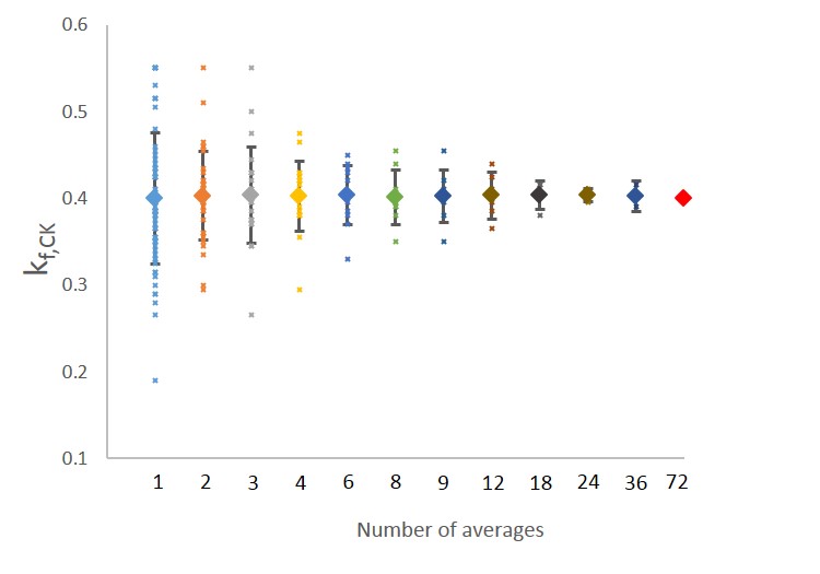
Figure 2 shows the representative fingerprints acquired from mouse hindlimb with 72 and 6 averages. The average kf,CK was 0.43 ± 0.032s-1, which is in good agreement with the literature reported kf,CK (0.40 s-1) for wildtype mice2. The estimated kf,CK using different signal averages is shown in Figure 3. The average value of kf,CK estimated using fewer signal averages showed no significant difference from that using higher number of signal averages. The kf,CK estimated from single-average fingerprint (i.e., 20-s acquisition time) showed a 19% variation. With 6 signal averages, parameter variation was consistently less than 10%, suggesting that kf,CK can be reliably determined within 10% error with 2-min acquisition. This is 25 times faster than the current MT-MRS method on in vivo mice.Discussion and conclusion
In this study, we demonstrated that CK-MRF method allowed efficient quantification of kf,CK on mouse hindlimb in vivo. The significantly improved acquisition efficiency by CK-MRF will enable the investigation of the role of CK in tissue metabolism and the development of metabolic disorder in laboratory animals and genetically manipulated mouse models of human diseases.Acknowledgements
This work was supported by grants from the U.S. National Institute of Health (R01 EB23704).References
1. Liu Y, Gu Y, Yu X. Assessing tissue metabolism by phosphorous-31 magnetic resonance spectroscopy and imaging: a methodology review. Quant Imaging Med Surg. 2017;7(6):707-716. doi:10.21037/qims.2017.11.03.
2. Nabuurs C, Huijbregts B, Wieringa B, Hilbers CW, Heerschap A. 31P saturation transfer spectroscopy predicts differential intracellular macromolecular association of ATP and ADP in skeletal muscle. J Biol Chem. 2010;285(51):39588-39596. doi:10.1074/jbc.M110.164665.
3. Wang CY, Liu Y, Huang S, Griswold MA, Seiberlich N, Yu X. 31P magnetic resonance fingerprinting for rapid quantification of creatine kinase reaction rate in vivo. NMR Biomed. 2017;30(12):1-14. doi:10.1002/nbm.3786.
Figures