2498
Compensation of Spectral Line Broadening in Proton-Echo-Planar-Spectroscopic-Imaging (PEPSI) using Dynamic Expansion of K-Space and Parallel Imaging1Neurology,Physics and Astronomy, University of New Mexico, Albuquerque, NM, United States
Synopsis
This study introduces a novel MRSI approach using dynamically expanding k-space acquisition during spectral encoding to compensate B0 inhomogeneity related signal losses and spectral line broadening, taking advantage of the sparsity of the spectral signal in the time domain. 2D PEPSI with segmented increases in k-space to a maximum 6x6-fold expansion in kx and ky using spectral time domain and ky undersampling was implemented on a clinical scanner. The study characterizes signal gains and resulting spectral line narrowing in regions with B0 inhomogeneity in phantoms and in vivo. This approach complements emerging hardware solutions for improving higher-order shimming.
INTRODUCTION
Spectral line broadening due to inhomogeneity of the static magnetic field (B0) remains remains a major challenge in MR spectroscopic imaging (MRSI), severely limiting applications and hampering clinical acceptance. Extending the capability of the existing field coil design using larger number of higher order shim coils1, dynamic shimming matching the current acquisition slice1-5, and integration of multi-coil B0 shimming into the design of RF array coils6 have improved B0 shimming performance over standard second-order spherical-harmonics shimming. However, these hardware intensive solutions are not widely available and cannot fully compensate magnetic field inhomogeneity in critical regions. Alternatively, decreasing the voxel size in MRSI to reduce spectral line broadening7 is very costly in terms of SNR. We have previously developed Proton-Echo-Planar-Spectroscopic-Imaging (PEPSI) with interleaved shimming using alternating shim gradient blips to simultaneously acquire data from two brain regions, which, however, was limited to regions with relatively uniform local gradient amplitudes and directions8.
In the present study, we generalize interleaved shimming for MRSI by dynamically expanding k-space during spectral encoding, taking advantage of the sparsity of the spectral signal in the time domain, and characterize signal gains and resulting spectral line narrowing in regions with B0 inhomogeneity in phantoms and in vivo.
THEORY
The effect of B0 inhomogeneity in MRSI can be described using the formalism of group spin-echo shift in k-space9. Local gradients Gl shift local k-space with increasing spectral encoding time t: Dk= -g*Gl*t, resulting in spectral line broadening Dn=1/teff where teff = kmax*g*Gl and kmax corresponds to the maximum encoded k-space vector 10. Expanding k-space by increasing readout gradient moments and progressive interleaving of phase encoding gradient blips with increasing gradient moments requires increasing undersampling of the spectral time domain, subject to the constraint imposed by the Nyquist criterion for sampling a spectral width large enough to separate (aliased) spectral peaks. While k-space expansion along the readout direction in PEPSI is accomplished by increasing the readout gradient duration, time efficient expansion along the phase encoding direction(s) requires time-dynamic increases in undersampling of the phase encoding domain(s) and reconstruction using parallel imaging.
METHODS
Implementation:
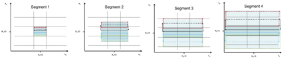
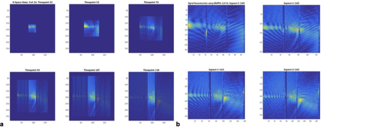
A 2D PEPSI pulse sequence with segmented increases in k-space (Fig.1) to a maximum 6x6 fold expansion in kx and ky was implemented on a Siemens Trio scanner equipped with 32 channel coil. Readout gradient durations at constant ADC readout bandwidth per pixel were increased linearly in 6 equidistant steps. Alternating blipped phase encoding gradients with linearly increasing moments were interleaved into the readout train in readout segments 2-6 to encode a kx line in the central fully encoded ky-space and a 2nd kx line in the outer expanded ky space. The blip gradient moments in all readout segments decreased linearly from one edge of conventionally encoded ky space to the other edge and changed sign in the center. The resulting ky-space data were undersampled 2-5 fold in readout segments 3-6 (Fig.2a).
Data acquisition and reconstruction:
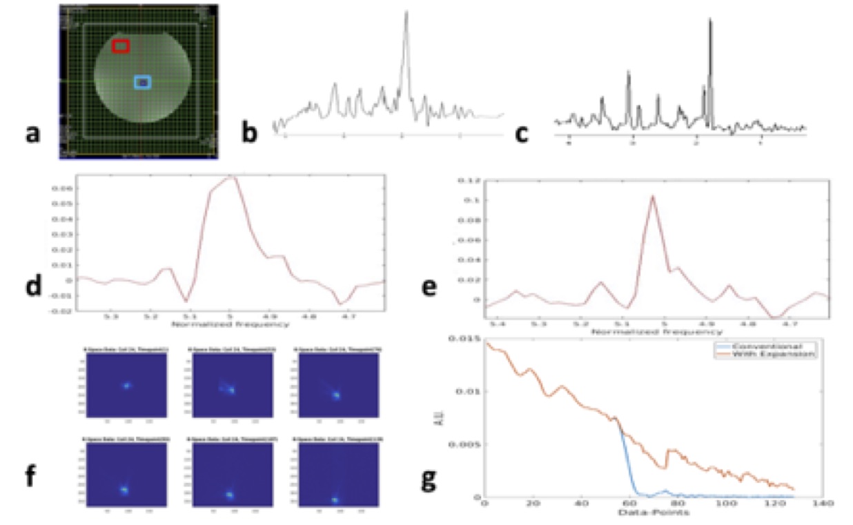
2D PEPSI data were acquired in a spherical phantom with physiological metabolite concentrations and in a healthy adult using TR/TE = 2200/15 ms, 32x32 spatial matrix, 8x8x20 mm3 nominal voxel size, 500 ms readout duration and 1:24 min scan time. Institutionally approved informed consent was obtained. Undersampled data were reconstructed using “OpenGRAPPA”11 (Fig.2b). Spectral reconstruction of nonuniformly sampled data was performed using the expanded Fourier Transform (https://www.mathworks.com/matlabcentral/fileexchange/11020-extended-dft).
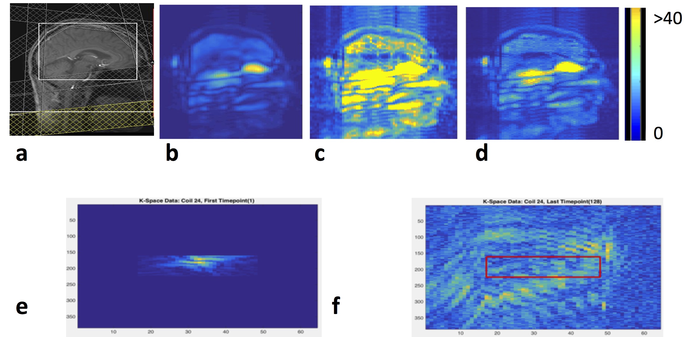
RESULTS
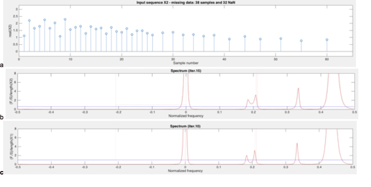
Fig.3 demonstrates the spectral reconstruction of simulated linearly undersampled time domain data with minor increase in spectral line width using the expanded Fourier Transform. Fig.4 shows the application of k-space expansion in a phantom with compensation of signal losses and spectral line broadening due to magnetic field inhomogeneity. Fig.5 shows considerable gains in signal magnitude in the last time slice of data acquired in the human brain.
DISCUSSION
This study demonstrates the recovery of signal lost and mitigation of spectral line broadening due to magnetic field inhomogeneity using dynamically expanding k-space in MRSI and spectral reconstruction of nonlinearly sampled time domain data. Current efforts are directed at characterizing SNR efficiency compared with conventional MRSI and hardware and temporal undersampling constraints associated with k-space expansion. Implementation of increased discretization of k-space expansion to minimize spectral artifacts due to intensity jumps at the edges of readout segments is in progress. Future work will inroduce target design of the expanding k-space trajectory based on magnetic field inhomogeneity mapping and generalization of this approach to 3D k-space expansion.
CONCLUSIONS
This k-space expansion approach when used in combination with volume prelocalization has the potential to complement emerging hardware solutions for improving higher-order shimming.
Acknowledgements
2P20GM103472-06, 1P30GM122734-01References
[1] D. H. Kim, E. Adalsteinsson, G. H. Glover, and D. M. Spielman, "Regularized higher-order in vivo shimming," Magnetic Resonance in Medicine, vol. 48, pp. 715-722, Oct 2002.
[2] A. M. Blamire, D. L. Rothman, and T. Nixon, "Dynamic shim updating: a new approach towards optimized whole brain shimming," Magn Reson Med, vol. 36, pp. 159-165, Jul 1996.
[3] L. M. Klassen and R. S. Menon, "Robust automated shimming technique using arbitrary mapping acquisition parameters (RASTAMAP)," Magnetic Resonance in Medicine, vol. 51, pp. 881-887, May 2004.
[4] E. Schneider and G. Glover, "Rapid Invivo Proton Shimming," Magnetic Resonance in Medicine, vol. 18, pp. 335-347, Apr 1991.
[5] K. M. Koch, L. I. Sacolick, T. W. Nixon, S. McIntyre, D. L. Rothman, and R. A. de Graaf, "Dynamically shimmed multivoxel 1H magnetic resonance spectroscopy and multislice magnetic resonance spectroscopic imaging of the human brain," Magn Reson Med, vol. 57, pp. 587-591, Mar 2007.
[6] J. P. Stockmann, T. Witzel, B. Keil, J. R. Polimeni, A. Mareyam, C. LaPierre, K. Setsompop, and L. L. Wald, "A 32-channel combined RF and B shim array for 3T brain imaging," Magn Reson Med, Feb 17 2015.
[7] A. Ebel and A. A. Maudsley, "Improved spectral quality for 3D MR spectroscopic imaging using a high spatial resolution acquisition strategy," Magn Reson Imaging, vol. 21, pp. 113-120, 2003.
[8] A. Caprihan, Li, T., Posse, S., "Single-Shot Interleaved Gradient Compensation of Susceptibility Induced Spectral Line Broadening in Proton Spectroscopic Echo-Planar Imaging (PEPSI)," in Proc. Int. Soc. Magn. Reson. Med., Seattle, WA, 2006, p. 70.
[9] S. Posse, "Direct imaging of magnetic field gradients by group spin-echo selection," Magn Reson Med, vol. 25, pp. 12-29, May 1992.
[10] S. Posse, R. Otazo, S. R. Dager, and J. Alger, "MR spectroscopic imaging: Principles and recent advances," J Magn Reson Imaging, Nov 27 2012.
[11] M. A. Griswold, P. M. Jakob, R. M. Heidemann, M. Nittka, V. Jellus, J. Wang, B. Kiefer, and A. Haase, "Generalized autocalibrating partially parallel acquisitions (GRAPPA)," Magn Reson Med, vol. 47, pp. 1202-1210, Jun 2002.
[12] K. P. Pruessmann, M. Weiger, P. Bornert, and P. Boesiger, "Advances in sensitivity encoding with arbitrary k-space trajectories," Magnetic Resonance in Medicine, vol. 46, pp. 638-651, Oct 2001.
[13] B. Jiang, X. Jiang, N. Xiao, X. Zhang, L. Jiang, X. A. Mao, and M. Liu, "Gridding and fast Fourier transformation on non-uniformly sparse sampled multidimensional NMR data," J Magn Reson, vol. 204, pp. 165-168, May 2010.
[14] D. Marion, "Fast acquisition of NMR spectra using Fourier transform of non-equispaced data," J Biomol NMR, vol. 32, pp. 141-150, Jun 2005.
Figures