2496
Effects of Point Spread Function and Regularization Information on the MRSI with Compressed Sensing1Department of Computer Science and Engineering, National Sun Yat-sen University, Kaohsiung, Taiwan, 2Department of Computer Science and Information Engineering, National Cheng-Kung University, Tainan, Taiwan, 3MOST AI Biomedical Research Center, Tainan, Taiwan
Synopsis
Compressed Sensing can be very useful in accelerating Phase-encoded Proton MRSI. The sampling functions and the reconstruction settings have been known as critical factors in recovering the data of the accelerated acquisition. The present work compared the choices of sampling functions and the regularization information in the reconstruction in a hope to optimize the framework of Compressed Sensing based MRSI. The results suggest that the spectral quality can be retained for as high as five-fold acceleration with an appropriate undersampling and reconstruction setting.
Introduction
Phase-encoded Proton MRSI has been extensively used to map the distribution of the metabolites but it is also known for taking long scan time. For the past few years, Compressed Sensing has proven its potential in accelerating MRI acquisition and is compatible with MRSI.1-4 The questions about how the settings in Compressed Sensing affect the quality of MRSI on a clinical scan have not yet been fully investigated. The present work compares the undersampling scheme for different acceleration factors and the regularization strategies in a hope to setup an optimized protocol for human MRSI scan with Compressed Sensing.Materials and Methods
A volunteer data was acquired on a 3.0T scanner (GE Discovery MR750, Waukesha, WI) using an 8 channel head coil with TR = 1.5s, TE = 30ms, FOV = 22 cm, Matrix Size = 24x24 and 20mm slice thickness. A pseudo-random sampling function with a centrally weighted variable density distribution 5,6 on the k-space was used to subsample the data at the acceleration of 2, 3, and 5 respectively. In addition to using any random sampling pattern, a series of subsampling masks were generated for the designated acceleration factors. The mask featuring the point spread function (PSF) with highest main peak to side lobe ratio in the image domain of the given acceleration was selected for the simulated undersampling. In the reconstruction, Fourier transformation was firstly performed along the time domain. Subsequently the k-space of each frequency was reconstructed with Compressed Sensing algorithm:
$$\min_{\rho}|s-E\rho|_2+\lambda_1|Ψ\rho|_1+\lambda_2|TV \rho|_1$$
where ρ is the MRSI, s : the k-space signal, E : the 2D Fourier Transformation operator, $$$Ψ$$$ : the sparsity transformation and TV : the total variation operator. The following experiments also compare three different reconstruction settings: (1) $$$Ψ$$$ is a Daubechies Wavelet transformation with λ1=0.005, λ2=0.002; (2) $$$Ψ$$$ is an identity matrix with λ1=0.005, λ2=0.002; (3) $$$Ψ$$$ is an identity matrix with λ1=0.005, λ2=0. The images were reconstructed with non-linear conjugate gradient algorithm. The reconstruction generally converges after 60 iterations. Each coil data was reconstructed separately then combined for the analysis by the LCModel.7
Result
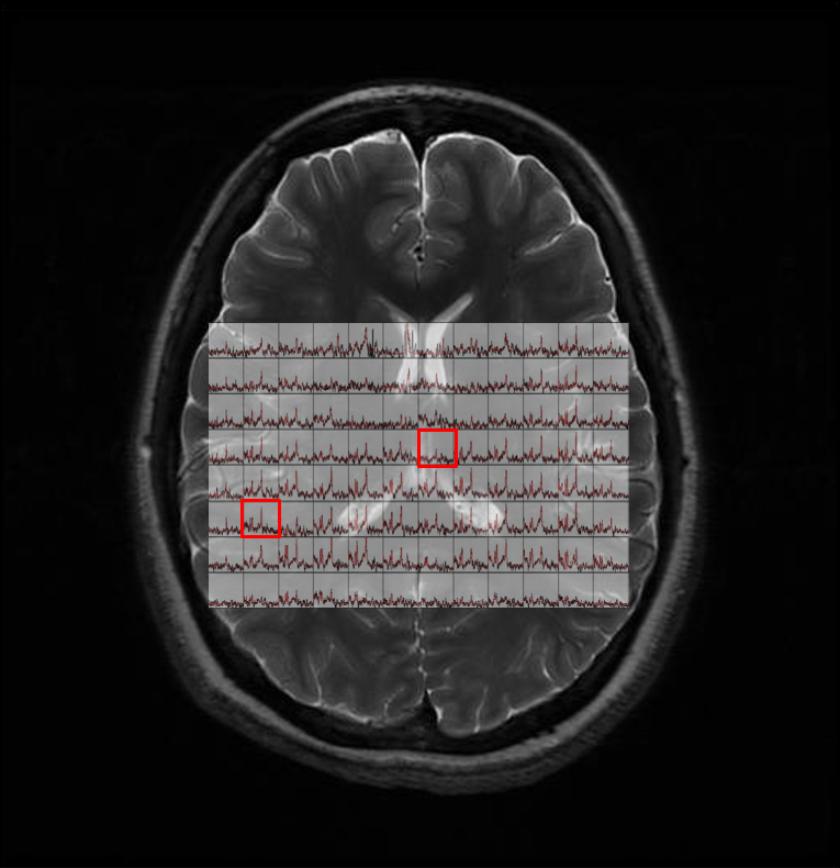
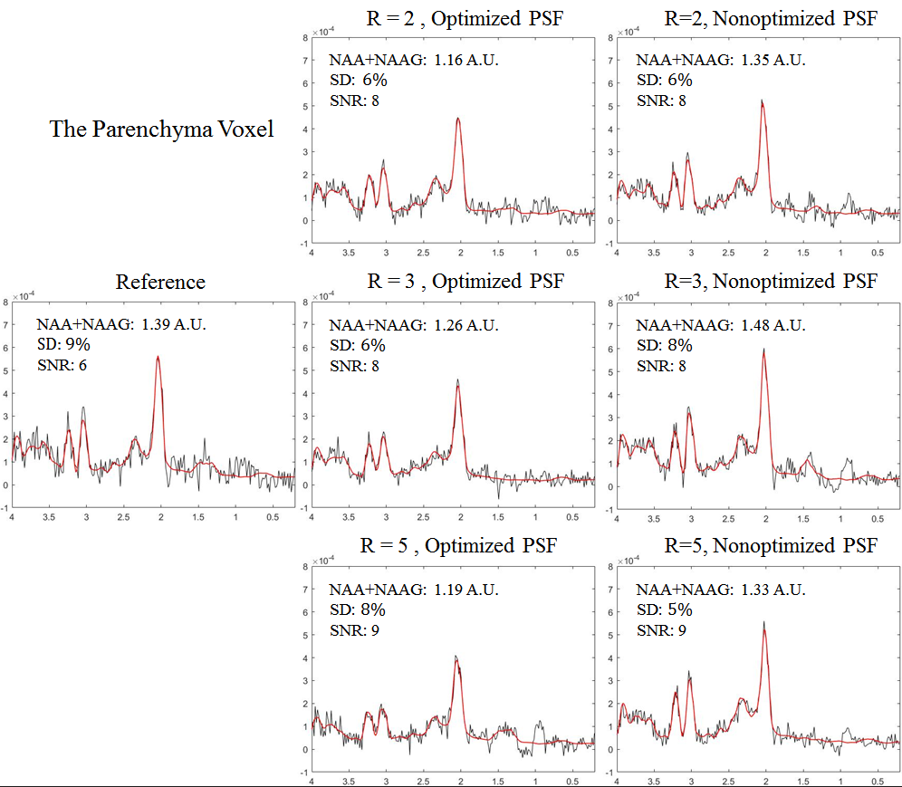
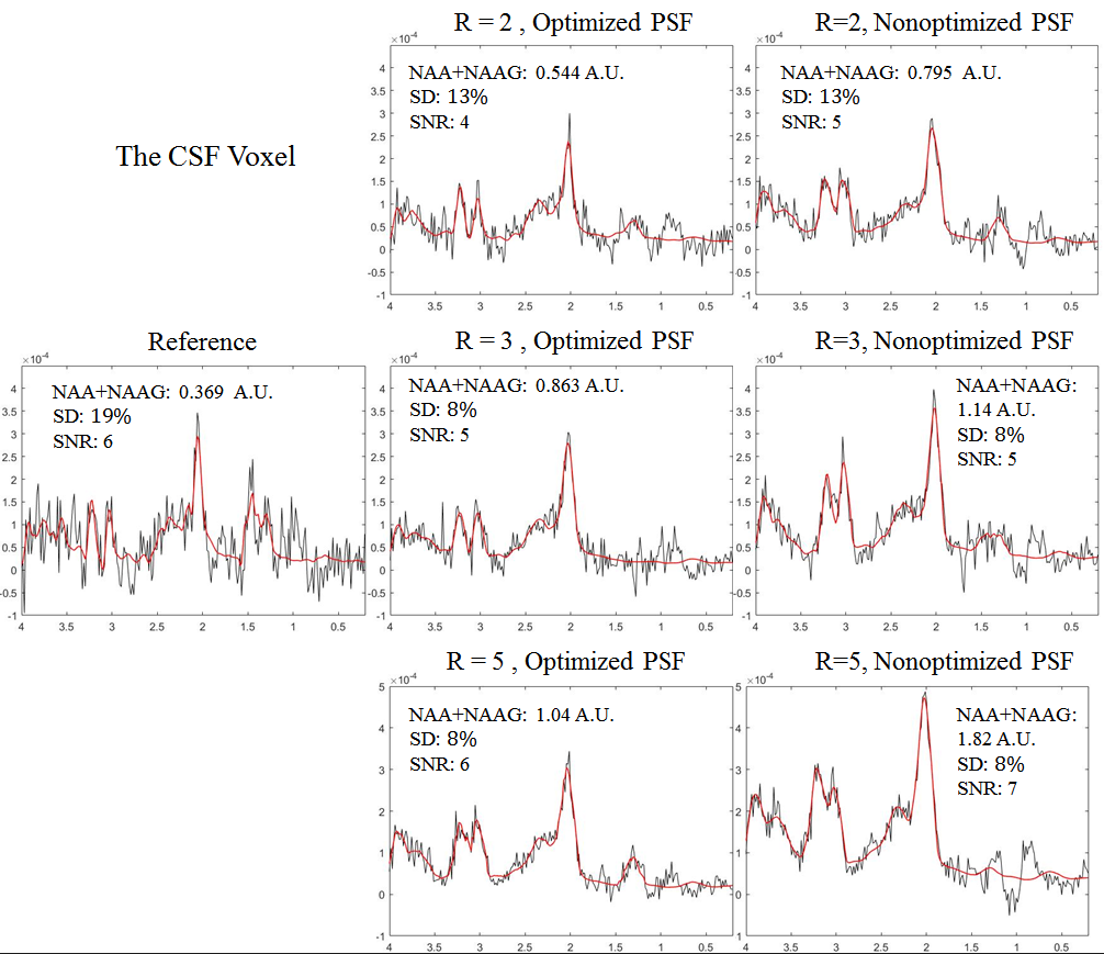
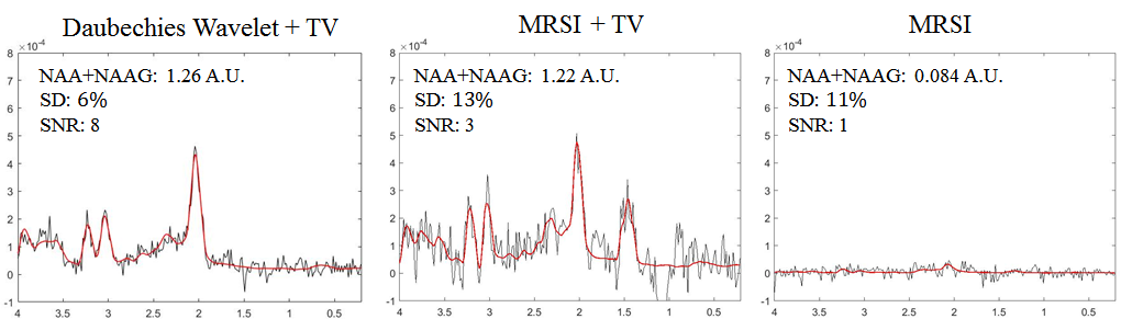
Figure 1 shows the original MRSI overlay on the corresponding T2W image. Two voxels, one at the parenchyma and the other containing cerebral-spinal-fluid (CSF), are selected for comparison. The spectra of three different acceleration settings with and without the optimized PSF are shown in Figure 2 for the parenchyma voxel and in Figure 3 for the CSF voxel respectively. Slightly lower NAA concentration was found in the parenchyma voxel after reconstruction with the optimized PSFs. In the CSF voxel, on the other hand, the aliasing artifacts become prominent if the PSF is not optimized. The components of the metabolites increase significantly as the acceleration increases which could influence the interpretation of the spectral data. A further investigation on the effect of regularization information was performed. Figure 4 shows the spectra reconstructed with different regularization settings for the three-fold acceleration. The removal of the Daubechies Wavelet transformation reduces SNR substantially while the spectrum is heavily distorted if neither of the wavelet transformation or total variation is included.Discussion and Conclusion
The present work has compared the undersampling and the reconstruction schemes used by Compressed Sensing to accelerate in vivo phase-encoded MRSI data. The results suggest that the PSF optimization will be essential in order to prevent from spectrum leakage to the other locations. In addition, the sparse transformation must be chosen properly to maintain the quality of the reconstruction. The results also suggest that the spectral quality can be well preserved at an acceleration factor of five with proper settings.Acknowledgements
Support from MOST grants, 107-2634-F-006-007 and 106-2221-E-110-024, and Mind Research and Imaging Center is acknowledged.References
- Lustig, M., D. Donoho, and J.M. Pauly. Sparse MRI: The application of compressed sensing for rapid MR imaging. Magnetic Resonance in Medicine. 2007;58(6):p.1182-1195.
- Lustig, M., et al. Compressed sensing MRI. IEEE Signal Processing Magazine. 2008;25(2):p.72-82.
- Geethanath, S., et al. Compressive Sensing Could Accelerate 1H MR Metabolic Imaging in the Clinic. Radiology. 2012;262(3):p.985-994.
- Donoho, D.L. Compressed sensing. IEEE Transactions on Information Theory. 2006;52(4):p.1289-1306.
- Chauffert, N., P. Ciuciu, and P. Weiss. Variable density compressed sensing in MRI. Theoretical vs Heuristic sampling strategies, in 2013 IEEE 10th International Symposium on Biomedical Imaging. 2013. p.298-301.
- Vujic, J.L. Monte Carlo Sampling Methods. 2008; Available from: http ://www.nuc.berkeley.edu/All-Courses.
- Stephen Provencher, LCModel&LCMgui User’s Manual. 2018; Available from: http://s-provencher.com/lcm-manual.shtml.
Figures