2494
Ultra high-field, high-resolution semi-LASER MRSI of the brain1University of California, San Francisco, CA, United States, 2University of Maryland, Baltimore, MD, United States, 3GE Healthcare, Munich, Germany
Synopsis
The purpose of this study was to implement and optimize multi-voxel semi-LASER MRSI in brain regions that are frequently used in clinical studies, such as deep gray structures and motor cortex, within a clinically feasible time.
Introduction
MRS data obtained with the use of ultra high-field strength (>=7T) MR scanners have considerable advantages over 3T data, including significantly higher SNR, better spectral resolution and more accurate quantification for both short TE single voxel methods and multi-voxel MRSI. These improved capabilities allow for the detection of separate glutamate (Glu), glutamine (Gln) and glutathione (GSH) resonances and/or obtaining higher spatial resolution spectral data. Semi-LASER localization, which can provide more uniform excitation, has been applied in the multi-voxel MRSI mode to evaluate brain metabolism in the center of the frontal brain at 7T (1, 2). The purpose of this study was to implement and optimize multi-voxel semi-LASER MRSI in brain regions that are frequently used for clinical studies, such as deep gray structures and motor cortex, within a clinically feasible scan time (5-10 minutes).Methods
Five healthy controls had MR scans using a 32-channel receive-only array with a volume transmit head coil (NOVA Medical, Wilmington, MA, United States) on a whole-body 7T GE MR950 scanner (GE Healthcare, Waukesha, WI, United States). Before spectral acquisition, the manufacturer’s higher-order shimming procedure was performed. Multi-voxel MRSI data were obtained using VAPOR water suppression and semi-LASER localization with TR/TE=2500-3000/30 ms, matrix=48x48, FOV=220x220 mm, slice thickness 15 mm, voxel size=5x5x15 mm, and an interleaved flyback applied in the anterior/posterior (A/P) direction. To eliminate lipid signal, we applied very selection suppression outer volume suppression (OVS) (3) and/or a spectrally selective adiabatic inversion recovery pulse (4). The spectral data were combined and processed as described previously (4), and then quantified by LCModel (5) using a simulated basis-set. Metabolite levels were expressed relative to the creatine (Cr) peak.Results
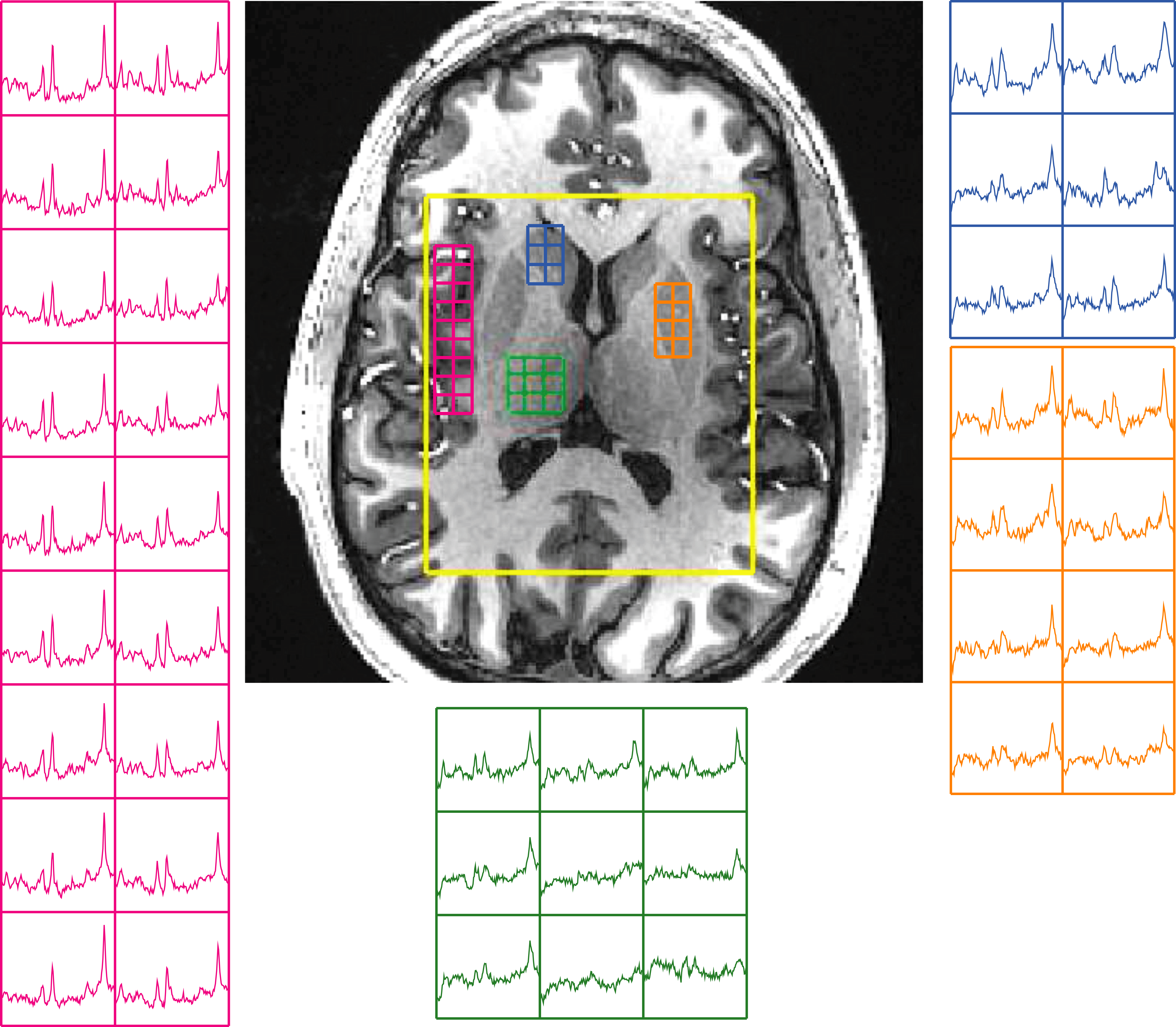
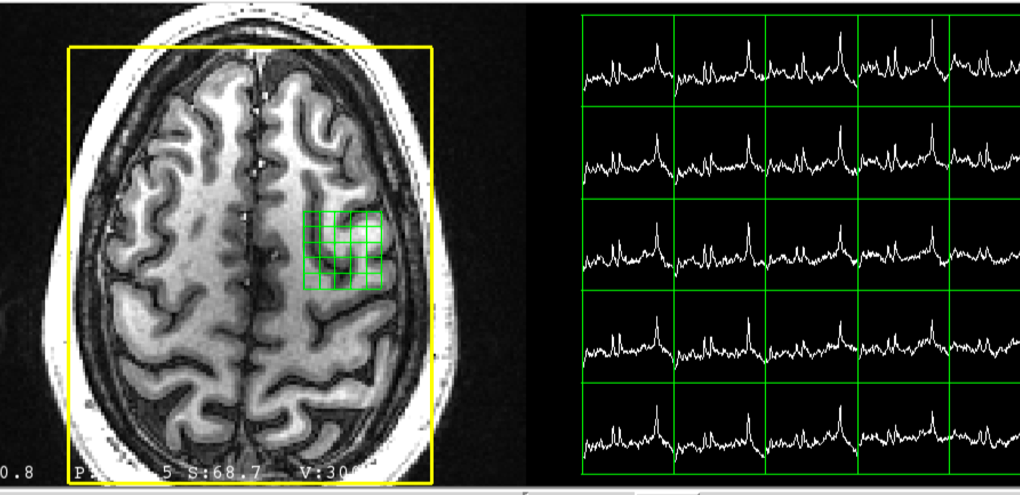
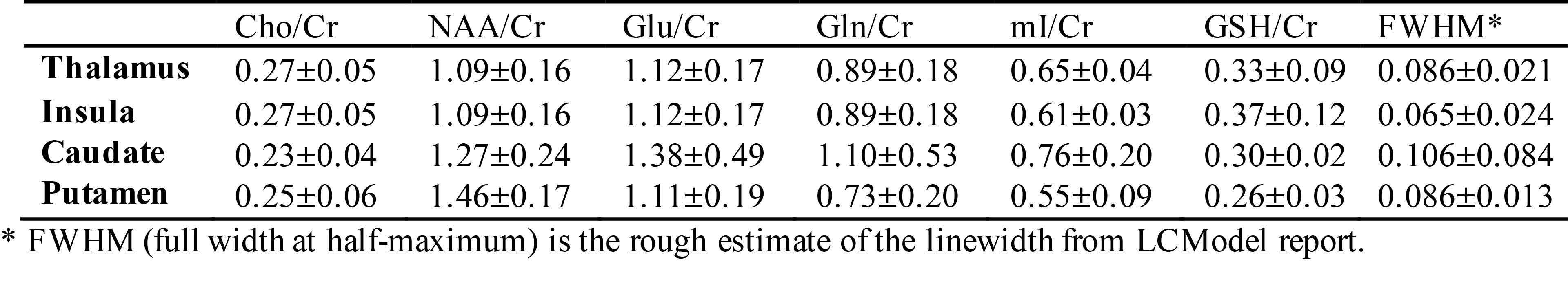
Figure 1 illustrates an example of multi-voxel MRSI data from a healthy volunteer. Table 1 summarizes the ratios of choline-containing compounds/Cr (Cho/Cr), N-acetyl-aspartate/Cr (NAA/Cr), Glu/Cr, Gln/Cr, myo-inositol/Cr and GSH/Cr in the deep brain regions from all the healthy controls. Because of the limited number of voxels with relative CRLB < 20% for GABA within these structures, the ratio of GABA/Cr was not included in the table. The localization approach used in this example excited a rectangular volume of interest, which could have suppressed metabolic signals that were close to the skull. In this case, the application of inversion recovery pulses can be beneficial for measuring brain metabolism in the cortex (see Figure 2).Discussion
Improving the sensitivity of MRSI and increasing the number of metabolites that can be assessed is an important advance for evaluating patients with neurological and psychiatric diseases. This study has demonstrated the application of high-resolution multi-voxel MRSI at 7T to obtain Glu, Gln, mI and GSH at the spatial resolution of 0.5x0.5x1.5cm (0.375 cm3) within a clinical feasible acquisition time. Among the deep brain structures, the caudate was the most difficult to shim and had a larger linewidth, which may require more advanced methods to achieve B0 homogeneity (6, 7). In future, we will evaluate differences in deep brain metabolism related to age and gender, as well as optimize our methods to better assess brain metabolism within the motor cortex.Conclusion
This study demonstrates the feasibility of using a fast and high spatial resolution multi-voxel MRSI acquisition at ultra high-field strength, which is an important advance in the application of MRSI for evaluating patients with psychiatric and neurodegenerative diseases, such as Huntington’s Disease.Acknowledgements
This work was supported by NIH R01 CA127612, NIH R21 HD092660, NIH R01 R01NS099564 and a technology development research grant from GE Healthcare.References
1. Coello E, Noeske R, Burns BL, Gordon JW, Jakary A, Menze B, Haase A, Larson PEZ, Li Y, Schulte RF. High-resolution echo-planar spectroscopic imaging at ultra-high field. NMR Biomed. 2018;31(11):e3950. Epub 2018/07/28. doi: 10.1002/nbm.3950. PubMed PMID: 30052300.
2. Scheenen TW, Heerschap A, Klomp DW. Towards 1H-MRSI of the human brain at 7T with slice-selective adiabatic refocusing pulses. MAGMA. 2008;21(1-2):95-101. Epub 2008/01/23. doi: 10.1007/s10334-007-0094-y. PubMed PMID: 18210177; PMCID: PMC2798032.
3. Tran TK, Vigneron DB, Sailasuta N, Tropp J, Le Roux P, Kurhanewicz J, Nelson S, Hurd R. Very selective suppression pulses for clinical MRSI studies of brain and prostate cancer. Magn Reson Med. 2000;43(1):23-33. Epub 2000/01/22. PubMed PMID: 10642728.
4. Li Y, Larson P, Chen AP, Lupo JM, Ozhinsky E, Kelley D, Chang SM, Nelson SJ. Short-echo three-dimensional H-1 MR spectroscopic imaging of patients with glioma at 7 Tesla for characterization of differences in metabolite levels. J Magn Reson Imaging. 2015;41(5):1332-41. Epub 2014/06/18. doi: 10.1002/jmri.24672. PubMed PMID: 24935758; PMCID: PMC4269580.
5. Provencher SW. Estimation of metabolite concentrations from localized in vivo proton NMR spectra. Magn Reson Med. 1993;30(6):672-9. Epub 1993/12/01. PubMed PMID: 8139448.
6. Pan JW, Lo KM, Hetherington HP. Role of very high order and degree B0 shimming for spectroscopic imaging of the human brain at 7 tesla. Magn Reson Med. 2012;68(4):1007-17. Epub 2012/01/04. doi: 10.1002/mrm.24122. PubMed PMID: 22213108; PMCID: PMC3323711.
7. Shen J, Rycyna RE, Rothman DL. Improvements on an in vivo automatic shimming method [FASTERMAP]. Magn Reson Med. 1997;38(5):834-9. Epub 1997/11/14. PubMed PMID: 9358459.
Figures