2492
Retrospective motion compensation for edited MRSI data1Department of Biomedical Engineering, The Johns Hopkins University School, Baltimore, MD, United States, 2Russell H. Morgan Department of Radiology and Radiological Science, The Johns Hopkins University School of Medicine, Baltimore, MD, United States, 3F. M. Kirby Research Center for Functional Brain Imaging, Kennedy Krieger Institute, Baltimore, MD, United States
Synopsis
A retrospective motion compensation method for edited MR spectroscopic imaging (MRSI) data of the human brain is presented. The algorithm identifies movement-affected data by comparing the residual water and lipid peaks at the same k-space point, and either phase corrects, replaces or removes motion-affected FIDs. The method was applied to in vivo GABA-edited MRSI data: relative to uncorrected spectra, the corrected spectra had significantly less subtraction artifacts. The method was also demonstrated for correction of glutathione-edited MRSI data.
Purpose
J-difference editing requires that spectra recorded with the editing pulses ‘on’ and ‘off’ have the same phase and frequency, otherwise subtraction artifacts will occur. Phase and frequency variation can occur due to scanner instability (field drift etc.) as well as due to subject head motion during the pulse sequence. Shot-by-shot retrospective phase and frequency correction (and discarding of severely corrupted spectra) has been shown to significantly reduce subtraction artifacts and improve spectral quality in single voxel acquisitions (1-3). To our knowledge, however, these methods have not been applied to edited MRSI data, since the presence of phase-encoding gradients confounds most common correction schemes. This abstract presents a retrospective compensation method for edited MRSI data based on comparing residual water and lipid peaks in ‘on’ and ‘off’ spectra at each point in k-space.Methods
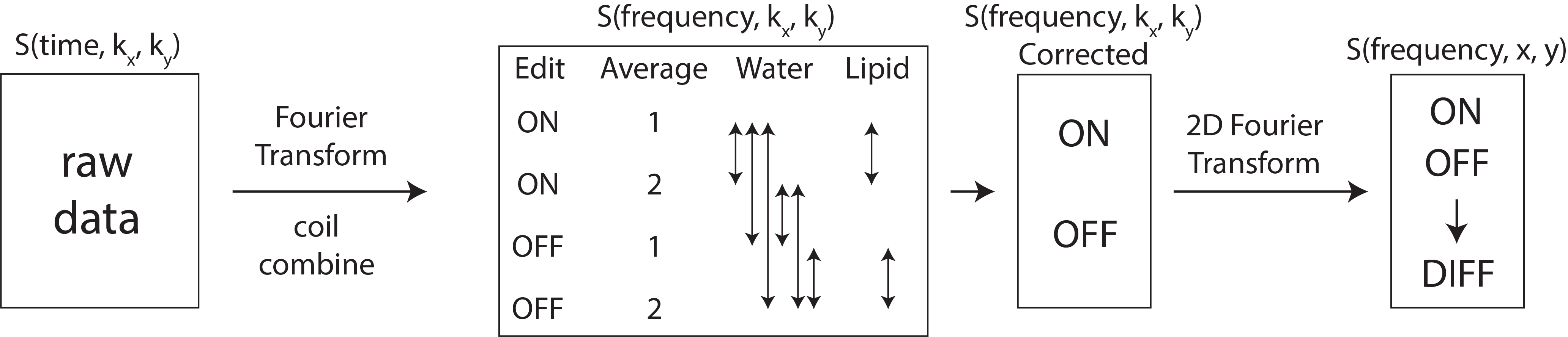
Figure 1 shows the data processing pipeline including motion correction in k-space. The algorithm identifies movement-affected data points in k-space by calculating correlation coefficients (CCs) for residual water and lipid signals on each point on k-space. For GABA-editing, it is expected that residual water will be the same in both ‘on’ and ‘off’ spectra; lipid spectra will be different since the ‘on’ editing pulse also affects the lipid signal. However, if more than one average is collected for each point in k-space, then lipid signals can be compared for each shot. Motion-affected FIDs with low CCs are preferentially phased to match the other FIDs at the same k-space point by optimizing the applied phase until CC > 0.9. If the phasing threshold is not met, the FID is replaced with another of the same sub-type; if CC < 0.6 across all FIDS, that point in k-space point is replaced with zeroes. This correction is repeated for each point in k-space.
Experiments were performed on a Philips Achieva 3T scanner with a 32-channel head coil. Edited spin-echo MRSI data (4) were acquired in 7 healthy adults (5 male, age: 30 ± 8 years) in a 20-mm slice at the level of the lateral ventricles with a field-of-view of 180 x 210 mm2, HGDB for dual-band water and lipid suppression (5), and 8 saturation bands. Scan parameters were TR 2s, TE 80 ms, four FIDs (2 ‘on’, 2 ‘off’) at each phase-encoding step, a nominal voxel size of 4.5 cm3. 20 ms sinc-Gaussian editing pulses were applied at 1.9 ppm (‘on’) and 7.5 ppm (‘off’). GSH-edited data were acquired in 1 healthy adult (male, age 26) with the same parameters as the GABA-edited acquisitions except with the ON-editing pulse applied at 4.56 ppm. Edited spectra with and without motion compensation were quantified by fitting peaks at 3.2 ppm (residual Cho) and 3.0 ppm (either GABA, or residual Cr).
Results
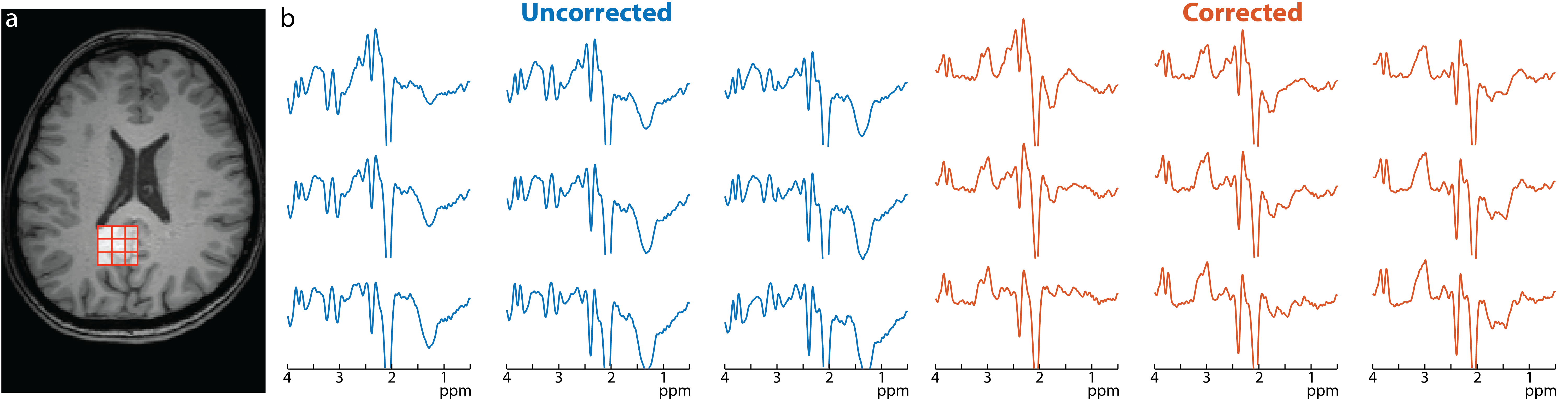
A motion-corrupted GABA-edited dataset is shown in figure 2. Large subtraction artifacts manifest in this example as negative Cho and Cr signals (at 3.2 and 3.0 ppm) and residual lipids at 1.3 ppm, but after compensation these are substantially reduced allowing a GABA peak to be observed at 3 ppm. Over all data sets the residual Cho signal was reduced by about a factor of 3 using motion compensation with a median (interquartile range) of 0.065 (0.062) for corrected spectra versus 0.2 (0.3) for uncorrected spectra.
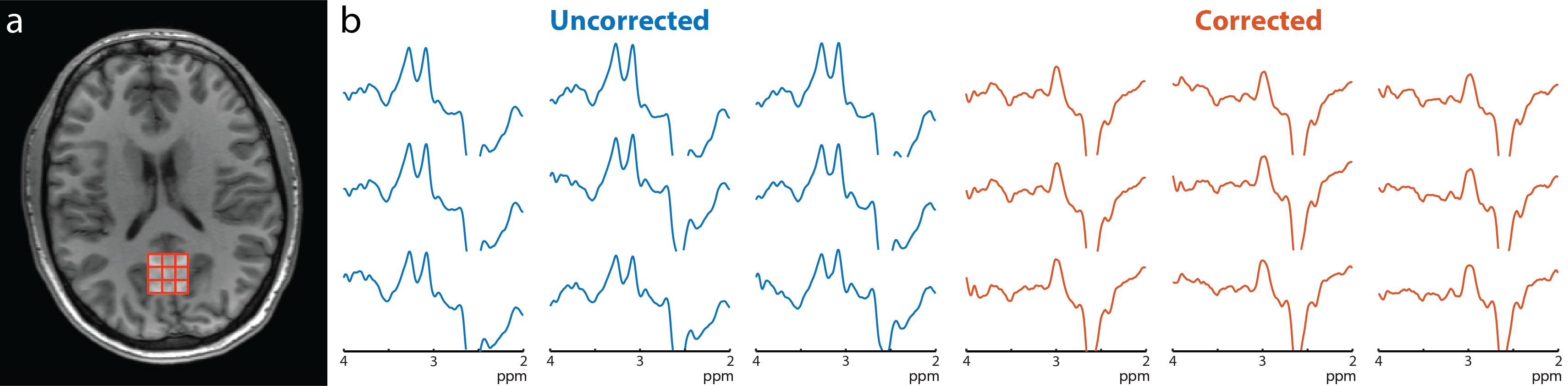
For the GSH-edited MRSI dataset, again substantial Cho and Cr subtraction artifacts are apparent in the uncorrected data, but are largely removed with the motion compensation scheme, allowing a GSH peak to be observed (Figure 3).
Discussion
A retrospective motion compensation approach for edited MRSI scans is presented. J-difference edited MRSI is very sensitive to head motion, since subtraction artifacts on just a few points in k-space may propagate throughout the whole spectroscopic image after spatial Fourier transformation. The scheme relies on similarity between residual water (and lipid) signals from multiple FIDs recorded at each point in k-space, and is relatively simple to implement. Unlike prospective motion correction schemes, the method does not require any additional hardware or acquisition software (6, 7) which are currently not widely available on commercial MR scanners. The method should also be applicable to conventional MRSI if more than one FID per k-space point is acquired.
It should be noted that the method will only compensate for intermittent motion or other instability, and will fail if there is sustained and/or large head motion. In addition, residual water and lipid signals need to be large enough to be estimated on individual points in k-space; the method therefore works best with moderate water and lipid suppression factors.
Acknowledgements
This work was supported by P41 EB015909.
References
1. Evans CJ et al. J. Magn. Reson. Imaging. 2013;975:970–975.
2. Mullins PG et al. Neuroimage 2014;86:43–52.
3. Waddell KW et al. 2007;25:1032–1038.
4. Zhu H et al. Magn. Reson. Med. 2011;65:603–609.
5. Zhu H et al. Magn. Reson. Med. 2010;63:1486–1492.
6. Bogner W et al. Neuroimage 2014;103:290–302.
7. Bogner W et al. Neuroimage 2014;88:22–31.
Figures