2486
Accelerated J-Resolved 1H-MRSI with Limited and Sparse Sampling of (k, tJ)-Space1School of Biomedical Engineering, Shanghai Jiao Tong University, Shanghai, China, 2Department of Electrical and Computer Engineering, University of Illinois at Urbana Champaign, Urbana, IL, United States, 3Beckman Institute for Advanced Science and Technology, Urbana, IL, United States, 4Gordon Center for Medical Imaging, Department of Radiology, Massachusetts General Hospital, Harvard Medical School, Boston, MA, United States
Synopsis
J-resolved 1H-MRSI is a powerful tool for mapping brain molecules, especially those with large spectral overlaps (e.g., glutamate, glutamine
Introduction
Many challenges in conventional 1H-MRSI stem from the fact that most of the detectable resonances lie in a small chemical shift range and the spectra of several key molecules (e.g., glutamate (Glu), glutamine (Gln) and $$$\gamma$$$-aminobutyric acid (GABA)) have significant overlaps with other brain metabolites (e.g., NAA, Cr, and Cho). J-resolved 1H-MRSI addresses this issue by lifting the 1D NMR spectrum onto a two-dimensional space but is not practical due to the increased data acquisition time required to cover the high-dimensional (k, tJ)-space ($$$t_J$$$ is TE time for J-coupling encoding). To address this problem, acceleration methods of J-resolved 1H MRSI1-3 have been proposed. These methods were able to produce high-quality 2D spectra with 64, 20, and 6 J-encodings respectively. Building on these advances, this work further accelerates J-resolved 1H-MRSI with a limited and sparse sampling of (k, tJ)-space (3 J-encodings). The proposed sampling pattern is chosen based on a physics-based spectral model and Cramer-Rao lower bound (CRLB) analysis. A model-based processing scheme is also proposed, which performs spectral quantification directly from the (k, tJ)-space data. Simulation and in vivo experiments have been performed to evaluate the proposed method, which have produced very encouraging results.Method
Data acquisition
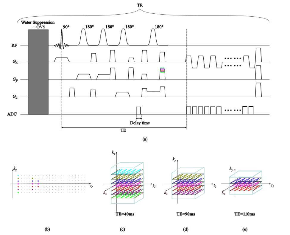
In this work, data were acquired using semi-LASER localization, which is robust to $$$B_1$$$ inhomogeneity, combined with echo-planar spectroscopic imaging readouts (Fig.1 a). The water signal is only weakly suppressed, enabling the tracking and correction of field inhomogeneity, field variations, and eddy current effects. Data acquisition was further accelerated by limited and sparse sampling in (k, tJ)-space (Fig.1 b-e). The $$$t_J$$$ dimension was highly undersampled, where the optimal selection of $$$t_J$$$ was found by CRLB analysis. To this end, we calculated the CRLB of measurement GABA concentration with all possible $$$t_J$$$ combinations (from 40 – 230 ms) and chose the one that lead to the minimal CRLB. In this work, we found the optimal $$$t_J$$$ combination ($$$t_J$$$ = 40, 90, and 110 ms). The scan FOV was 180x180 mm2 with a slice thickness of 10 mm and excitation volume of 90x90x10 mm3, leading to the in-plane resolution of 2.3x1.6 mm2. Other parameters were: TR=1250 ms, total acquisition time = 3.43 mins.
Data processing
For the data acquired at the very first $$$t_J$$$ value with high-resolution and high-SNR, nuisance signals can be effectively removed using a union-of-subspaces method4. For later $$$t_J$$$ values, the limited k-space coverage and poor SNR makes nuisance removal rather challenging. We address this challenge by using generalized series model5 to exploit the correlation between nuisance signals from different $$$t_J$$$ acquisitions to remove nuisance signal in later $$$t_J$$$'s. After nuisance signals removal, a physics-based spectral model is used to reconstruct the spatiospectral distribution of metabolites. The image function can be written as a partially separable (PS) function6 to perform spectral quantification directly from the (k, tJ)-space data:
$$\rho(x,t_J,t_2 )=\sum_{l=1}^{L}c_l(x)v_l(t_J,t_2),$$
where $$$t_J$$$ is the $$$TE$$$ time, $$$t_2$$$ is the chemical shift time, $$$\{c_l(x)\}$$$ are spatial coefficients, and $$$\{v_l(t_J,t_2)\}_{l=1}^{L}$$$ are the basis functions determined by quantum mechanical simulations and training data. The spatial coefficients are estimated from the solution to the following regularized least-squares problem
$$\hat{C}=\arg\min_{C}\| M\mathcal{F}B_0CV-d\|_2^2+\lambda\| WD(CV)\|_2^2,$$
where $$$M$$$, $$$\mathcal{F}$$$, and $$$B_0$$$ are the sampling, Fourier encoding, and the field inhomogeneity operators, $$$V$$$ is a row matrix of the basis functions, $$$d$$$ is the vector of water removed k-space data, $$$W$$$ is the edge weight matrix, $$$D$$$ is the gradient operator, and $$$\lambda$$$ is the regularization parameter. The $$$B_0$$$ map and edge weights in $$$W$$$ are predetermined using the data from the first $$$t_J$$$ encoding.
Results and Discussion
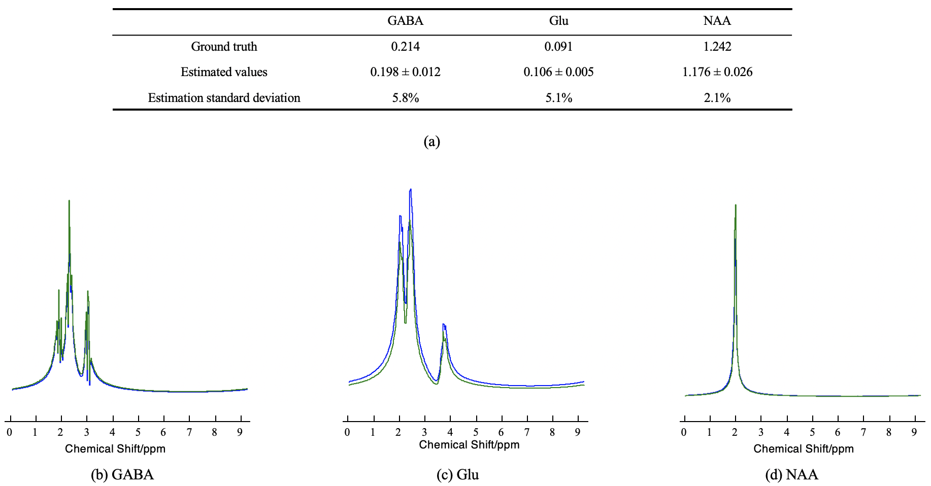
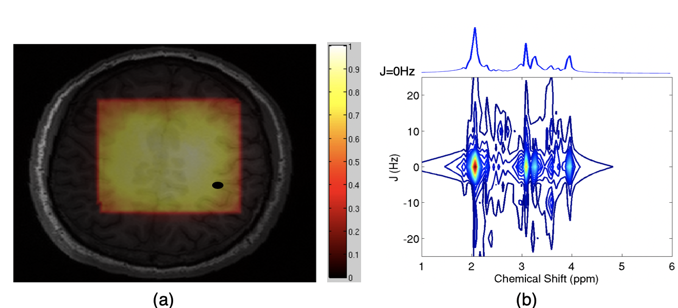
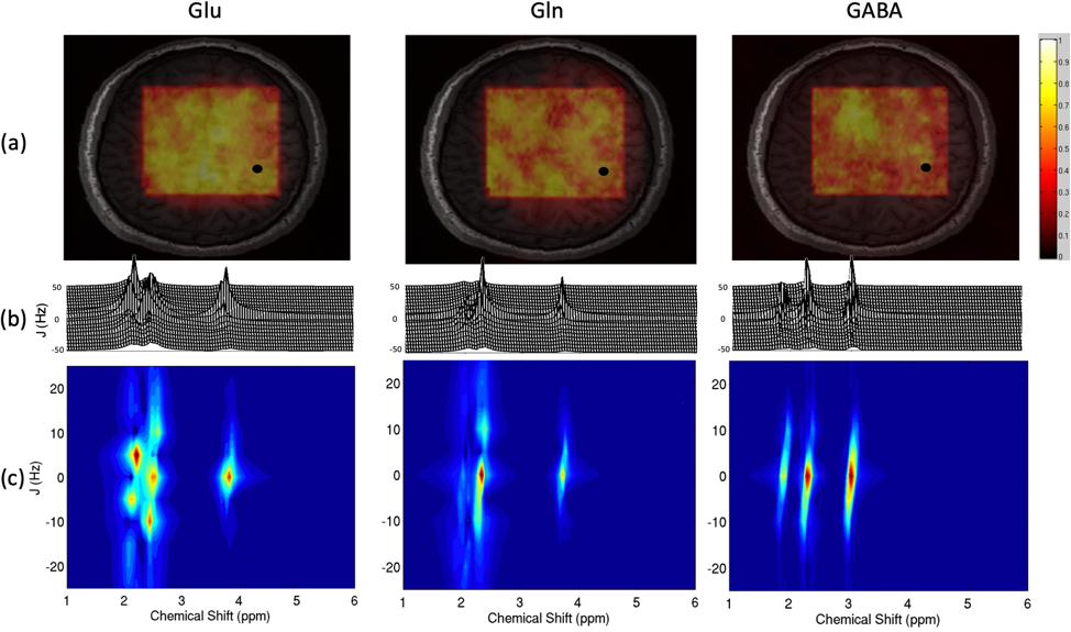
Simulation results (Fig. 2) show that the proposed reconstruction and quantification method can produce accurate reconstruction from sparse and limited data. In vivo data were acquired from a healthy volunteer (with IRB approval) on a 3 T Siemens Prisma scanner using a commercial 20-channel head and neck coil, with the parameters described in the data acquisition scheme. We obtained very encouraging in vivo experimental results (Fig.3., Fig.4.) with the proposed method using only 3 J-encodings (3.43 mins). This highly sparse acquisition method is enabled by the subspace model7. Since we have high-quality physics-based subspace structure, only a few spectral samplings in $$$t_J$$$ and $$$t_2$$$ are needed.Conclusion
A new method is proposed to accelerate J-resolved 1H-MRSI with limited and sparse sampling of (k, tJ)-space, which is enabled by subspace modeling and prior spectral knowledge derived from quantum mechanics simulation. Experimental results show high-quality 2D spectra can be reconstructed using as few as 3 J-encodings (3.43 mins). Our method may enable J-resolved 1H-MRSI for simultaneous mapping of brain neurotransmitters such as glutamate and GABA in clinical applications.Acknowledgements
This work is supported in parts by NIH-R21-EB021013, NIH-R21 EB023413, NIH-R01-EB023704 and NIH-P41-EB022544, MOST 2017YFC0109002.References
1. Wilson NE, Iqbal, Z., Burns B.L, et al., et al. Accelerated five-dimensional echo planar J-resolved spectroscopic imaging: Implementation and pilot validation in human brain. Magn. Reson. Med., 2016; 75(1):42-51.
2. Ma C, Lam F, and Liu Q, et al. Accelerated high-resolution multidimensional 1H_MRSI using low-rank tensors. Proc. Intl. Soc. Mag. Reson. Med., 2016.
3. Lam F, Cheng B, and Christodoulou, A. G. et al., Accelerated J-resolved MRSI using joint subspace and sparsity constraints. Proc. Intl. Soc. Mag. Reson. Med., 2017.
4. Ma C, Lam F, Johnson C. L, et al., Removal of nuisance signals from limited and sparse 1H MRSI data using a union‐of‐subspaces model. Magn. Reson. Med., 2015.
5. Liang ZP, Lauterbur PC. A generalized series approach to MR spectroscopic imaging. IEEE Trans on Med Imaging. 1991 Jun;10(2):132-7.
6. Liang ZP. Spatiotemporal imaging with partially separable functions. In Proc. IEEE Int Symp Biomed Imaging, USA, 2007; 988-991
7. Lam, Fan, and Liang ZP. A subspace approach to high‐resolution spectroscopic imaging. Magn. Reson. Med., 2014; 71:1349-1357.
Figures