2480
Parallel Imaging for Concentric Circle Readouts with GRAPPA Reconstruction for Full-Brain 3D-FID-MRSI at 7T1Department of Biomedical Imaging and Image-guided Therapy, High Field MR Centre, Medical University of Vienna, Vienna, Austria, 2Department of Radiology, Martinos Center for Biomedical Imaging, Massachusetts General Hospital, Harvard Medical School, Boston, MA, United States, 3Christian Doppler Laboratory for Clinical Molecular MR Imaging, Vienna, Austria
Synopsis
Non-Cartesian sampling methods for MRSI such as concentric ring trajectories (CRT) are highly suitable at field strenghts ≥7T while being SNR-efficient due to its self-rewinding property and low-pass k-space-weighting. However slewrate constraints enforce the CRT to sample the k-space periphery with 2-fold or 3-fold the time of the inner circles via more temporal interleaves (TI). The combination of variable density parallel imaging (PI) and CRT for MRSI allows high acceleration factors since the undesired but necessary variable TIs can be easily undersampled and allowing therefore higher accelerations in the k-Space periphery.
Introduction
Proton-MR-spectroscopic imaging (MRSI) is a non-invasive tool for imaging the concentrations of neurochemical compounds simultaneously, however low SNR-sensitivity and long acquisition times hinder widespread clinical applications[1]. Non-Cartesian sampling methods such as concentric ring trajectories (CRT)[2] are highly suitable at B$$$_0$$$≥7T while being SNR-efficient due to its self-rewinding property and low-pass k-space-weighting. Although CRT allows the acquisition of higher matrices compared to other trajectories, slewrate constraints enforce the CRT to sample the k-space periphery with 2-fold (matrizes>10×10) or 3-fold (matrizes>44×44) the time of the inner circles via more temporal interleaves (TI). The combination of CRT and parallel imaging (PI)[3] is allowing attractive sampling patterns since these time demanding and low-SNR outer k-Space circles can be easily undersampled and suppress the TA even further which could be of interest for inherently high SNR acquisition methods such as hyperpolarized MRSI. The purpose of this proof-of-concept study is therefore to develop a 3D-MRSI sequence with rapid parallel imaging CRT(PI-CRT) encoding for 7T and compare different variable density sampling patterns in phantom and simulate them invivo.Methods
The invivo scan was performed at 7T (Magnetom,Siemens Healthcare,Erlangen,Germany,Gmax=40mT/m,Smax=200mT/m/ms) with a 32-channel receive head coil. A previously described single-slice phase-encoded FID-MRSI sequence was extended by slab-selection and ellipsoidal encoding of a 3D-k-space. This was achieved via in-plane 2D-CRT readout using sine and cosine modulated gradients combined with through-plane phase encoding to minimize scan time requirements (Fig.1,top). The scan time was further shortened by employing variable temporal interleaves (TI) with only one TI for rings sampled in the center of the k$$$_x$$$/k$$$_y$$$-plane and up to two TIs in the periphery, thus doubling the sampling time there. Further scan parameters: Acquisition delay 1.3ms, TR 280ms, spectral bandwidth 2778Hz, acquisition time without PI 2:59min, matrix size 32×32×31, voxel size 6.9×6.9×4.2mm³. The interleaved MOSAIC[4] prescan (1 circle/TR) served for coil combination and as an ACS for the PI reconstruction. Phantom PI simulations were performed on a 80×80 oil phantom (40 circles) via zerofilling of each 2nd circle (Fig.1,bottom left) with additionally incorporating variable density sampling patterns such that the inner 2,4,6,...,34 circles out of 40 total for the central partition are fully sampled. Based on the phantom measurement the 3D-invivo variable density pattern was determined according to the difference-images to the ground-truth. For the 3D-invivo simulations we chose therefore that the inner 4 circles out of 16 (if the elliptical encoding allows it) are measured fully which corresponds to an acceleration of R=2 due to variable TIs (inner 5 circles had one TI, the other 11 two TIs) . Heidemann-Griswold-GRAPPA[5] reconstruction transformed the polar k-space to a Cartesian Circles×Samples grid (e.g. 16×550 for the central partition, Fig.1,bottom right) where conventional GRAPPA reconstructions was employed (Kernel 3×10). The underlying assumptions of a constant density k-space, thus equally weighted k-space samples, was however neglected.Results
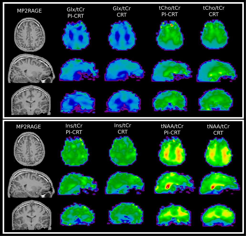
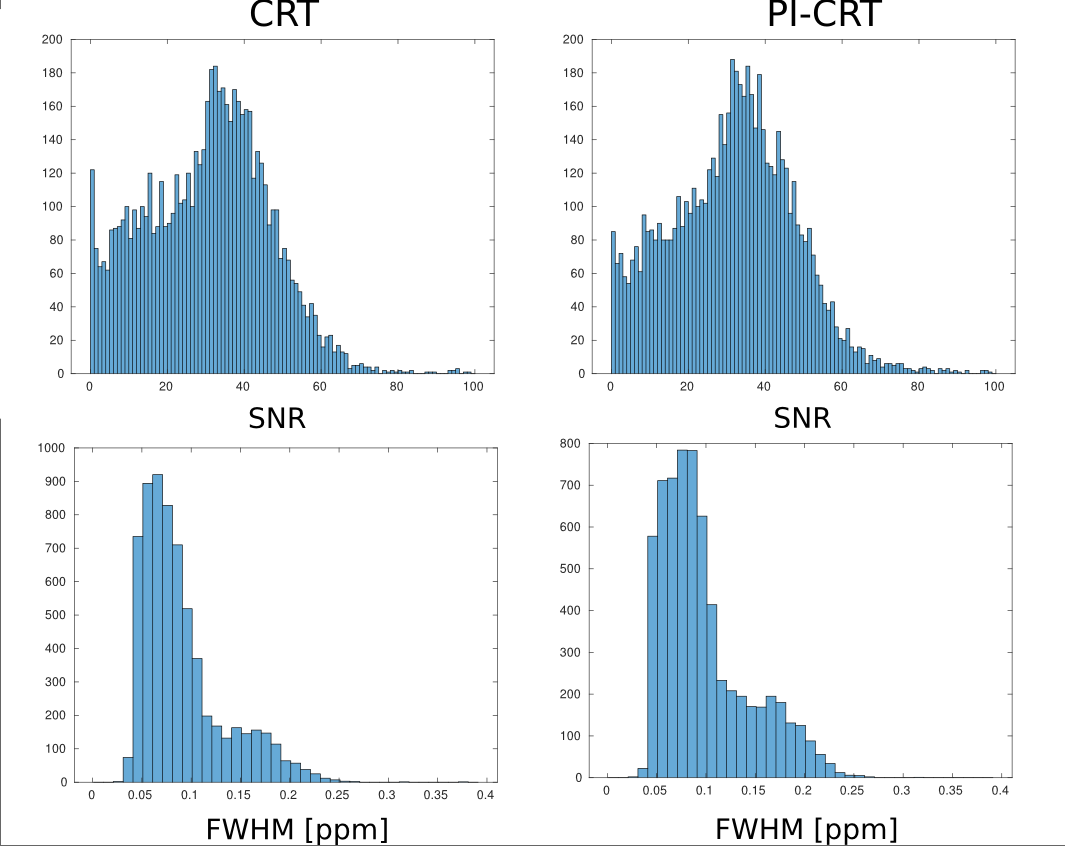
Fig.2 shows phantom results of a single-coil element out of 32 coils total. Three distinct sampling approaches are shown i) every 2nd circle undersampled ii) the inner 10 (1/40th of circle number) fully sampled, rest two-fold undersampled iii) fully sampled k-space. While the GRAPPA reconstruction fails for the 1st case, the variable density pattern reveals good spectral and localization quality. Other variable density patterns together with difference images are shown in Fig.3, with the three cases of Fig.2 highlighted in red for the zerofilled and the GRAPPA reconstruction. Invivo results of the 32×32×31 matrix (measured TA: 2:59min,simulated TA: 1:30min) are displayed in Fig.4 showing Glx/tCr; tCho/tCr, Ins/tCr and tNAA/tCr ratio maps for PI-CRT and fully sampled CRT. Histograms in Fig.5 report that there was no major SNR-loss inflicted by the PI undersampling, however PI-CRT had worse spectral linewidths than CRT.Discussion and Conclusion
The combination of variable density PI and CRT for MRSI allows acceleration factors of R=2 (without major SNR-losses), thus making this method more efficient than conventional PI since the undesired but necessary variable TIs can be easily undersampled and allowing therefore higher accelerations in the k-Space periphery. The fact that the kSpace center is sampled with one TI, thus enabling fast variable density patterns, can be attributed to the CRT encoding and stands out compared to other non-Cartesian methods. Compared to spherical phase encoding (TR 280ms) PI-CRT results in an acceleration factor of ×35. The usage of the interleaved MOSAIC scan for the ACS and coil combination may result in less movement artifacts compared to different methods but further investigation will be necessary as well as additional adjustments for improving the quality of the metabolic maps. We think that this method could be of high interest in cases where the SNR is not a limiting factor, e.g. hyperpolarized MRSI, or in cases where speed/low resolutions are highly desired, e.g. dynamic MRSI.Acknowledgements
Research support by the Austrian Science Fund (FWF): KLI 646, KLI 718 and P 30701.References
[1] Oz, G., Alger, J. R., Barker, P. B., et al., MRS Consensus Group (2014). Clinical proton MR spectroscopy in central nervous system disorders. Radiology, 270(3), 658-79.
[2] Hingerl, L. , Bogner, W. , Moser, P. , Považan, M. , Hangel, G. , Heckova, E. , Gruber, S. , Trattnig, S. and Strasser, B. (2018), Density‐weighted concentric circle trajectories for high resolution brain magnetic resonance spectroscopic imaging at 7T. Magn. Reson. Med., 79: 2874-2885. doi:10.1002/mrm.26987
[3] Strasser, B. , Považan, M. , Hangel, G. , Hingerl, L. , Chmelik, M. , Gruber, S. , Trattnig, S. and Bogner, W. (2017), (2 + 1)D‐CAIPIRINHA accelerated MR spectroscopic imaging of the brain at 7T. Magn. Reson. Med., 78: 429-440. doi:10.1002/mrm.26386
[4] Moser, P. , Strasser, B. , Hingerl, L. , Považan, M. , Hangel, G. , Heckova, E. , Gruber S. , Trattnig, S. and Bogner, W. (2018) MOSAIC - a generalized multi-channel coil combination for 1H-MRSI via interleaved calibration scans , ISMRM 2018
[5] Heidemann, R. M., Griswold, M. A., Seiberlich, N. , Krüger, G. , Kannengiesser, S. A., Kiefer, B. , Wiggins, G. , Wald, L. L. and Jakob, P. M. (2006), Direct parallel image reconstructions for spiral trajectories using GRAPPA. Magn. Reson. Med., 56: 317-326. doi:10.1002/mrm.20951
Figures
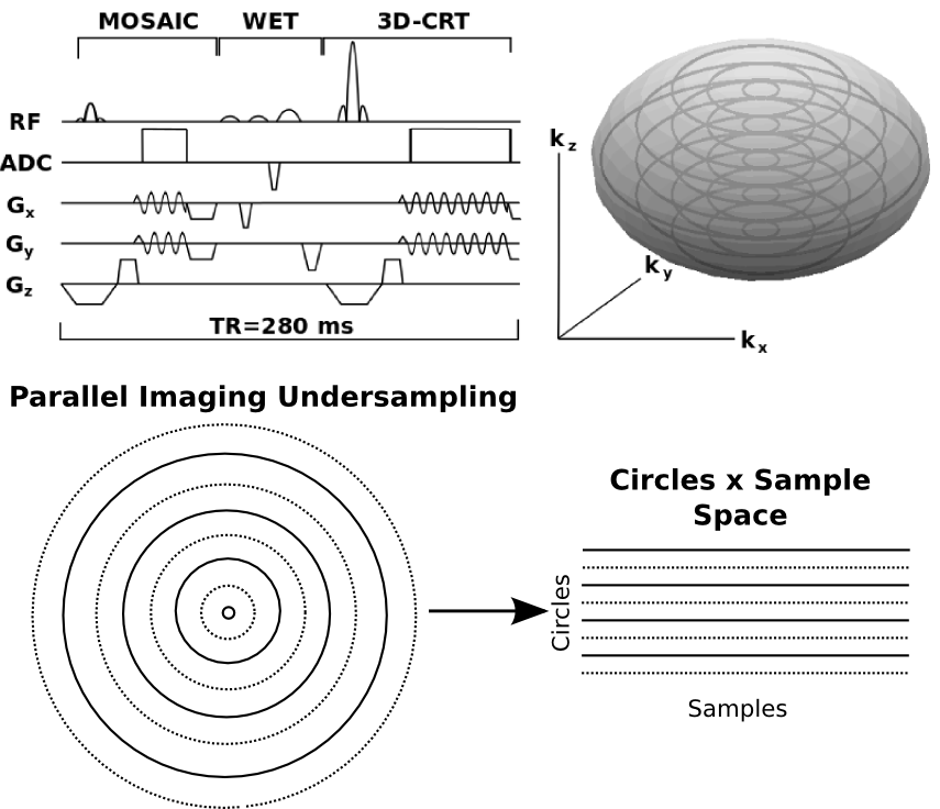
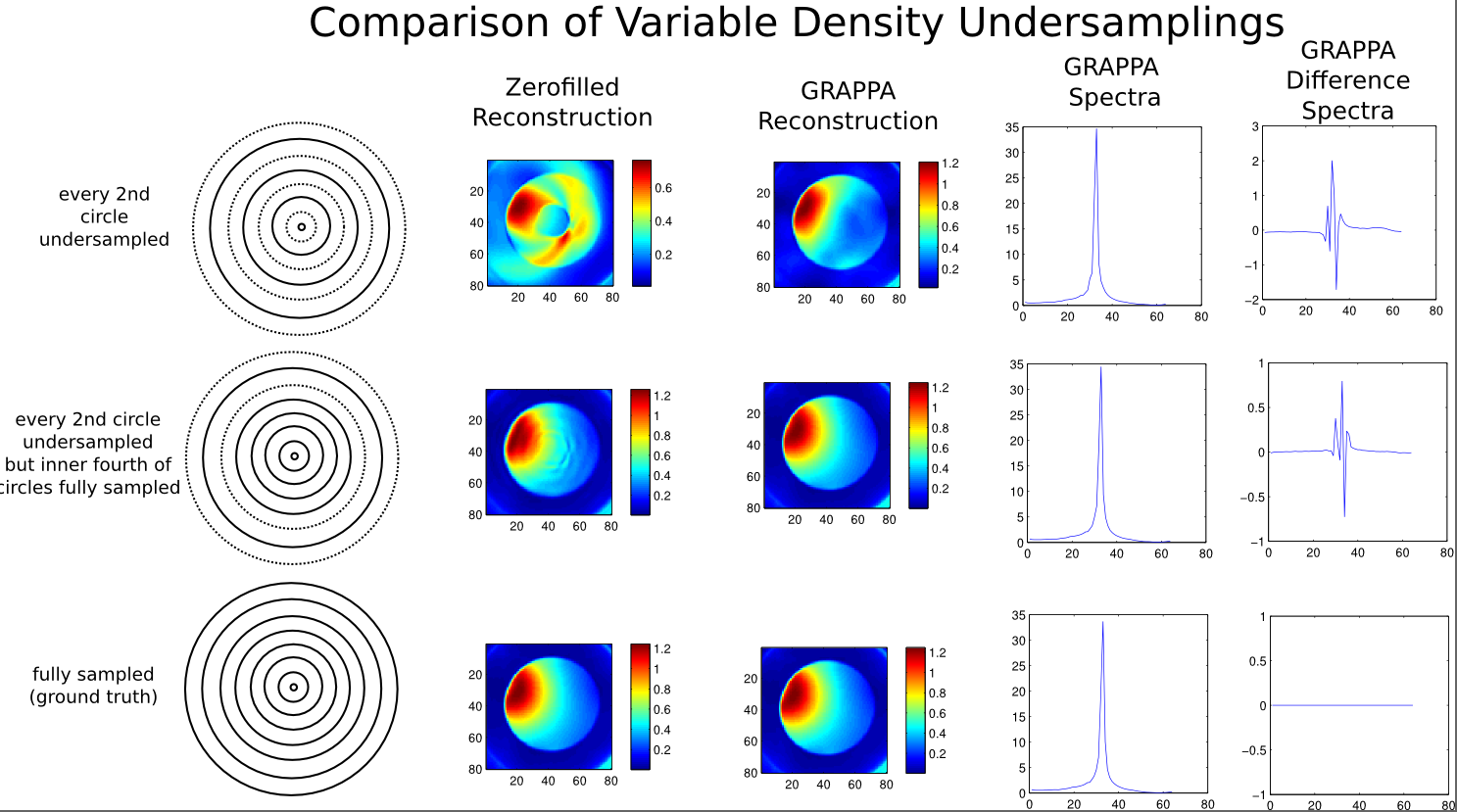
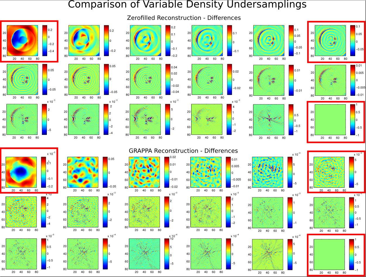
Figure 3. Difference images of different variable density sampling approaches. The red boxes highlight the three cases displayed in Figure 2. Always every 2nd circle is undersampled with the additional incorporation that the inner 2,4,6, ... 34 circles out of 40 total are fully sampled.