2474
Feasibility of 7T 31P MRSI in lung tumors1Radiology, University Medical Center Utrecht, Utrecht, Netherlands, 2Oncology, University Medical Center Utrecht, Utrecht, Netherlands
Synopsis
In this study we show the feasibility of 31P MRSI acquisition from a lung carcinoma tumor in a patient using a 31P whole body birdcage coil at 7T. We showed that even without B0 shimming, 31P spectra could be aligned and averaged to differentiate several metabolites, related to membrane metabolism, in the lung tumor. 31P MRSI has great potential for the detection of therapy response in lung tumor cancer, as often the tumor is still relatively large to obtain sufficient spectral signal.
Introduction
In the recent years, many new expensive cancer therapies have been introduced, including molecular therapies (such as immunotherapy) for lung cancer therapy. Immunotherapy is a new, promising but costly therapy for patients with non-small cell lung carcinoma. Immunotherapy works by boosting the body natural defenses to kill cancer cells, resulting in inhibition of tumor growth without necessarily a decrease of the tumor. At this moment it is difficult to predict which patients show a good response to immunotherapy. Therefore, the need for a method that can detect the tumor metabolic changes is crucial for early response evaluation. Phosphorous (31P) spectroscopic imaging is a technique that allows the detection of cell membrane metabolism and already studies have shown its use in therapy follow up1. In this study we show the feasibility of 31P MRSI acquisition in a lung carcinoma tumor patient.Methods
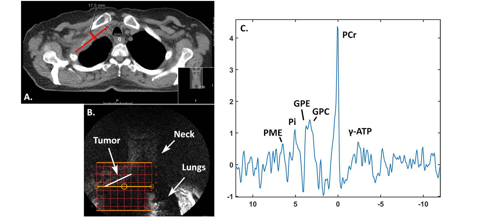
31P MRSI was performed using an in-house designed 31P whole body birdcage coil integrated in a 7T MR system (Philips Healthcare, Best, Netherlands) in combination with two 31P receive coils in quadrature mode. The body coil, tuned at 120Mhz was powered by a 25kW amplifier. Two fractionated dipole antennas were driven in quadrature as transceivers to acquire anatomy localization MRI2. No B0 shimming was performed. Setup consistency was assessed in a group of volunteer using a pulse acquire with increasing flip angles. One patient with a 1.75x7.25 cm lung carcinoma was positioned supine with the 31P receiver coils on the right upper part of the chest. The patient received three sessions of chemotherapy and the tumor was in remission. The patient gave informed consent prior scanning. Phosphor (31P) spectra were acquired using a 3D 31P chemical shift imaging protocol (TE/TR, 0.44ms/60ms; FA, 20o; 26mm isotropic nominal resolution; BW, 4800Hz; 12x8x8 matrix, NSA, 320; 256 sample points, 23 min. total acquisition time) with Hamming weighted acquisition. Data were processed in Matlab 2017b. All voxels within the lung carcinoma were aligned to PCr prior to averaging.Results
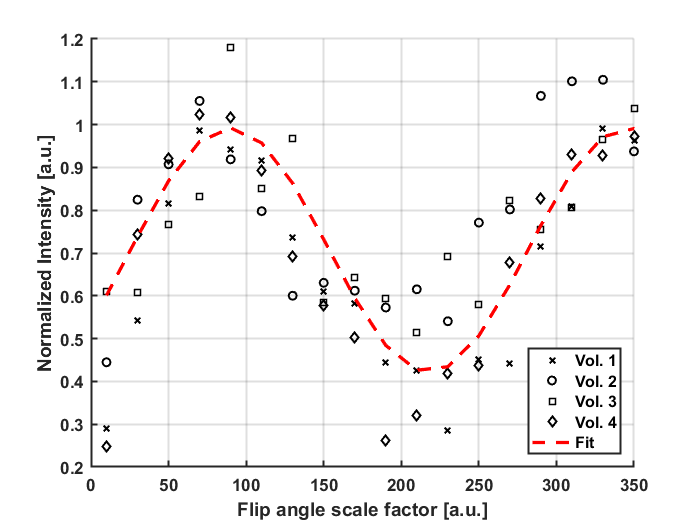
Figure 1 shows that the 31P transmit level is consistent over multiple subjects with only a minor inter-subject variation of 14%. Tumor localization was performed using both the CT acquired one day prior to the MR session and the MR image as shown in figure 2 A and B with the CSI overlay. Figure 2C shows the resulting spectrum after alignment and averaging of 20 voxels located in the tumor. B0 shimming did not improve the B0 field homogeneity, thus was discarded for following acquisitions. Even without B0 shimming, the linewidth of the peaks was sufficient to differentiate the phosphomonoesters, inorganic phosphate, glycerophosphocholine and glyceroethanolamine. For PCr the linewidth at full width half maximum was 30Hz. The close proximity to the muscles in the chest may cause a PCr peak (0 ppm) leakage.Discussion and Conclusion
We showed that without B0 shimming 31P spectra could be
aligned and averaged to differentiate several metabolites, related to membrane
metabolism, in the lung tumor. 31P MRSI has great potential for the
detection of therapy response in lung tumor cancer, when the tumor is still
relatively large to obtain sufficient spectral signal. In the presence of large
susceptibility differences, such as the lungs and the moving heart, spectral
quality seemed sufficient for the discrimination of the metabolites.Acknowledgements
No acknowledgement found.References
1. van der Kemp et al., Detection of Alterations in Membrane Metabolism During Neoadjuvant Chemotherapy in Patients with Breast Cancer Using Phosphorus Magnetic Resonance Spectroscopy at 7 Tesla. SpringerPlus. 2014;3:634-. PubMed PMID: 25932360.
2. Raaijmakers et al., The Fractionated Dipole Antenna: A New Antenna for Body Imaging at 7 Tesla. Magn Reson Med. 2016 Mar;75(3):1366-74. PubMed PMID: 25939890.
Figures