2468
Development of a Quantitative Assessment tool for Peripheral Artery Feature Extraction (pCafe)1University of Washington, Seattle, WA, United States, 2Thammasat University, Bangkok, Thailand
Synopsis
Peripheral artery disease is a relatively common disease, normally caused by reduced blood flow to the limbs due to atherosclerosis in the arteries supplying them. Peripheral arteries’ anatomy, including collateral circulation, and flow information enable disease status assessment. We developed pCafe to semi-automatically trace peripheral arteries from 3D magnetic resonance angiography and measure both morphometry (anatomy) and intensity features (velocity). pCafe was validated on subjects with, and without peripheral artery occlusion, showing excellent agreement with human reviewer’s measurement (intra-class coefficient of 0.998). pCafe may be a useful tool to quantitatively characterize peripheral vascular structures in peripheral artery disease research.
INTRODUCTION
Peripheral artery disease (PAD) is a relatively common disease due to reduced blood flow to the limbs. PAD caused by atherosclerosis affects more than 200 million persons worldwide, and these patients are at a significantly higher risk of myocardial infarction, cardiovascular events and amputation1,2. The physiological adaptations to restore blood flow to occluded regions include increasing the diameter and the number of vessels within the collateral blood vessel circuit2,3. To fully evaluate and monitor PAD status and progression, it is important to conduct quantitative assessment of peripheral arteries, including collateral arteries. Current techniques to assess the severity of PAD in the lower limbs are focused on arterial pressure4,5, blood flow6, and the degree of stenosis7 of the larger arteries, but a comprehensive quantification of structural features of the peripheral arterial circulation is lacking.
Therefore the aim of this study is to develop a peripheral artery feature extraction (pCafe) technique8 to quantitatively assess the morphometry and intensity features of the peripheral vasculature using 3D contrast enhanced (CE) magnetic resonance angiography (MRA).
METHODS
MR imaging
Two subjects with peripheral artery disease were scanned using a 3.0T Philips (Best, The Netherlands) Ingenia CX MR scanner using torso phased array coil covering the legs. Study procedures followed local IRB guidelines and informed consent was obtained for all subjects. After single dose Prohance injection, single station first pass 3D CE-MRA was obtained covering the lower part of the thigh and knees bilaterally. Imaging parameters were as follows: TR/TE = 4.56/2.195 ms, flip angle = 20°, in-plane resolution = 0.81 mm×0.81 mm, slice thickness = 3 mm, field of view = 430 mm*430 mm.
Feature extraction
MRA images were resampled to isotropic resolution of 0.81 mm in 3D space and image intensities were normalized using the Nyul9 method to allow comparable intensity features from different cases in the dataset. Artery regions were then automatically traced and reconstructed using an improved open-curve active contour model10. Several key landmark points need to be added manually so that all the arteries can be labeled as one of the following peripheral artery types: superficial femoral artery (SFA), profunda femoris artery (PFA) and their collateral arteries, both left and right. In the case of occluded SFA, collateral arteries can be further labeled as SFA collateral arteries proximal and distal to the occluded region (labeled upper and lower collaterals, respectively). An experienced vascular surgeon supervised the tracing and labeling process and corrections were made when needed.
A group of representative features (listed in Table 1) reflecting typical PAD arterial characteristics can be quantified using pCafe.
Validation
An experienced human reviewer measured SFA/PFA length using Philips Intellispace Portal Software (Philips Healthcare, Best, the Netherlands) to indirectly validate the accuracy of traces and vessel length measurements. Reference vessel length was measured by manually selecting centerline points along the arteries. After the centerline was drawn, length was measured on the corresponding curved planar reformatted image and compared to the length measurements using pCafe.
RESULTS
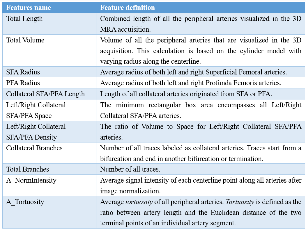
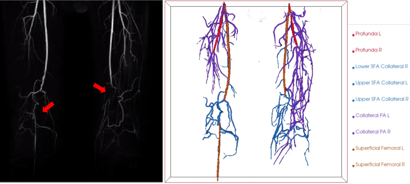
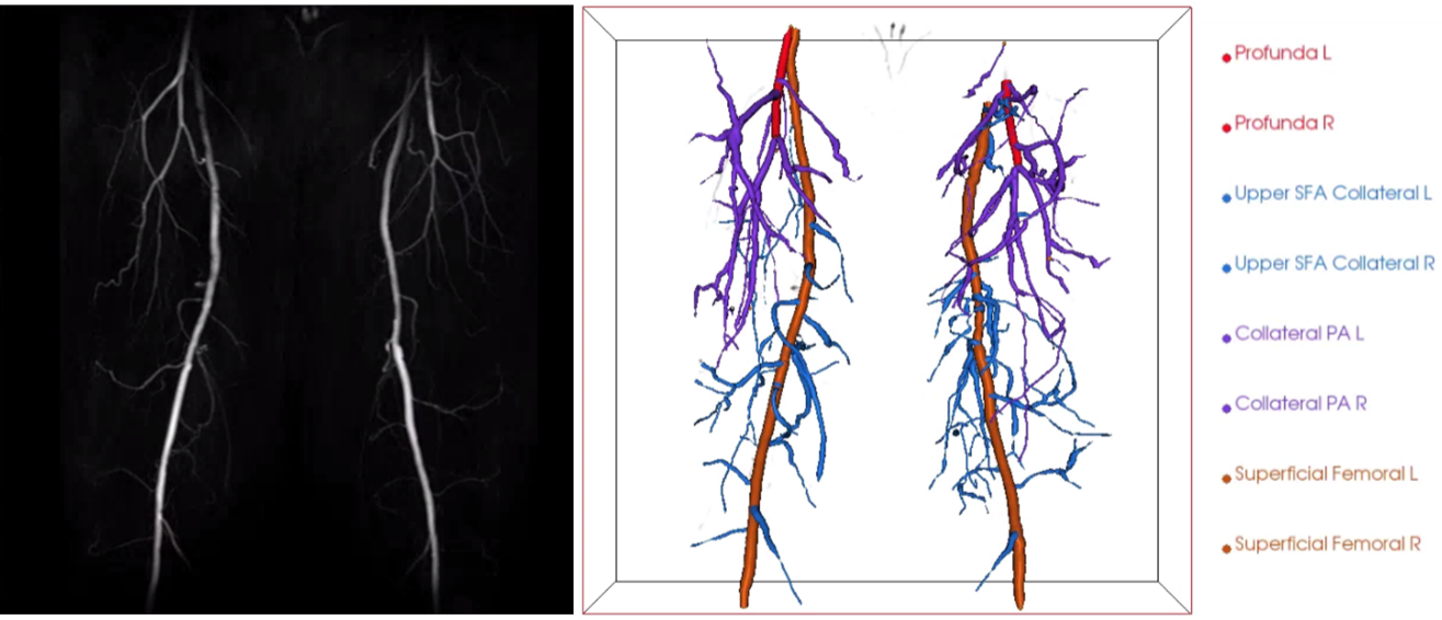
Original maximum intensity projection images, along with reconstructed collateral arteries using pCafe from subjects with and without occluded SFA are shown in Figure 1 and Figure 2, respectively.
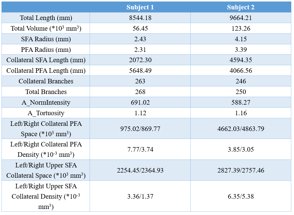
A group of representative features extracted from pCafe for the subject with peripheral artery occlusion (subject 1) and the subject without occluded SFA (subject 2) is shown in Table 2.
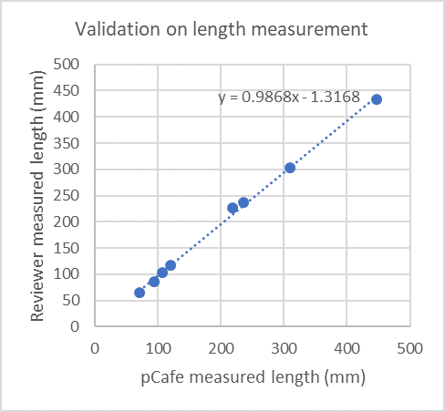
The pCafe-measured length of eight arteries (both sides of SFA and PFA for two subjects) is in agreement with the reviewer-measured length. The mean absolute difference of the artery length is 6.24mm, and the intra-class coefficient is 0.998. A plot with the arterial length measurements is shown in Figure 3.
DISCUSSION
pCafe was shown to reconstruct peripheral arteries and extract features from subjects with and without SFA occlusions. Fourteen representative features were shown in this study, but with all peripheral arteries reconstructed, new custom features can be designed and added as needed. The quantitative information provided by pCafe is promising for a comprehensive description of peripheral artery status.
We used a human operator to verify pCafe artery tracings, as small arteries may be incorrectly connected due to the relatively weak signal intensity. Further improvement of pCafe is ongoing to reduce human input.
The validation of collateral arteries and radius of peripheral arteries is currently difficult to achieve, as it is challenging for human reviewers to segment and trace all the arteries in 2D image slices. To further validation, a reproducibility study on a larger study cohort is needed in the future.
CONCLUSION
A semi-automated quantitative measurement tool for peripheral arteries based on 3D CE MRA has been developed and may provide a novel set of vascular features for assessing PAD status and development.Acknowledgements
This research is supported by grants from Philips Healthcare.References
1. Kullo IJ, Rooke TW. Peripheral Artery Disease. Solomon CG, ed. N Engl J Med. 2016;374(9):861-871. doi:10.1056/NEJMcp1507631.
2. Murrant CL. Structural and functional limitations of the collateral circulation in peripheral artery disease. J Physiol. 2008;586(24):5845. doi:10.1113/jphysiol.2008.166298.
3. Schirmer SH, Royen N van. Stimulation of collateral artery growth: a potential treatment for peripheral artery disease. Expert Rev Cardiovasc Ther. 2004;2(4):581-588. doi:10.1586/14779072.2.4.581.
4. Traupe T, Ortmann J, Stoller M, Baumgartner I, de Marchi SF, Seiler C. Direct Quantitative Assessment of the Peripheral Artery Collateral Circulation in Patients Undergoing Angiography. Circulation. 2013;128(7):737-744. doi:10.1161/CIRCULATIONAHA.112.000516.
5. Stoller M, Stoller D, Seiler C. Physical exercise and quantitative lower limb collateral function. Open Hear. 2016;3(1):e000355. doi:10.1136/openhrt-2015-000355.
6. Wuestenfeld JC, Herold J, Niese U, et al. Indocyanine green angiography: A new method to quantify collateral flow in mice. J Vasc Surg. 2008;48(5):1315-1321. doi:10.1016/J.JVS.2008.06.049.
7. Sacks D, Robinson ML, Marinelli DL, Perlmutter GS. Peripheral arterial Doppler ultrasonography: Diagnostic criteria. J Ultrasound Med. 1992;11(3):95-103. doi:10.7863/jum.1992.11.3.95.
8. Chen L, Mossa-Basha M, Balu N, et al. Development of a quantitative intracranial vascular features extraction tool on 3D MRA using semiautomated open-curve active contour vessel tracing. Magn Reson Med. 2018;79(6):3229-3238. doi:10.1002/mrm.26961.
9. Nyul LG, Udupa JK, Zhang X. New variants of a method of MRI scale standardization. IEEE Trans Med Imaging. 2000;19(2):143-150. doi:10.1109/42.836373.
10. Wang Y, Narayanaswamy A, Tsai CL, Roysam B. A broadly applicable 3-D neuron tracing method based on open-curve snake. Neuroinformatics. 2011;9(2-3):193-217. doi:10.1007/s12021-011-9110-5.
Figures