2458
Signal Enhancement and Optimum Receiver Arrays For Human Hyperpolarized 13C MR Spectroscopic Imaging1Radiology and Biomedical Imaging, University of California, San Francisco, San Francisco, CA, United States, 2Center for Cancer Research, National Cancer Institute, National Institutes of Health, Bethesda, MD, United States, 3Berkeley Institute for Data Science, Berkeley, CA, United States, 4GE Healthcare, Chicago, IL, United States
Synopsis
A data-driven processing framework was proposed for dynamic hyperpolarized 13C-MR Spectroscopic Imaging to maximally extract diagnostic information from existing datasets and techniques that utilized whitened-SVD2 to optimally combine array data, and tensor-low-rank denoising3,4 to enhance SNR. The framework was applied and evaluated on brain, abdomen, and pelvic datasets acquired using multi-channel arrays or single-element receivers. Substantial improvement in quality of low-SNR lactate and alanine was observed with 30+ fold apparent SNR gain, whereas high-SNR pyruvate remained largely artifact-free. Correlation of high kPL with biopsy-confirmed cancer strongly indicated that this recovered important pathological information.
Purpose
Hyperpolarized 13C is a powerful emerging stable-isotope imaging technology to probe metabolism in human cancer. As multicenter HP-13C trials begin, new coil development and acquisition methods are still work in progress, and there is a key unmet need to maximally extract diagnostic information1 from datasets acquired with existing techniques. This report presents a new HP-MRSI processing framework at UCSF. It summarizes our attempt to optimally combine phased data from receiver arrays using whitened-SVD(WSVD)2, and to enhance signal against noise using tensor-low-rank denoising(TD)3,4. Optimization was evaluated on dynamic MRSI datasets from brain, abdomen and pelvis using array and single-element receiver configuration.Methods
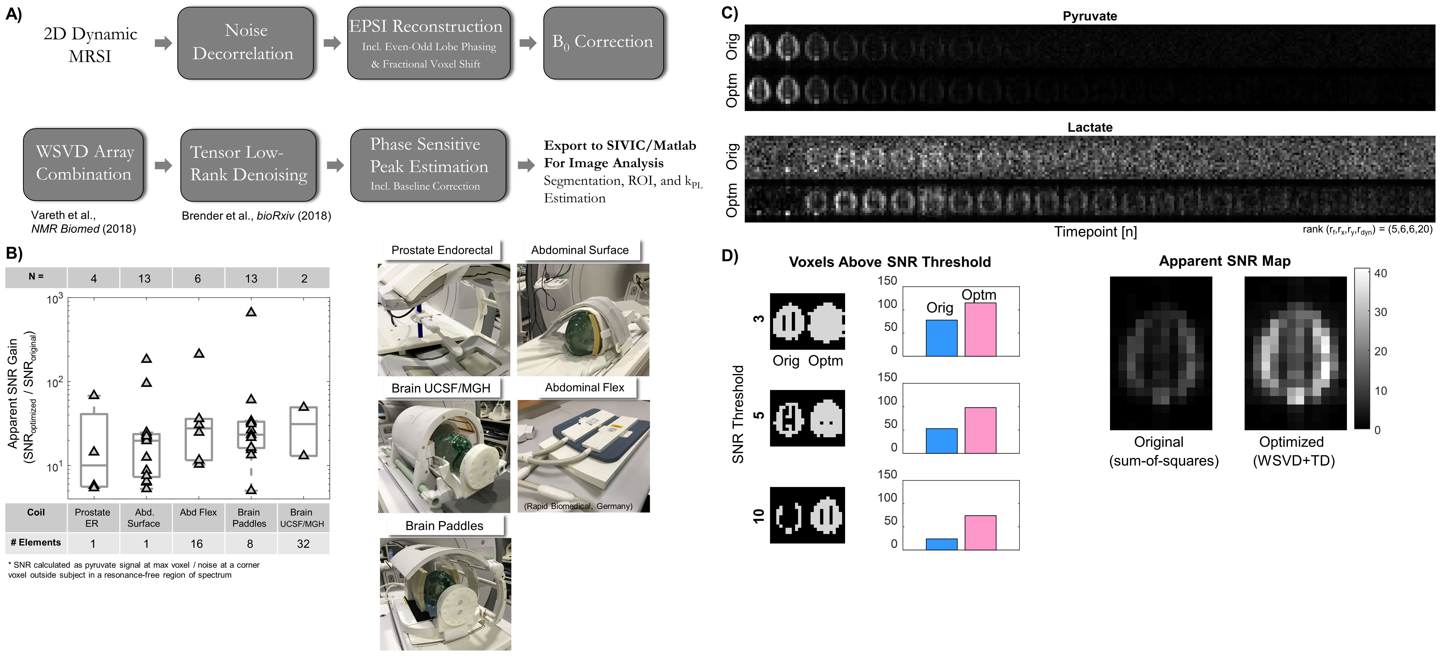
Patient Studies: The patient data (N=38) used was acquired with a 2D MRSI sequence with EPSI readout(TR/TE=130ms/3.5ms, resolution temporal=2-5s, spatial = 1-4cc), following injection of 250mM HP-[1-13C]pyruvate5 polarized using a 5T SPINlab(GE Healthcare). Fig.1B summarized the 13C receivers in this study, including 8 and 32-channel brain, 16-channel and surface abdominal, and an endorectal prostate coil5,6,7. All human studies were IRB-approved at UCSF.
Image Processing Framework: Image processing workflow of the 2D MRSI data is summarized in Figure 1A, where noise decorrelation and SVD combination are unique to multichannel datasets. The processing and visualization are realized on MATLAB and SIVIC8. Pyruvate-to-lactate conversion rate(kPL) was evaluated using an inputless kinetic model9.
WSVD Array Combination: The WSVD algorithm2 first decorrelates the receiver channels, followed by SVD decomposition to extract the voxel-wise complex coil sensitivity weighting from a principal eigenvector. Sum-over-time was used, assuming coil profile is temporally-invariant.
Tensor Low-Rank Denoising: TD3,4 utilizes Tucker decomposition to separate the spectral-spatial-dynamic components into factor matrices in D dimensions and a core tensor X = G×1A×2B×3C (conceptually similar to eigenvectors and singular values in SVD). Exploiting the spatiotemporal correlation, noise is reduced by decreasing rank in each dimension. Selecting the ideal set of ranks presents a classical bias-variance tradeoff problem10. One strategy is to formulate the problem as
$$\DeclareMathOperator*{\argmin}{arg\,min}\argmin_{r_{1},r_{2},..,r_{i},..,r_{D}} \frac{1}{K}\sum_k^{}|S_{pyr,orig}(k)-S_{pyr,TD}(k)|^{2}+w_{0}\cdot\sigma_{noise,TD}^2$$
where ri is the rank in ith dimension, Spyr(k) is the pyruvate signal from kth highest SNR voxel, σ2noise,TD is estimated at the FID tail or from a noise voxel outside subject, and w0 is a weighting factor. An alternative strategy that the authors used, is to empirically decrease the rank in each dimension and visually inspect the dynamic pyruvate series. Rank should be set slightly higher before artifacts become visible on pyruvate images, or right when reliable kPL fits are attained in tumor ROI11, whichever happens first.
Results and Discussions
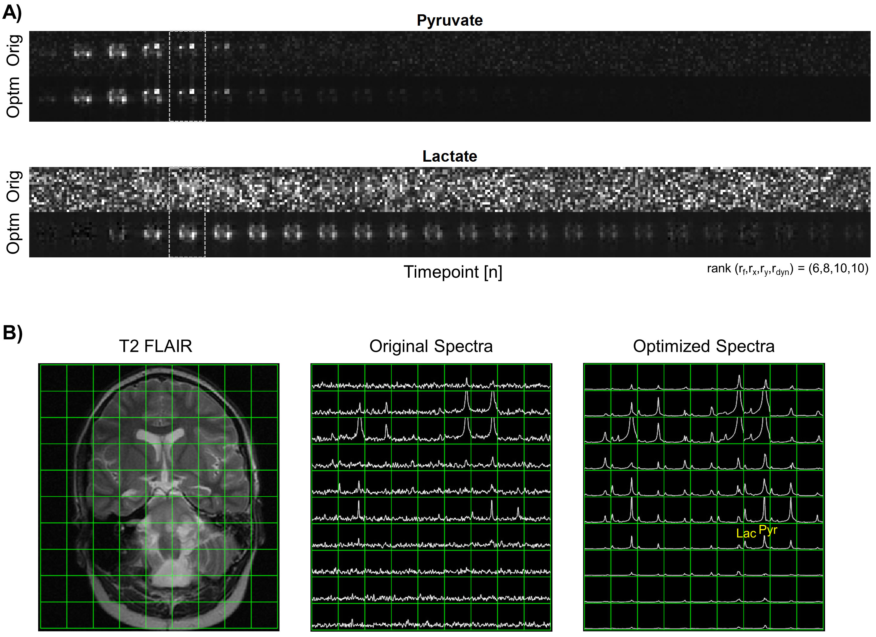
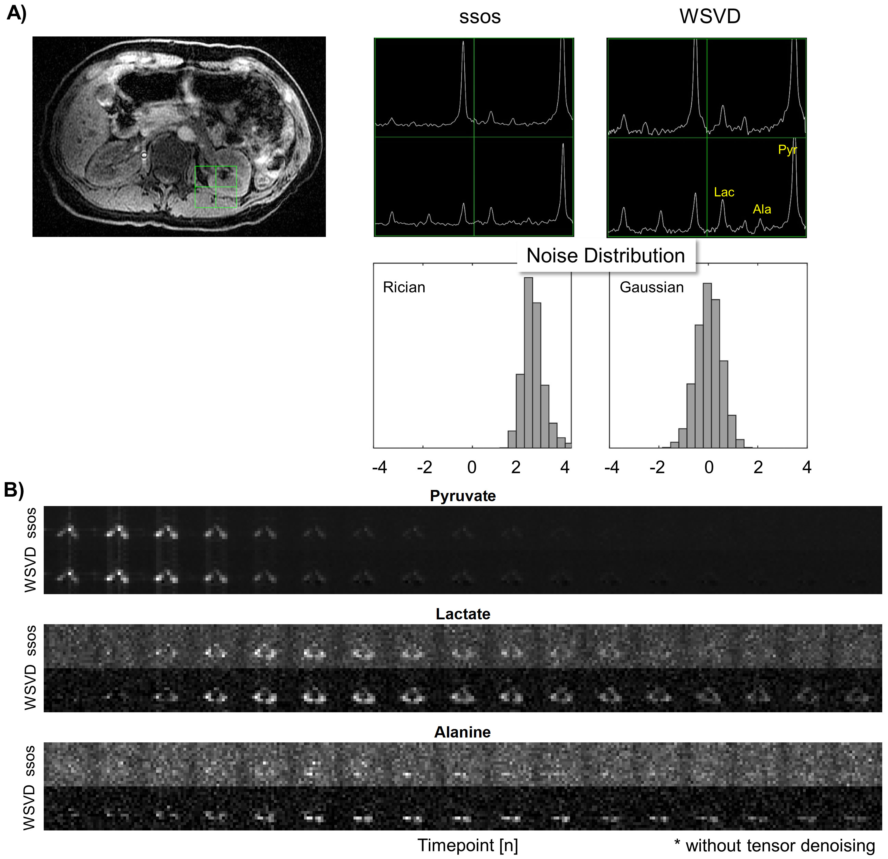
Brain data (Figs.1C-D and 2) showed the overall framework recovered low-SNR lactate while high-SNR pyruvate remained largely artifact-free, enabling reliable quantification of cerebral metabolism in more voxels. Abdominal volunteer exam (Fig.3) highlights that WSVD array combination reduced baseline and improved noise statistics, substantially enhancing lactate and alanine in kidney/muscle over background.
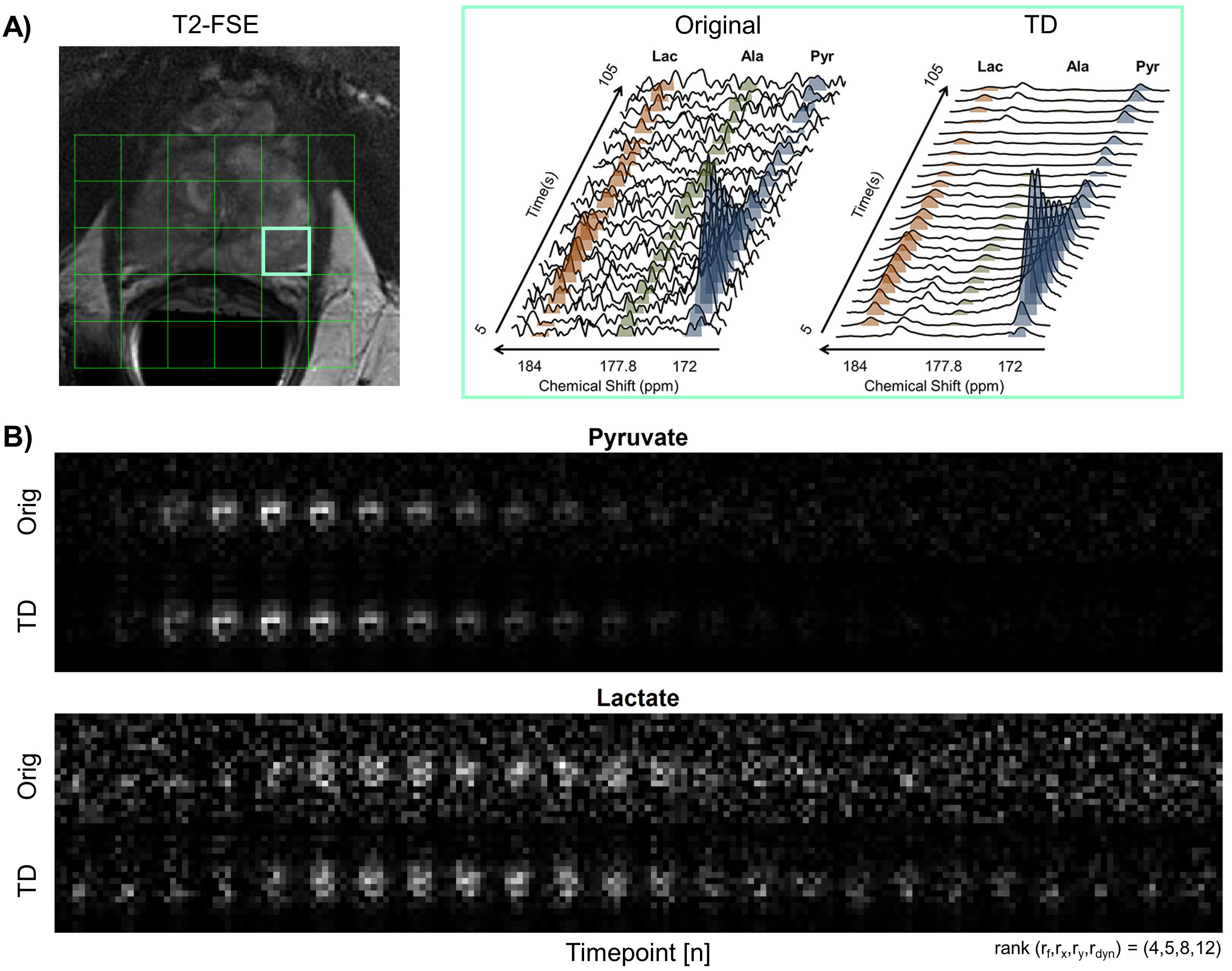
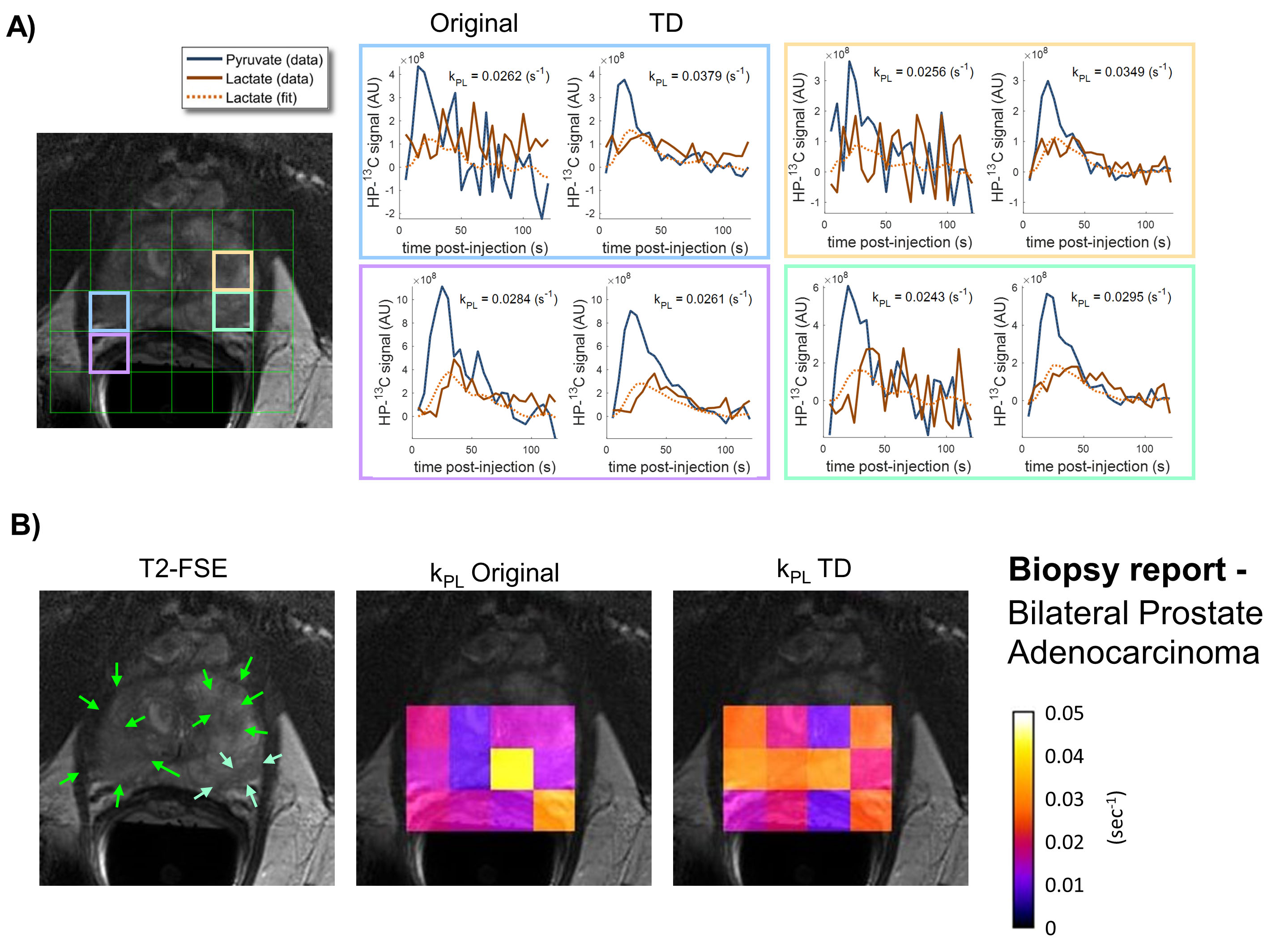
Figures 4 and 5 illustrated a patient diagnosed with bilateral biopsy-confirmed prostate cancer. Dynamic spectroscopy (Fig.4A) observed 67-fold apparent SNR gain, and recovery of the otherwise undetectable pyruvate-hydrate and alanine. High kPL (Fig.5B, orange and magenta) showed good agreement with T2 lesions (Fig.5B, green arrows) and the biopsy finding of bilateral midgland cancer. This strongly indicates that TD recovered quantitative pathological information rather than created artifacts.
The nature of the HP-13C spectroscopy – finite and known number of discrete resonances, enables one to aggressively drive the spectral rank down and truly unleash the TD denoising power. Of note, no assumptions are made about the chemical shift or lineshape of each resonance. Recovery of low SNR resonances suggested that this framework may also benefit HP probe development12 where proof-of-concept studies could be made possible despite low polarization, slow conversion, or short T1. One general concern of denoising methods – spatiotemporal “imprinting” of high SNR resonances onto low SNR one, was not observed. Pyruvate and lactate have distinct spatial distribution and dynamics upon detailed inspection(Figs.1C,2A&4B).
Overall, this data-driven framework is versatile across imaging targets and receiver configurations. Mean apparent SNR gain was 63-fold for arrays, and 31-fold for single-element receivers, where at least 10-fold can typically be expected for arrays (Fig.1B). The ultimate power of signal enhancement and optimal array combination will depend not only on the design/geometry of array coils, but also the anatomy, pathophysiology and pharmacokinetics of the target, which determines the spatiotemporal complexity, and therefore the rank of the underlying data.
Conclusions
The HP-13C MRSI processing framework utilized optimal receiver-array combination and SNR enhancement to substantially improve extraction of diagnostic information from human cancer. This could benefit ongoing and future multicenter trials, and also new HP probe discovery.Acknowledgements
This work was supported by grants from the NIH (R01EB017449, R01EB013427, R01CA166655, and P41EB013598).References
[1] Shannon CE, A Mathematical Theory of Communication; Bell Syst. Tech. J. 1948
[2] Vareth M et al., A comparison of coil combination strategies in 3D multi-channel MRSI reconstruction for patients with brain tumors; NMR Biomed. 2018; e3929
[3] Tucker LR, Some mathematical notes on three-mode factor analysis; Psychometrika 1966; 31(3), pp.279-311.
[4] Brender JR et al., PET by MRI: Glucose Imaging by 13C-MRS without Dynamic Nuclear Polarization by Noise Suppression through Tensor Decomposition Rank Reduction; bioRxiv. 2018;
[5] Nelson SJ et al., Metabolic Imaging of Patients with Prostate Cancer Using Hyperpolarized [1-13C]Pyruvate. Sci Transl Med. 2013;5(198)
[6] Park I et al., Development of methods and feasibility of using hyperpolarized carbon‐13 imaging data for evaluating brain metabolism in patient studies; Proceedings of ISMRM. 2018
[7] Autry A et al., Comparison between 8- and 32-channel phased-array receive coils for in vivo hyperpolarized C-13 brain imaging; Magn Reson Med. 2018; 80(3), 864-873
[8] Crane JC et al., SIVIC: open-source, standards-based software for DICOM MR spectroscopy workflows; Int J Biomed Imaging. 2013;169526
[9] Larson PEZ et al., Investigation of Analysis Methods for Hyperpolarized 13C-pyruvate Metabolic MRI in Prostate Cancer Patients; NMR in Biomed. 2018; DOI:10.1002/nbm.3997
[10] Geman S et al., Neural networks and the bias/variance dilemma; Neural computation. 1992
[11] Mammoli D et al., Modeling In Vivo Metabolism of Hyperpolarized Pyruvate in Human Brain Tumor Patients; Proceedings of ISMRM. 2018;
[12] Chaumeil MM et al., Studies of metabolism using 13C MRS of hyperpolarized probes; Methods in enzymology. 2015;
Figures