2448
A fully automated method for concealing patient identity in 3D multi-contrast brain MR images1Department of Imaging and Interventional Radiology, The Chinese University of Hong Kong, Hong Kong, China
Synopsis
In addition to personally identifying information (PII) commonly found in metadata of medical images, superficial anatomical features contained in 3D brain MR images pose a unique challenge to medical privacy, and this place a serious obstacle for data sharing in large-scale collaborative efforts. A fully automated method for concealing patient identity in 3D multi-contrast brain MR images is presented. The proposed method is training-free and can be applied to automatically conceal patient’s identity information in the 3D brain MR images, which makes this approach particularly useful for handling brain MR images in large neuroimaging databases.
Introduction
Data sharing has been promoted as an important step in neuroimaging studies. This fact has led to the creation of large-scale online brain MR image databases. However, the skin surface features in the brain MR images, which are particularly relevant to patients’ identity, pose a unique challenge to medical privacy. Therefore, it’s crucial to have in place measures to share the 3D brain MR images in compliance with medical privacy regulation[1][2]. Approaches employed to date usually require: (1) using an image registration method to pre-align the novel image with the standard template image, (2) employing a training procedure to identify particular anatomical structures, or (3) using an iterative procedure to remove all non-brain voxels during de-identification[3-6]. Such approaches are useful but are computationally intensive, and often require significant manual intervention to complete the de-identification procedure. These limitations substantially complicate the process of making image data sets available to other investigators. This paper describes a fully-automated approach for concealing patient identity in 3D multi-contrast brain MR images.Methods
A two-stage procedure is employed to de-identify the 3D brain MR images. (1) Using an automated method to extract the skin surface of the patient’s head; (2) Modifying the skin surface of the patient head to conceal patient identity. The proposed method will only remove a small portion of image voxels and preserve most of the image content unchanged.
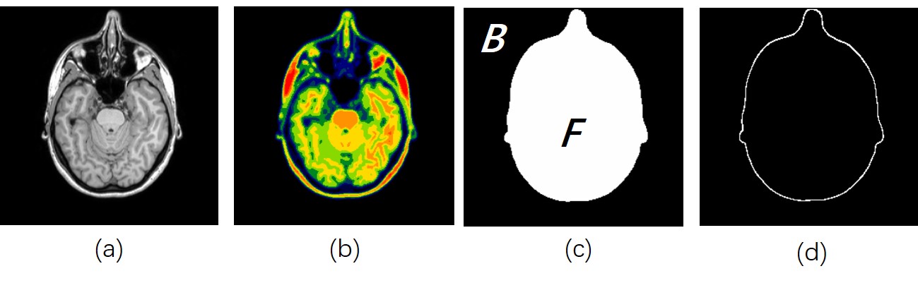
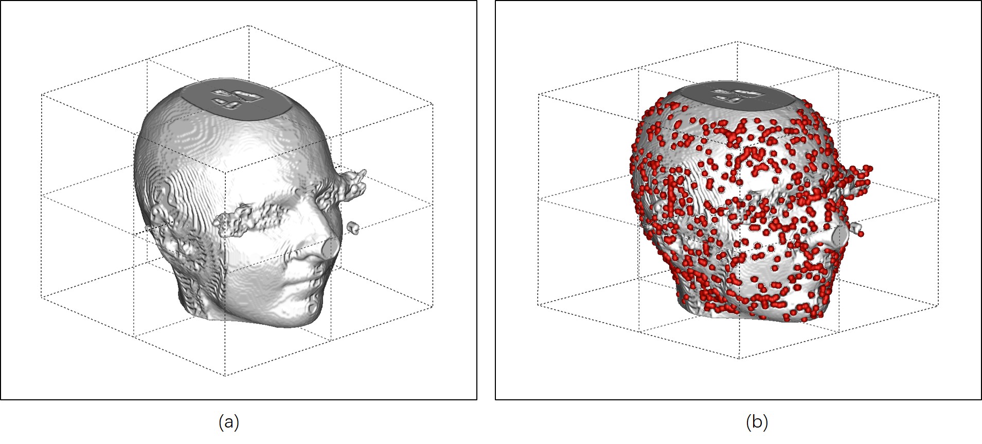
In most structural MR images, the image background appears as a dark connected region (The black region in Fig 1a). Therefore, determining the skin surface of patient’s head is handled by tracking the boundary between the image background region (denoted as $$$B$$$, as shown in Fig.1c) and other non-background regions (represented by $$$F$$$, as shown in Fig.1c). The following steps were employed to estimate the skin surface of the patient’s head. An intensity threshold $$$t$$$ that roughly separate the background and non-background regions in the brain MR image is determined. Here we modeled the intensity distribution of the brain MR image as a finite mixture of four Gaussian distributions, each of which represents the intensity distribution of one tissue type. K-means clustering algorithm is used to segment the image into a labeled image (Fig.1b) with different image voxel types (including background voxels), the mean values for each type are calculated. The threshold $$$t$$$ is determined by the smallest mean value, denoted as $$$L$$$, $$$t=L/5$$$. For image voxels with intensity value smaller than $$$t$$$ were set to zero and classified as background voxels, otherwise set to 255 as non-background voxels. However, the threshold $$$t$$$ determined by the k-means clustering algorithm is imperfect, in some situation, a small number of image voxels may be misclassified. Image morphological operations as well as super-voxel based analysis were performed to correct misclassification errors. Finally, to obtain the skin surface of patient’s head, the boundary voxels between the background region $$$B$$$ and the non-background region $$$F$$$ were extracted. The extracted skin surface of the head was denoted as $$$S$$$(Fig.1d). Fig 2a depicts a 3D rendering of the extracted skin surface $$$S$$$.
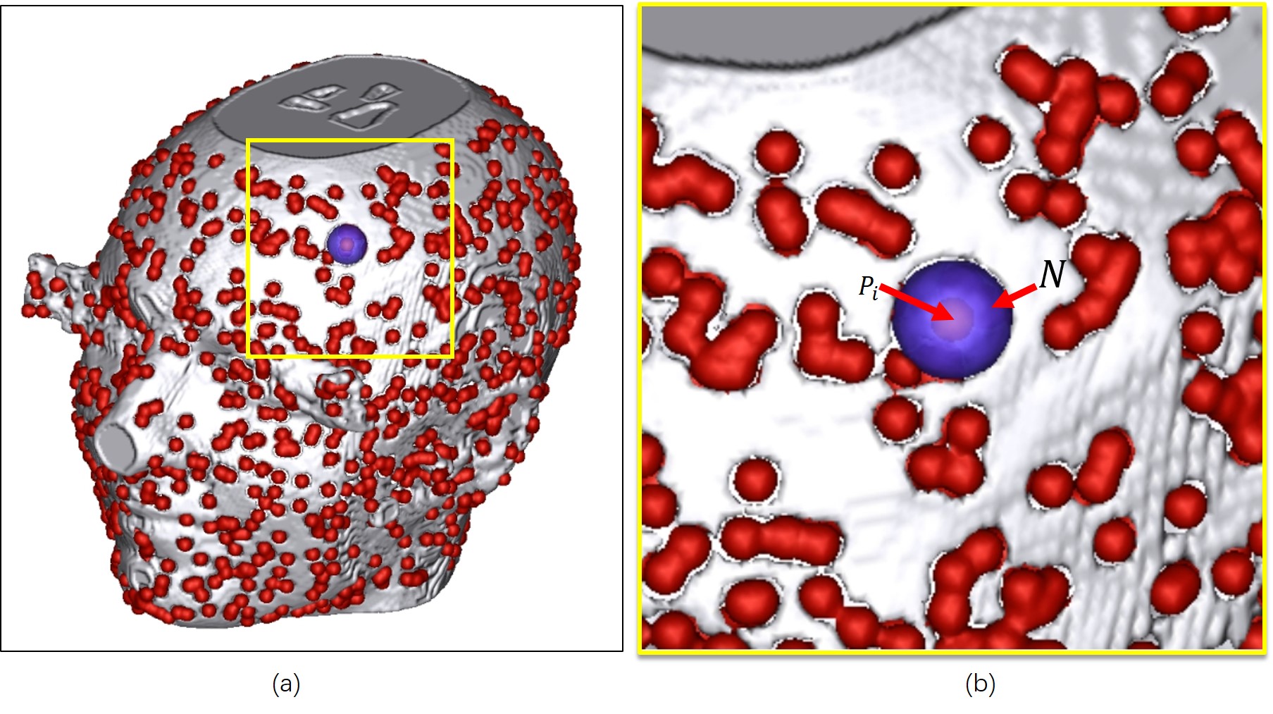
To de-identify the brain MR image, an efficient method was adapted to modify the surface anatomy of $$$S$$$. Firstly, seeds points were randomly placed on the skin surface $$$S$$$ (Fig.2b). Secondly, the image voxels adjacent to the seed point were removed by setting the voxel value to 0 (Fig.3a,b). This operation will create a surface deformation on the skin surface during 3D surface reconstruction and can be applied to conceal the patient's identity in the MR image.
Results
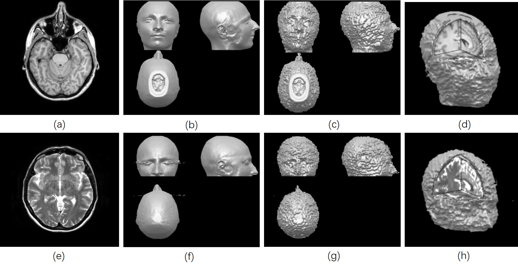
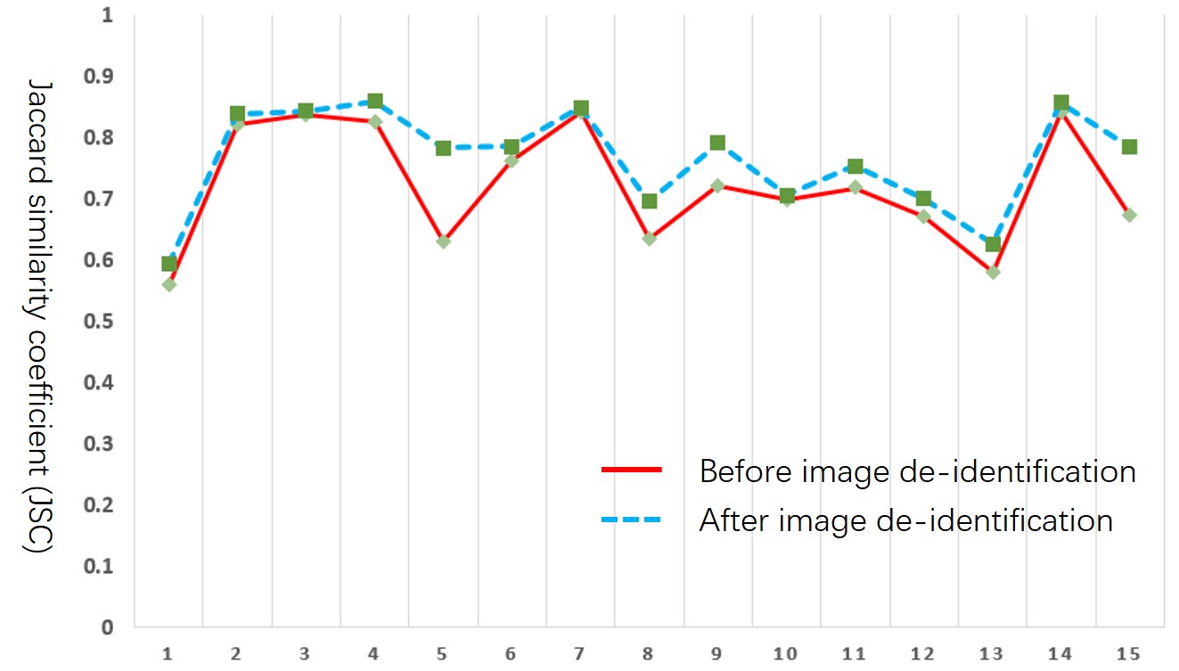
To evaluate the performance of the proposed method, we have applied the method to de-identify 30 T1-w and 30 T2-w brain MR images from IXI database[7]. Visual inspection indicated that the subject’s skin features in all the tested images were adequately removed while preserving the internal brain voxel intact for future analysis. We also compared the brain extraction accuracy for BET [4], quantitative assessment suggested that the proposed method improved the performance of brain extraction procedure, which is a routine workflow in brain MR post-processing (Fig 5).Conclusion
A fully-automated de-identification method for multi-contrast 3D brain MR images is presented. The proposed method is computationally efficient, easy-to-implement, registration-free and can be applied to brain MR images of multiple imaging contrast. Visual inspection suggested that the proposed method can adequately obscure the skin features in brain MR images. Quantitative comparison indicated that the proposed method does not appreciably alter the outcomes of the routinely-used brain MR post-processing workflows. The proposed method is an effective solution for privacy protection in multi-contrast 3D brain MR images.Acknowledgements
This study is supported by the grant from the Research Grants Council of the Hong Kong SAR (Project No. SEG CUHK02).References
[1] Chen, J.J.-S., et al., Implications of Surface-Rendered Facial CT Images in Patient Privacy. American Journal of Roentgenology, 2014. 202(6): p. 1267-1271.
[2] Schimke, N. and J. Hale. Neuroimage data sets: rethinking privacy policies. in Proceedings of the 3rd USENIX conference on Health Security and Privacy, USENIX Association. 2012.
[3] Schimke, N. and J. Hale. Quickshear defacing for neuroimages. in Proceedings of the 2nd USENIX conference on Health security and privacy. 2011. USENIX Association.
[4] Smith, S.M., Fast robust automated brain extraction. Human brain mapping, 2002. 17(3): p. 143-155.
[5] Bischoff‐Grethe, A., et al., A technique for the deidentification of structural brain MR images. Human brain mapping, 2007. 28(9): p. 892-903.
[6] Budin, F., et al., Preventing facial recognition when rendering MR images of the head in three dimensions. Medical image analysis, 2008. 12(3): p. 229-239.[7] IXI database. http://brain-development.org/ixi-dataset/
Figures