2444
Robust Cardiac and Respiratory Self-Gating Using an Adapted Singular Spectrum Analysis (SSA-FARI): Application to Simultaneous-Multi-Slice Imaging1Institut für Diagnostische und Interventionelle Radiologie, University Medical Center Göttingen, Göttingen, Germany, 2Partner site Göttingen, German Centre for Cardiovascular Research (DZHK), Göttingen, Germany
Synopsis
In cardiac MRI we have to deal with both cardiac and respiratory motion. Nowadays, breath holds and the use of external devices such as ECG are clinical practice to deal with this motion. However, not all patients can hold their breath and external devices are error-prone. Therefore, self-gating techniques have been developed to extract the respiration and cardiac signal from the k-space data itself. Many of those require various pre- and post-processing steps like filtering or averaging and lack robustness. Here, we present a novel and robust, yet easy to implement self-gating approach based on Singular Spectrum Analysis.
Introduction and Purpose
In cardiac imaging breath-holds, navigator sequences or the use of external tools such as an ECG are required in clinical practice [1-3]. However, external devices demand patient preparation and are error-prone [4], navigator sequences destroy the steady state and prolong acquisitions and breath-holds can be difficult for sick patients. Therefore, self-gating techniques have been proposed to extract cardiac and respiratory motion from the k-space data itself [5-8]. However, the methods generally lack robustness and the motion signlas cannot always be separated [9].
To overcome these limitations, we propose to use an adapted Singular Spectrum Analysis (SSA) [10], a simple, popular and powerful tool for time-series analysis [11-13]. This SSA For Advanced Reduction of dImensionality (SSA-FARI) can separate trend, noise and oscillatory components and is therefore perfectly suited for self-gating MRI. Here, we apply SSA-FARI to in-vivo radial cardiac Simultaneous-Multi-Slice imaging.
Theory
We use the central k-space line along the slice-dimension $$$k_z$$$ ($$$k_x=0$$$, $$$k_y=0$$$) for autocalibration [14,15]. All $$$N_p$$$ partitions and all $$$N_c$$$ coils are stacked into one dimension to yield the multi-channel time-series $$$\boldsymbol{X}$$$ of size [$$$(N_p \times N_c) \times N_t$$$], with $$$N_t$$$ being the number of acquired central k-space lines.
SSA-FARI
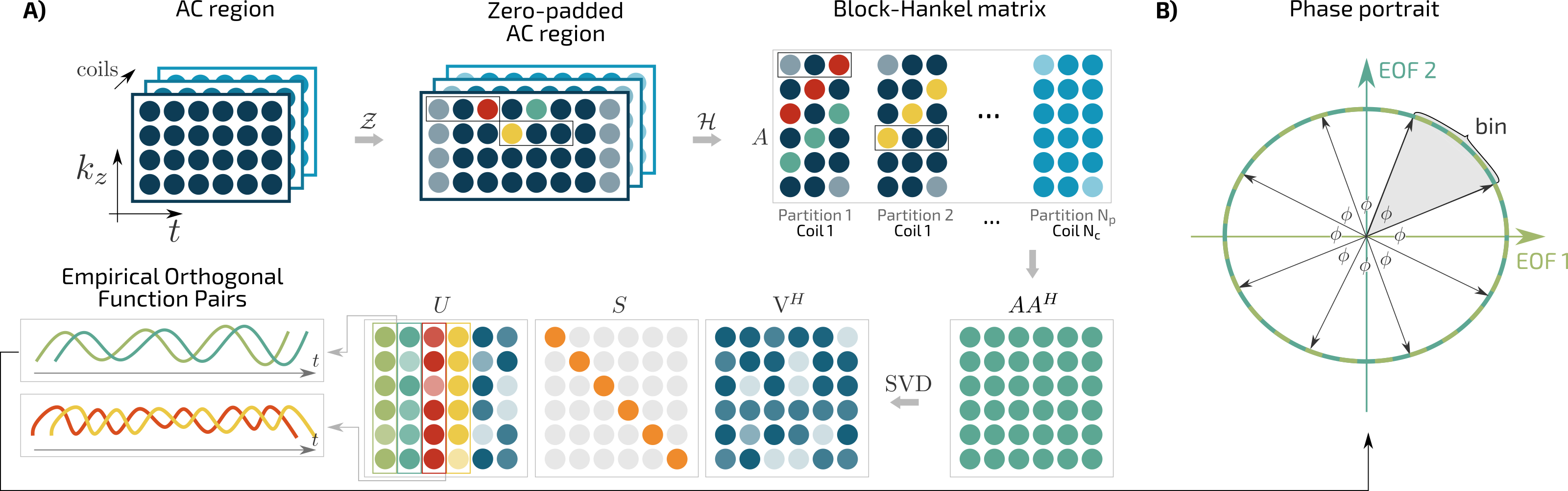
The SSA-FARI method consists of four elementary steps (Figure 1A):
1. Zero padding. We zero-pad $$$\boldsymbol{X}$$$ to obtain matrix $$$\tilde{\boldsymbol{X}}$$$ of size [$$$(N_p\times N_c)\times (N_t+ W-1)$$$],
$$\tilde{\boldsymbol{X}} = \mathcal{Z}\boldsymbol{X}.$$
This step is necessary to obtain full temporal information in the decomposition (step 4).
2. Hankelization. We construct the Block-Hankel calibration matrix $$$\boldsymbol{A}$$$ of size [$$$N_t\times ((N_p\times N_c)\times W)$$$] by sliding a window of size [$$$1\times W$$$] through the channels of the zero-padded AC region $$$\tilde{\boldsymbol{X}}$$$,
$$ \boldsymbol{A} = \mathcal{H}\tilde{\boldsymbol{X}}.$$
This operation is similar to the construction of the calibration matrix used in ESPIRiT [16].
3. Singular Value Decomposition. We decompose $$$\boldsymbol{A}$$$ using an SVD
$$ \boldsymbol{A} = \boldsymbol{U}\boldsymbol{S}\boldsymbol{V}^H.$$
$$$\boldsymbol{U}$$$ consists of Empirical Orthonormal Functions (EOFs) which are data-adaptive weighted moving averages of the original time series $$$\boldsymbol{X}$$$ [11]. In particular, each (quasi) periodic oscillation contained in $$$\boldsymbol{X}$$$ yields an EOF quadrature-pair [12]. Hence, cardiac and respiratory motion are represented by an EOF pair, respectively. Since these EOF pairs can directly be used for binning, the grouping and diagonal averaging steps of classical SSA do not have to be performed.
4. Binning. We divide the amplitude-amplitude scatter plot, i.e. the phase portrait of the EOF pair, into N circular sectors with central angle $$$\varphi=360^\circ / N$$$, and assign the samples according to their circular sector (Figure 1B).
Relation to Principle Component Analysis (PCA)
SSA-FARI can be seen as a PCA applied to a time-delayed embedding of the AC region, which exploits the locally low-rankness of dynamic time-series. By choosing $$$W>1$$$, not only spatial but temporal correlations can be exploited. For $$$W=1$$$, SSA-FARI applied to $$$\boldsymbol{A}$$$ corresponds exactly to a PCA applied to $$$\boldsymbol{X}^T$$$,$$A|_{W=1} = \mathcal{H}\tilde{\boldsymbol{X}}=\mathcal{H}\mathcal{Z}\boldsymbol{X}=\boldsymbol{X}^T.$$
Methods
We perform 45 second in-vivo cardiac Simultaneous-Multi-Slice FLASH scan (TE/TR=1.47/2.30 ms, FOV 256x256 mm², base resolution 160) with three slices on a SIEMENS Skyra 3T. We correct gradient delays using RING [17] and filter artifacts in the AC region caused by system imperfections using an orthogonality constraint based on the projection angle. We perform self-gating with SSA-FARI ($$$W=400$$$) and bin the data into 30 cardiac and 12 respiratory bins and thereby distinguish inhalation and exhalation [18,19]. Extending XD-GRASP [20], we use in-plane wavelet-regularization and total variation on the cardiac and respiratory dimensions. For comparison, we determine the self-gating signal using PCA [8]. Self-gating and image reconstruction is performed using BART [21].Results and Discussion
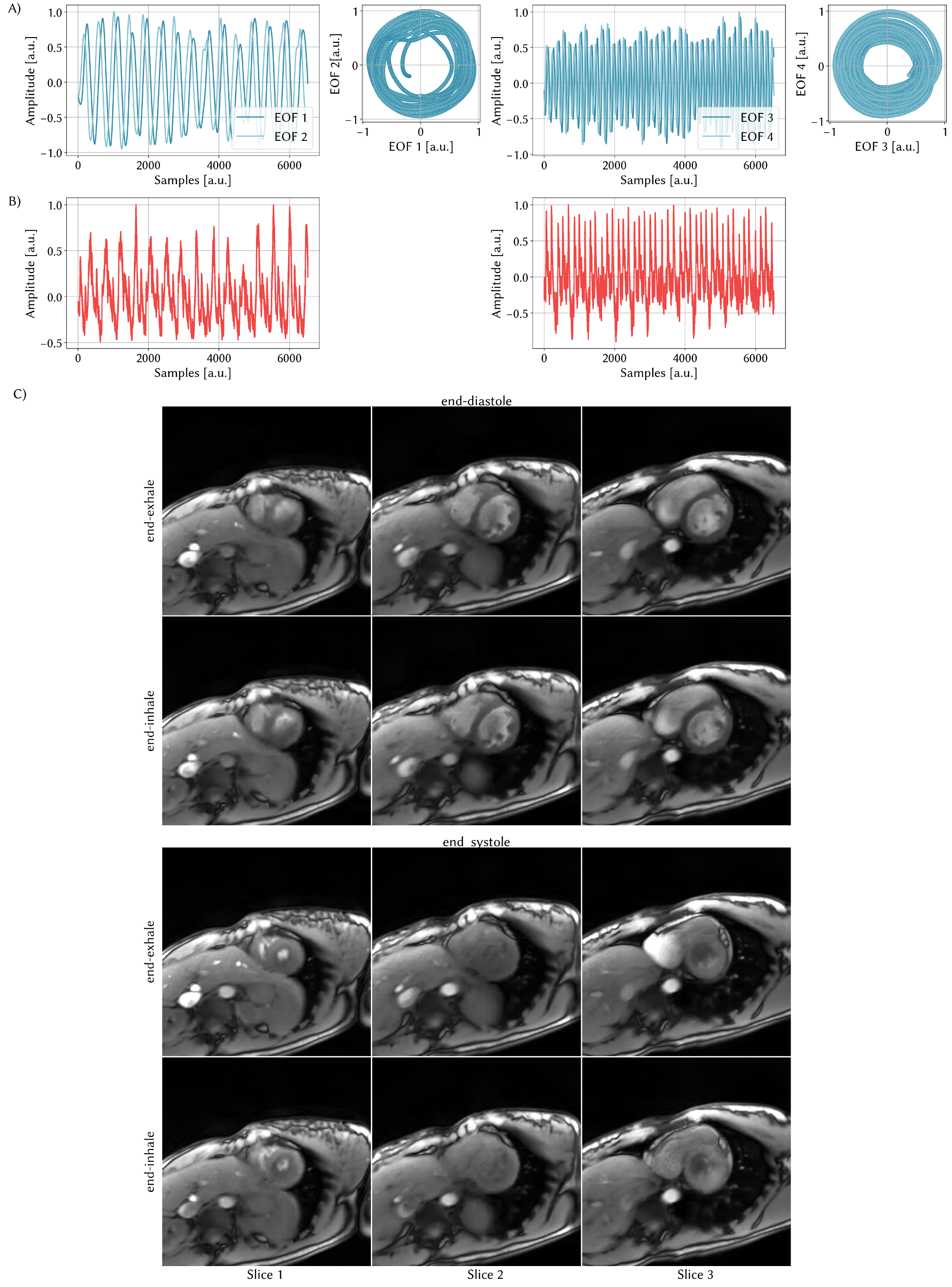
Figure 2A shows the SSA-FARI self-gating plots of the first EOF pairs representing respiratory (left) and cardiac motion (right). In the phase portrait the quadrature nature of the EOF pairs can be appreciated. Compared to the SSA-FARI self-gating signal, the PCA result (Figure 2B) is spoiled by noise and would require further post-processing.
Figure 2C shows representative frames of the reconstruction based on SSA-FARI self-gating. All cardiac and respiratory phases have been correctly detected and clearly separated.
PCA does not eliminate noise, requires further processing and, as a linear dimensionality reduction technique, cannot capture the possibly non-linear manifold on which the self-gating signal lives. Non-linear dimensionality reduction techniques are often limited to 2D-imaging or the detection of respiratory motion only [22-24]. SSA-FARI proofs to be an easy to implement, yet powerful and robust alternative for self-gating.
Conclusion
We introduced a novel and robust self-gating method based on Singular Spectrum Analysis (SSA-FARI). It extracts reliably cardiac and respiratory motion, eliminates noise and thereby outperforms the conventional PCA method, which in practice requires further processing [25-27]. Although not shown here, SSA-FARI can also be used for self-gating in Single-Slice and Stack-of-Stars imaging (FLASH and bSSFP).Acknowledgements
No acknowledgement found.References
[1] Ehman RL, McNamara M, Pallack M, Hricak H, Higgins C. Magnetic resonance imaging with respiratory gating: tech-niques and advantages. Am J Roentgenol 1984; 143:1175–1182.
[2] Liu YL, Riederer SJ, Rossman PJ, Grim RC, Debbins JP, Ehman RL. A monitoring, feedback, and triggering system forreproducible breath-hold MR imaging. Magn. Reson. Med. 1993; 30:507–511.
[3] Higgins CB, Hricak H. Magnetic resonance imaging of the body. 1987; DOI: 10.1016/c2009-0-52819-x.
[4] Rokey R, Wendt RE, Johnston DL. Monitoring of acutely III patients during nuclear magnetic resonance imaging: Use of aTime-varying filter electrocardiographic gating device to reduce gradient artifacts. Magn Reson Med 1988; 6:240–245.
[5] Larson AC, White RD, Laub G, McVeigh ER, Li D, Simonetti OP. Self-gated cardiac cine MRI. Magn. Reson. Med. 2004;51:93–102.
[6] Uribe S, Muthurangu V, Boubertakh R, Schaeffter T, Razavi R, Hill DLG, Hansen MS. Whole-heart cine MRI using real-time respiratory self-gating. Magn. Reson. Med. 2007; 57:606–613.
[7] Paul J, Divkovic E, Wundrak S, Bernhardt P, Rottbauer W, Neumann H, Rasche . High-resolution respiratory self-gatedgolden angle cardiac MRI: comparison of self-gating methods in combination with k-t SPARSE SENSE. Magn. Reson. Med.2015; 73:292–298.
[8] Pang J, Sharif B, Fan Z, Bi X, Arsanjani R, Berman DS, Li D. ECG and navigator-free four-dimensional whole-heart coro-nary MRA for simultaneous visualization of cardiac anatomy and function. Magn. Reson. Med. 2014; 72:1208–1217.
[9] Gao Y, Zhou Z, Han F, Finn PJ, Hu P. Improved respiratory motion self-gating in cardiovascular MRI. J. Cardiov. Magn.Reson. 2016; 18:P7.
[10] Rosenzweig S, Scholand N, Holme HCM, Uecker M. Robust Cardiac and Respiratory Self-Gating using an adapted Singular Spectrum Analysis. In Proc. 22nd SCMR, Bellevue 2019; accepted.
[11] Vautard R, Yiou P, Ghil M. Singular-spectrum analysis: A toolkit for short, noisy chaotic signals. Physica D 1992; 58:95–126.
[12] Vautard R, Ghil M. Singular spectrum analysis in nonlinear dynamics, with applications to paleoclimatic time series. Phys-ica D 1989; 35:395–424.
[13] Groth A, Ghil M. Multivariate singular spectrum analysis and the road to phase synchronization. Phys. Rev. E 2011;84:036206.
[14] Larson AC, White RD, Laub G, McVeigh ER, Li D, Simonetti OP. Self-gated cardiac cine MRI. Magn. Reson. Med. 2004;51:93–102.
[15] Feng L, Axel L, Chandarana H, Block KT, Sodickson DK, Otazo R. XD-GRASP: Golden-angle radial MRI with reconstruc-tion of extra motion-state dimensions using compressed sensing. Magn. Reson. Med. 2016; 75:775–788.
[16] Uecker M, Lai P, Murphy MJ, Virtue P, Elad M, Pauly JM, Vasanawala SS, Lustig M. ESPIRiT—an eigenvalue approach toautocalibrating parallel MRI: where SENSE meets GRAPPA. Magn. Reson. Med. 2014; 71:990–1001.
[17] Rosenzweig S, Holme HCM, Uecker M, Simple Auto-Calibrated Gradient Delay Estimation From Few Spokes Using Radial Intersections (RING), Magn. Reson. Med. 2018, 10.1002/mrm.27506.
[18] Nehrke K, Bornert P, Manke D, Bock JC. Free-breathing cardiac MR imaging: study of implications of respiratory mo-tion—initial results. Radiology 2001; 220:810–815.
[19] Burger I, Meintjes EM. Elliptical subject-specific model of respiratory motion for cardiac MRI. Magn Reson Med 2013;70:722–731.
[20] Feng L, Axel L, Chandarana H, Block KT, Sodickson DK, Otazo R. XD-GRASP: Golden-angle radial MRI with reconstruc-tion of extra motion-state dimensions using compressed sensing. Magn. Reson. Med. 2016; 75:775–788.
[21] Uecker M, Virtue P, Ong F, Murphy MJ, Alley MT, Vasanawala SS, Lustig M. Software toolbox and programming library forcompressed sensing and parallel imaging. In: ISMRM Workshop on Data Sampling and Image Reconstruction, Sedona,2013.
[22] Usman M, Vaillant G, Atkinson D, Schaeffter T, Prieto C. Compressive manifold learning: Estimating one-dimensionalrespiratory motion directly from undersampled k-space data. Magn Reson Med 2014; 72:1130–1140.
[23] Chen X, Usman M, Baumgartner CF, Balfour DR, Marsden PK, Reader AJ, Prieto C, King AP. High-Resolution Self-GatedDynamic Abdominal MRI Using Manifold Alignment. IEEE Trans. Med. Imaging 2017; 36:960–971.
[24] Usman M, Atkinson D, Kolbitsch C, Schaeffter T, Prieto C. Manifold learning based ECG-free free-breathing cardiac CINEMRI. J. Magn. Reson. Imaging 2015; 41:1521–1527.
[25] Paul J, Divkovic E, Wundrak S, Bernhardt P, Rottbauer W, Neumann H, Rasche V. High-resolution respiratory self-gated golden angle cardiac MRI: comparison of self-gating methods in combination with k-t SPARSE SENSE. Magn. Reson. Med. 2015; 73:292–298.
[26] Feng L, Axel L, Latson LA, Xu J, Sodickson DK, Otazo R. Compressed sensing with synchronized cardio-respiratory sparsity for free-breathing cine MRI: initial comparative study on patients with arrhythmias. J. Cardiov. Magn. Reson. 2014; 16:O17.
[27] Zhang T, Cheng JY, Chen Y, Nishimura DG, Pauly JM, Vasanawala SS. Robust self-navigated body MRI using dense coil arrays. Magn. Reson. Med. 2016; 76:197–205.
Figures