2442
Navigated Steady-State Free Precession with Water-Excitation for Real-Time Cardiac Imaging at 3 Tesla1Beckman Institute for Advanced Science and Technology, University of Illinois at Urbana-Champaign, Urbana, IL, United States, 2Paul C. Lauterbur Research Center for Biomedical Imaging, Shenzhen Institutes of Advanced Technology, Shenzhen, China, 3Department of Bioengineering, University of Illinois at Urbana-Champaign, Urbana, IL, United States
Synopsis
Real-time imaging offers the opportunity to be performed without the need for ECG synchronization and breath holding while requiring good contrast and high spatiotemporal resolution to resolve the myocardium dynamics. We propose a novel method employing bSSFP acquisition with partial separable model for high quality real-time cardiac imaging. For acquisition, the new method interleaves a self balanced spiral-in and spiral-out navigator with Cartesian acquisition for temporal basis estimation. A (1-1) binomial water excitation pulse is adopted to suppress lipid signal and achieve steady-state of the water. For reconstruction, the new method exploits the partial separable model with "soft" SENSE and sparsity constraints. In vivo experiments have been conducted and results show that the proposed method is able to produce high quality dynamic cardiac images.
Introduction
Real-time (RT) MRI provides dynamic imaging of the heart without the need for ECG synchronization and breath holding, which is especially essential for patients with arrhythmia1. Previous method2-3 exploit low-rank and sparse model to achieve high spatiotemporal resolution but the contrast is suboptimal due to the use of FLASH sequence. To suppress the lipid signal, fat saturation is commonly used4 but it will interrupt the steady-state. In this work, we propose a new method employing balanced Steady-State Free Precession (bSSFP) acquisition5 with partial separable (PS) model for high quality real-time cardiac imaging. Water excitation6 using a binomial series (1-1) of spatially-selective RF pulse train is employed to suppress the lipid signal. Real-time spatiotemporal function is reconstructed from highly undersampled imaging data using PS model with "soft" SENSE and sparsity constraints. Results from in vivo experiment demonstrated the potential of the proposed method in producing high quality cardiac images.Method
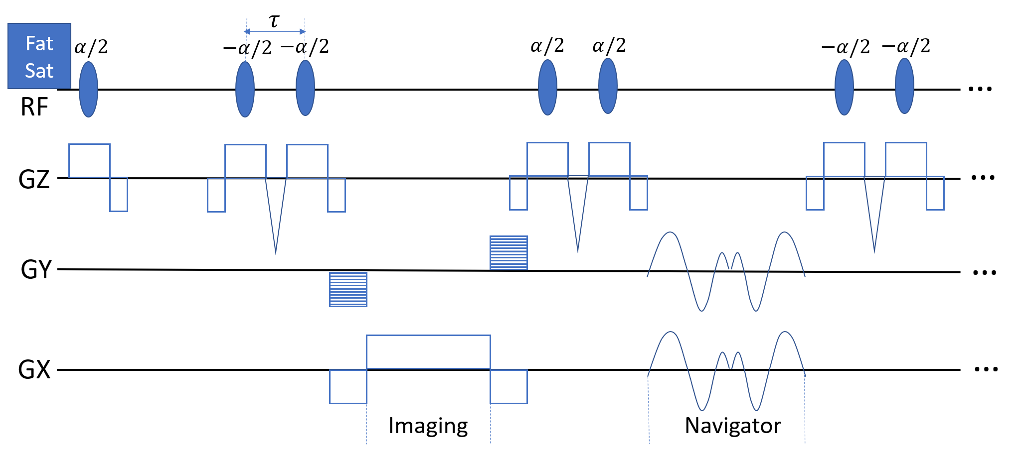
Navigated bSSFP with Water Excitation:
The designed bSSFP pulse sequence is illustrated in Fig. 1. The initial α/2 pulse is applied right after a standard fat saturation module to suppress the lipid signal at the very beginning. Afterwards, binomial pulse is employed for all the dummy, imaging and navigating acquisitions to continuously suppress any fat signal remaining or recovered during the process in the longitudinal direction. In this work, we use the simplest 1-(180°)-1 binomial pulse, which consists of two α/2 pulses with inter-pulse delay (τ) chosen to allow 180° of phase evolution between water and fat spins (τ = 1.1ms at 3 Tesla). Increasing the order of binomial pulse will improve spectral selectivity but will further compromise TR.
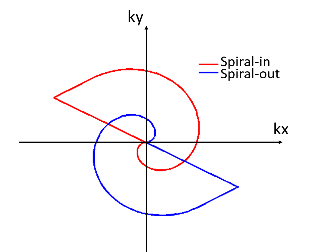
The trajectory of spiral-in and spiral-out navigator is shown in Fig. 2., which is interleaved with conventional Cartesian acquisition for estimation of temporal basis functions. The designed navigator is expected to be able to efficiently capture temporal variations along both directions (i.e., x and y) with good SNR. The navigator is self-balanced owing to the symmetry of the spiral-in and spiral-out trajectory, thus it can be well accommodated with the steady-state acquisition. Note that the apply frequency of the navigator is rather flexible that it is adjustable according to the desired frame rate.
Image Reconstruction using PS Model with "soft" SENSE and Sparsity Constraints:
After obtaining the undersampled k-space data, the spatiotemporal function can be reconstructed via solving the following optimization problem:
$$\hat{\mathbf{U}}=\underset{\mathbf{U}}{\operatorname{argmin}}\sum_{c=1}^{C}||\Omega F\mathbf{S}_c\mathbf{U}V-d_c||_2^2+\lambda_1||\Psi U||_1+\lambda_2||UV_f||_1$$
where $$$d_c$$$ denotes the acquired k-space data vector from the $$$c$$$-th coil, $$$\Omega$$$ selects the acquired locations in k-space, $$$F$$$ represents the Fourier encoding operator. Rows in $$$V\in \mathbb{C}^{r\times Nt}$$$ stores the temporal basis functions estimated from the navigator signal with $$$r$$$ the chosen rank and $$$N_t$$$ the number of time points. $$$\mathbf{S}_c=[S_c^1,S_c^2,...,S_c^L]$$$ denotes the set of coil sensitivity functions for the $$$c$$$-th coil calibrated from the complete time averaged k-space data using ESPIRiT7. A "soft" SENSE formulation7 (i.e., L>1) is employed here to improve robustness to motion. For L>1, there exists L spatial coefficient maps need to be estimated (i.e., $$$\mathbf{U}=[U^1;U^2;...;U^L]\in \mathbb{C}^{NL\times r}$$$) with $$$\mathbf{S}_c\mathbf{U}=\sum_{l=1}^{L}S_c^lU^l$$$ and N the total number of image pixels. In this study, we use L=2. The regularization terms impose sparsity of the coefficient maps in transform domain $$$\Psi $$$ and spatiospectral sparsity of the desired spatiotemporal function respectively. Half-quadratic regularization with continuation strategy is adopted to solve the optimization problem.
Experiment and Results
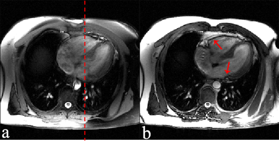
Free breathing cardiac data were collected from a healthy human subject on a 3T MR scanner using a spinal coil and a flexible phase array coil with (TR/TE 4.04/2.02 ms) and without (TR/TE 3.24/1.62) water excitation (FOV 340$$$\times$$$340 mm$$$^2$$$, resolution 1.3$$$\times$$$1.3 mm$$$^2$$$, slice thickness 8mm, FA $$$\alpha$$$ 43°). Navigator signals were acquired twice every 8 TRs, enabling a frame rate of 40ms for water excitation and 33 ms for non water excitation cases.
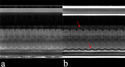
Reconstructed images are shown in Fig. 3 and Fig. 4. Both cases achieved high spatial and temporal resolution in resolving the dynamics of the heart. With water excitation incorporated, significant lipid signal attenuation in epicardial fat (red arrows) can be observed.
Conclusion
This work developed a new bSSFP sequence with interleaved spiral-in and spiral-out navigator acquisition and binomial water excitation. Results from in vivo experiments have been obtained to demonstrate the potential of the proposed method.Acknowledgements
This work was supported in part by National Key R&D Program of China 2016YFC0100103 and NSFC-61671441.References
- Zhang S., Joseph A.A., Voit D., et al. Real-time magnetic resonance imaging of cardiac function and flow—recent progress. Quant Imaging Med Surg. 2014 Oct; 4(5): 313–329.
- Zhao B, Haldar JP, Christodoulou AG ,Liang ZP. Image reconstruction from highly undersampled (k, t)-space data with joint partial separability andsparsity constraints. IEEE Transactions on Medical Imaging 31 (9), 1809-1820
- Christodoulou AG, Zhang H, Zhao B, Hitchens TK, Ho C, Liang Z-P. High-Resolution Cardiovascular MRI by Integrating Parallel Imaging WithLow-Rank and Sparse Modeling. IEEE transactions on bio-medical engineering. 2013;60(11):3083-3092.
- Li Feng Simone Coppo Davide Piccini Jerome Yerly Ruth P. Lim Pier Giorgio Masci Matthias Stuber Daniel K. Sodickson Ricardo Otazo. 5D whole‐heart sparse MRI. Magn Reson Med. 2018 Feb;79(2):826-838
- Schär, M., Kozerke, S., Fischer, S. E. and Boesiger, P. (2004), Cardiac SSFP imaging at 3 Tesla. Magn. Reson. Med., 51: 799–806.doi:10.1002/mrm.20024
- Hung-Yu Lin, Subha V Raman, Yiu-Cho Chung, Orlando P Simonetti. Rapid phase-modulated water-excitation steady-state free precession for fat-suppressed cine cardiovascular MR. Journal of Cardiovascular Magnetic Resonance, 2008, Volume 10, Number 1, Page 1
- M. Uecker, P. Lai, M. J. Murphy, P. Virtue, M. Elad, J. M. Pauly,S. S. Vasanawala, and M. Lustig. Espirit-an eigenvalue approach toautocalibrating parallel mri: Where sense meets grappa. Magn ResonMed, vol. 71, no. 3, pp. 990–1001, 3 2014.
Figures