2441
Rapid Parallel MRI with Convolution-based Reconstruction (CORE) and Deblurring by Compressed Sensing1Department of Biomedical Engineering, Technion - Israel Institute of Technology, Haifa, Israel, 2Department of Radiology, Leiden University Medical Center (LUMC), Leiden, Netherlands
Synopsis
Methods combining Compressed Sensing (CS) and Parallel MRI (PI) for accelerated MRI have shown great promise, yet they are commonly hindered by heavy iterative computations. This work introduces the novel CORE-Deblur method for accelerated MRI, which integrates CS and PI and offers fast computations with very few iterations. CORE-Deblur utilizes the recently introduced CORE-PI technique and introduces the novel concept of using CS for image deblurring. Experiments with in-vivo data show that for highly subsampled k-space (R=5) CORE-Deblur reduces the number of CS iterations by 10-fold (from 95 to about 5-7) and improves the reconstruction accuracy by 5%-8%.
Introduction
Compressed Sensing (CS) and Parallel MRI (PI) have emerged as two useful approaches for accelerating MRI scans by k-space subsampling and subsequent reconstruction schemes. CS methods utilize image sparsity in the domains of non-Fourier transforms (e.g. the wavelet transform), and PI methods utilize multicoil acquisition and information about the coils sensitivity maps for image reconstruction. While methods combining CS and PI1–7 have shown great promise, they are hindered by a heavy computational burden, which is mainly due to massive iterative computations.
This work proposes a novel reconstruction method utilizing both CS and PI, with simple computations and very few iterations. The proposed method utilizes: (1) the recently introduced CORE-PI technique8, which computes the convolution of the unknown MR image with a known user-defined kernel, and (2) a unique implementation of CS for reconstruction by deblurring the convolved image.
Theory
The proposed reconstruction method utilizes the CORE-PI8 technique, which is a unique parallel MRI reconstruction method: it has as inputs highly undersampled parallel k-space data, estimated sensitivity maps and a user-defined kernel $$$g(x)$$$, and computes the convolution between the unknown MR image $$$f(x,y)$$$ and the kernel:
$$f^{conv}(x,y)=f(x,y)*g(x) \quad\quad\quad(1)$$
CORE-PI is suitable for a 2D Cartesian k-space subsampled with a 1D subsampling scheme, i.e. using a subset of columns or rows. The method offers subsampling flexibility: both regular and random sampling schemes are possible. Furthermore, CORE-PI offers simple linear computations, and a flexible kernel choice: any kernel that can be represented by a curve containing $$$N_c$$$ points, where $$$N_c$$$ is the number of coils, is suitable.
The proposed reconstruction method, which is coined CORE-Deblur, consists of two steps: (i) computation of the convolved image $$$f^{conv}(x,y)$$$ using CORE-PI, and (ii) image reconstruction by implementation of a CS reconstruction that is initiated from the convolved image. The CS reconstruction solves the following convex optimization problem:
$$min\parallel{\boldsymbol{\Psi} f(x,y)}\parallel_1 \quad s.t.\quad \parallel{\boldsymbol{F_s} C_nf(x,y)-y_n}\parallel_2<\epsilon \quad n[1,N_c] \quad\quad\quad (2)$$
where $$$\boldsymbol{\Psi}$$$ is a sparsifying transform, $$$y_n$$$ are the k-space samples acquired by coil , $$$C_n$$$ is the sensitivity map of coil $$$n$$$ and $$$\bf{F_s}$$$ is an operator describing the Fourier transform and the subsampling. Eq. (2) describes a coil-by-coil CS image reconstruction process with a multi-coil joint sparsity constraint. Once the individual coil images are reconstructed, $$$f(x,y)$$$ is obtained by merging them using Roemer’s optimal method9.
Since the process described in eq. (2) is initiated from the convolved (i.e. blurred) image $$$f^{conv}(x,y)$$$ and produces the deblurred image $$$f(x,y)$$$, it may be conceived as an image deblurring process.
Methods
The proposed reconstruction method was implemented on in-vivo brain data obtained with a 32-coil array from two T1-weighted-7T scans of healthy volunteers. Sensitivity maps were estimated from low-resolution pre-scans. K-space data acquired in high-resolution scans were retrospectively sub-sampled in one dimension using a regular scheme with a reduction factor of R=5. The CORE-Deblur method was implemented with a Gaussian kernel with $$$\sigma=0.5$$$. The CS problem was solved using the Projection Onto Convex Sets (POCS) approach10 with a Daubechies-2 wavelet. Computations were performed in Matlab on an HP-spectre-x360 PC.
The proposed method was compared to a coil-by-coil CS-MRI11 reconstruction. The latter also solves the convex optimization problem of eq. (2) using POCS, but it is initialized by the conventional method: k-space Zero Filling and an Inverse Fourier Transform. Reconstruction errors were computed regarding gold standard images obtained from the fully sampled k-space. Errors were measured using the Normalized Root Mean Square Error (NRMSE) measure.
Results & Discussion
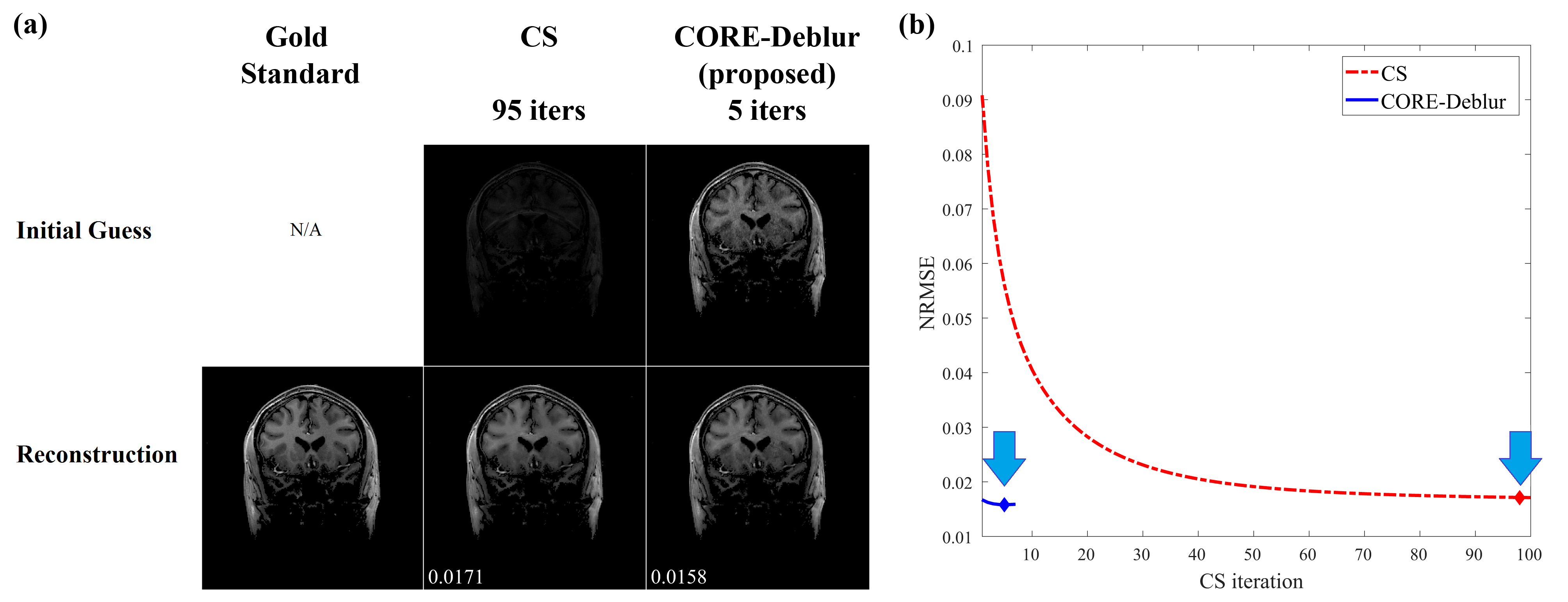
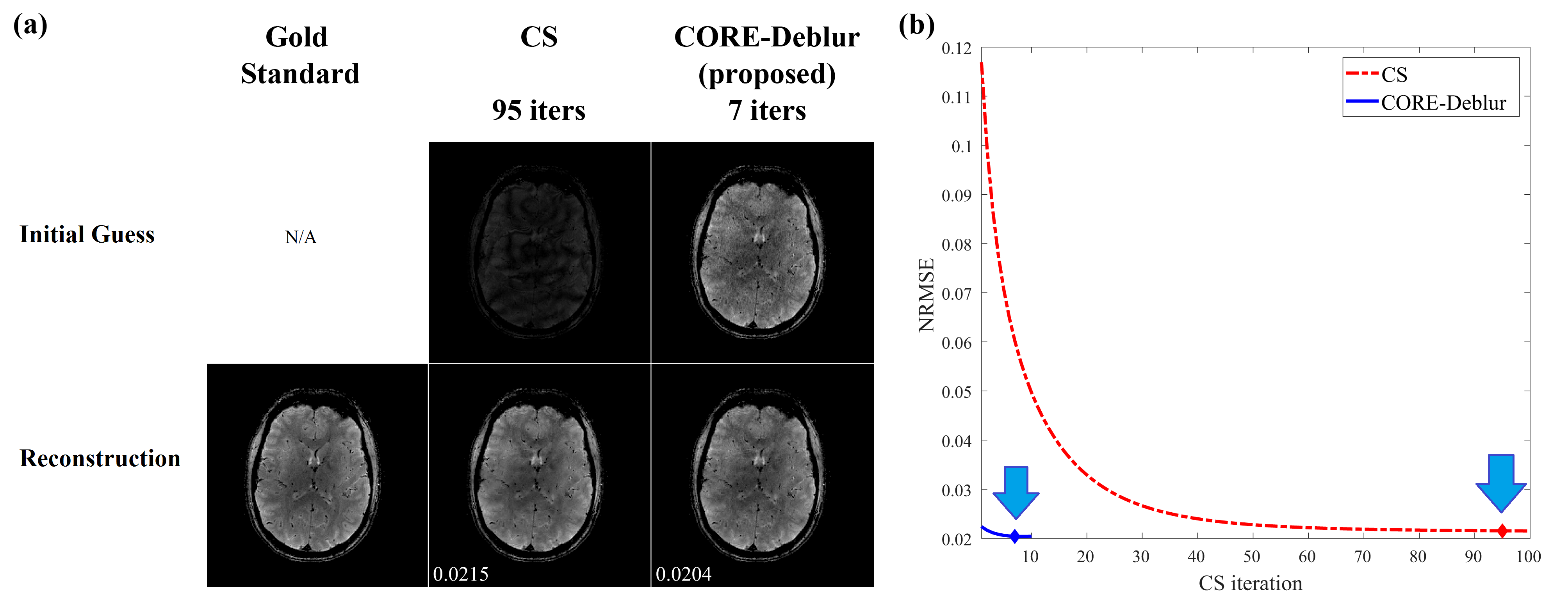
Figures 1 and 2 demonstrate the reconstructions obtained by the proposed CORE-Deblur method from only 20% of k-space data (R=5), and compare them to the gold standard images and the CS reconstructions. The results show that the CORE-Deblur technique produces initial guesses which are quite close to the gold standards, without discernible artifacts.
Strikingly, CORE-Deblur converges rapidly, within only 5-7 iterations, whereas CS requires about 95 iterations (Figs. 1,2). In both experiments, CORE-Deblur also obtains highly accurate final reconstructions, with errors lower than CS by 7.6% (Fig 1) and 5.1% (Fig. 2).
The results therefore demonstrate that CORE-Deblur offers a significantly lower computational burden than CS, and a better reconstruction quality.
Conclusion
This work introduces the novel CORE-Deblur method for accelerated MRI, and the concept of using CS for deblurring.
Results from in-vivo data show that: (1) CORE-Deblur produces high quality reconstructions, better than those of CS by 5%-8%, and (2) requires significantly fewer iterations (90% less). The method is therefore suitable for real-time MRI applications.
Acknowledgements
No acknowledgement found.References
1. Lustig, M. & Pauly, J. M. SPIRiT: Iterative self-consistent parallel imaging reconstruction from arbitrary k-space. Magn. Reson. Med. 64, 457–471 (2010).
2. Ying, L. & Sheng, J. Joint image reconstruction and sensitivity estimation in SENSE (JSENSE). Magn. Reson. Med. 57, 1196–1202 (2007).
3. She, H., Chen, R. R., Liang, D., Dibella, E. V. R. & Ying, L. Sparse BLIP: BLind Iterative Parallel imaging reconstruction using compressed sensing. Magn. Reson. Med. 71, 645–660 (2014).
4. Feng, L. et al. Golden-angle radial sparse parallel MRI: Combination of compressed sensing, parallel imaging, and golden-angle radial sampling for fast and flexible dynamic volumetric MRI. Magn. Reson. Med. 72, 707–717 (2014).
5. Otazo, R., Kim, D., Axel, L. & Sodickson, D. K. Combination of compressed sensing and parallel imaging for highly accelerated first-pass cardiac perfusion MRI. Magn. Reson. Med. 64, 767–776 (2010).
6. Wu, B., Millane, R. P., Watts, R. & Bones, P. Applying compressed sensing in parallel MRI. in Proceedings of the 16th Annual Meeting of ISMRM Vol. 1480 (2008).
7. Vasanawala, S. et al. Practical parallel imaging compressed sensing MRI: Summary of two years of experience in accelerating body MRI of pediatric patients. in IEEE International Symposium on Biomedical Imaging: From Nano to Macro 1039–1043 (IEEE, 2011).
8. Shimron, E., Webb G., A. & Azhari, H. CORE-PI: Non-iterative Convolution-based Reconstruction for Parallel MRI in the Wavelet Domain. Med. Phys. (2018). doi:10.1002/MP.13260
9. Roemer, P. B., Edelstein, W. A., Hayes, C. E., Souza, S. P. & Mueller, O. M. The NMR phased array. Magn. Reson. Med. 16, 192–225 (1990).
10. Daubechies, I., Defrise, M. & De Mol, C. An iterative thresholding algorithm for linear inverse problems with a sparsity constraint. Commun. Pure Appl. Math. 57, 1413–1457 (2004).
11. Lustig, M., Donoho, D. & Pauly, J. M. Sparse MRI: The application of compressed sensing for rapid MR imaging. Magn. Reson. Med. 58, 1182–1195 (2007).
Figures