2434
A novel feature based image reconstruction for neuro-interventional MRI1Biomedeical Engineering, Shanghai Jiao Tong University, Shanghai, China, 2KTH Royal Institute of Technology, Stockholm, Sweden, 3Functional Neurosurgery, Shanghai Jiao Tong University, Shanghai, China, 4Electrical and Computer Engineering, University of Illinois at Urbana-Champaign, Urbana, IL, United States
Synopsis
Interventional MRI (I-MRI) provides exceptional advantages to other imaging modalities in image-guided neurosurgery. However, real-time imaging presents great challenges for temporal/spatial resolution, image contrast, and SNR. We presented a novel feature based image reconstruction algorithm using golden-angle sampling and compressed sensing. Images were decomposed into the reference part and the novel feature reflecting the interventional process. Experiments of using porcine brain for biopsy showed the proposed method had better performance in terms of SNR and computational time. It demonstrated that the proposed method have potentials in applications of MR-guided intervention such as image-guided epilepsy treatment.
Introduction
Interventional MRI (I-MRI) plays a crucial role in MR guided therapy such as MR guided neurosurgery. For interventional procedures such as deep brain stimulation (DBS) in functional neurosurgery, MR guidance could provide exceptional advantages to other imaging modalities (1) To achieve better temporal and spatial resolutions in I-MRI, many methods have been proposed (2). Data acquisition schemes such as non-Cartesian sampling and keyhole imaging were used (3). Compressed sensing (CS) also showed to be effective in I-MRI (3-6). Recently, Golden-Angle Radial Sparse Parallel MRI (GRASP) (3) combing radial sampling and CS showed good performance in dynamic cardiac imaging. However, these methods may not be directly applicable to neuro I-MRI since it has relatively higher demand for image contrast and SNR. In this study, using radial sampling scheme, we proposed a novel feature based image reconstruction method for I-MRI. Results were compared with that from GRASP, keyhole, and NUFFT based on I-MRI experiments using porcine brain.
Methods
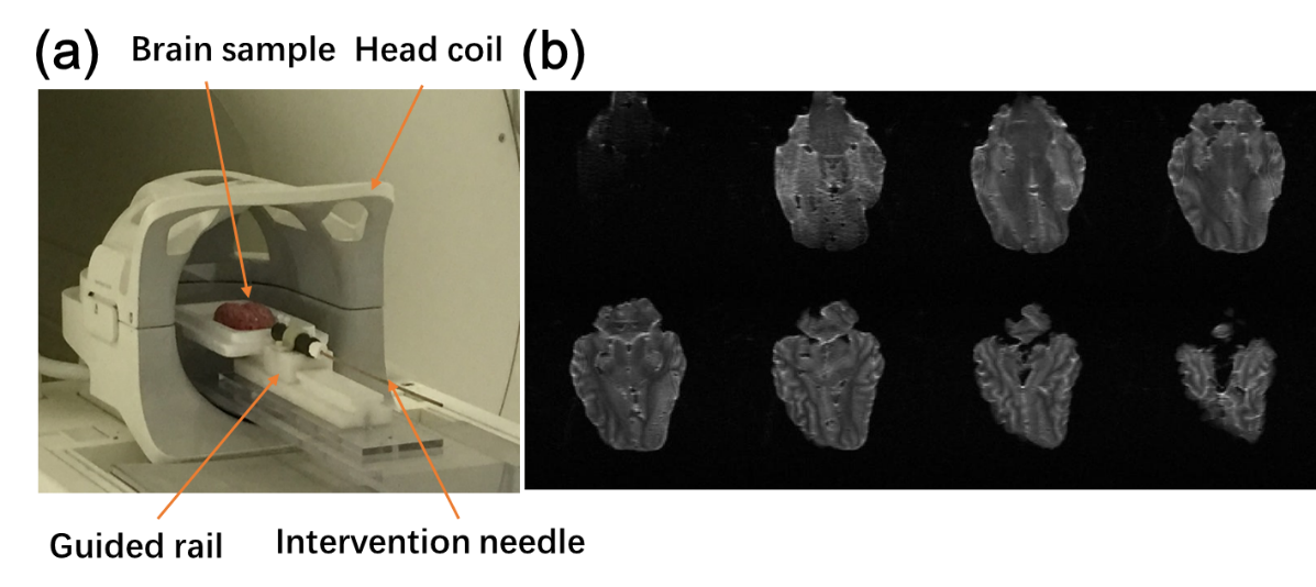
Porcine brains were acquired from local market. A custom-built interventional device was used for the I-MRI experiment (Figure 1). An interventional needle was fixed on a guided rail with a ruler onside showing the biopsy distance. A FLASH sequence with 3D Stack-of-Stars (SOS) golden angle radial acquisition scheme was used to image the interventional procedure. The 3D data consisted of 8 slices with a thickness of 3 mm, and TE/TR was 1.76/3.85 ms. All imaging were carried out in a 3.0T scanner (uMR780, United Imaging Healthcare, Shanghai, China) with a 24 channel head & neck coil. A total of 400 spokes were collected with 256 readout points for each spoke. We used 21 spokes to reconstruct each time frame, with a temporal resolution of 646.8 ms for a volume of 8 slices.
The dynamic image x can be decomposed into the undeformed reference xr and the novel feature dx . Let TV (temporal Total Variation) and W (Wavelet linear operator) denote sparsifying transforms where a and b are regularization weights. We reconstruct the novel feature by
$$arg \min_{dx} \parallel E\cdot (dx +x_{r})-y\parallel_2^2 +a\cdot\parallel TV(dx+x_{r})\parallel_{1} + b\cdot\parallel W(dx+x_{r})\parallel_{1}$$
Where E is the under-sampled non-uniform fast Fourier transform (NUFFT) corresponding to the radially sampled trajectory, y is the measurement. The final reconstructed image is $$x = x_{r}+dx$$
For neuro intervention, signal-to-noise ratio (SNR) and image contrast are important to trace the needle position. Therefore, we evaluate the reconstructed images by comparing the SNR values. In addition, computational time and local contrast values were also compared.
Results
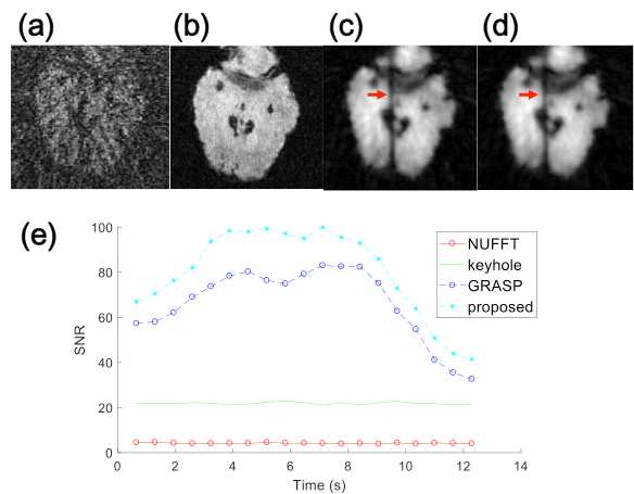
Reconstruction results showed that both NUFFT
and keyhole methods could not capture the intervention process using 21
sampled
spokes. While GRASP and the proposed method showed the clear position of
the
needle, the proposed method had a better SNR throughout the time frames
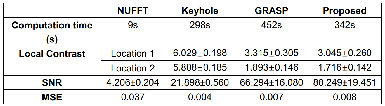
(Figure 2). The reconstruction time were evaluated using a PC with Intel
i5-2520M
core, 2.50 GHz CPU, and 4GB RAM. Local contrast values were estimated at
a
location close to the needle and away from the needle. Results showed
that the proposed
method had better performance computational time and SNR but no
significant
differences in other metrics.Discussion
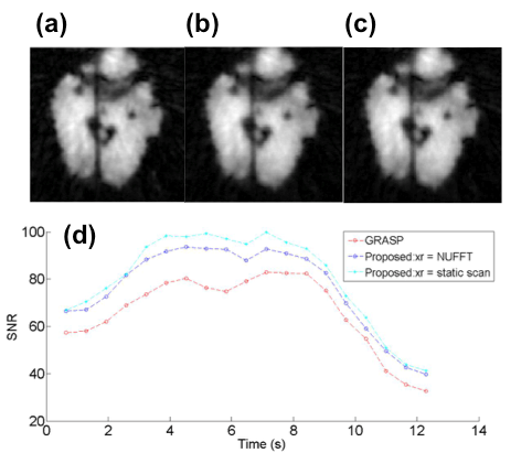
In this study, we proposed a novel feature based reconstruction scheme for I-MRI. By reconstruction with respect to the interventional feature, we achieved a faster reconstruction with better SNR compared with GRASP. However, the reference image xr is crucial for this method. We compared the different xr input and found that anatomical images with better resolution and contrast benefit the final reconstruction results (Figure 3). Future studies include using the FISP sequence to get more details of brain.Acknowledgements
Funding is provided by grant 31870941 (YF) from National Natural Science Foundation of China. Start-up grant from SJTU is also acknowledged.References
1. Peters TM, Cleary K. Image-Guided Interventions. Tecnhology and Applications. Boston: Springer; 2008.
2. Campbell-Washburn AE, Faranesh AZ, Lederman RJ, Hansen MS. Magnetic Resonance Sequences and Rapid Acquisition for MR-Guided Interventions. Magn Reson Imaging Clin N Am. 2015;23(4):669-79.
3. Feng L, Grimm R, Block KT, Chandarana H, Kim S, Xu J, et al. Golden-angle radial sparse parallel MRI: Combination of compressed sensing, parallel imaging, and golden-angle radial sampling for fast and flexible dynamic volumetric MRI. Magnetic Resonance in Medicine. 2014;72(3):707-17.
4. Ajit Shankaranarayanan B, Michael Wendt et al. Radial Keyhole Sequences for Low Field Projection Reconstruction Interventional MRI. J Magn Reson Imaging. 2001;142-151(12).
5. Song HK, Dougherty L. Dynamic MRI with projection reconstruction and KWIC processing for simultaneous high spatial and temporal resolution. Magn Reson Med. 2004;52(4):815-24.
6. Christodoulou AG, Brinegar C, Haldar JP, Zhang H, Wu YJL, Foley LM, et al. High-resolution cardiac MRI using partially separable functions and weighted spatial smoothness regularization. international conference of the IEEE engineering in medicine and biology society; 2010.
Figures