2431
Single-lung dynamics assessed using XD-GRASP MRI and automatic segmentation1Department of Radiology, NYU School of Medicine, New York, NY, United States
Synopsis
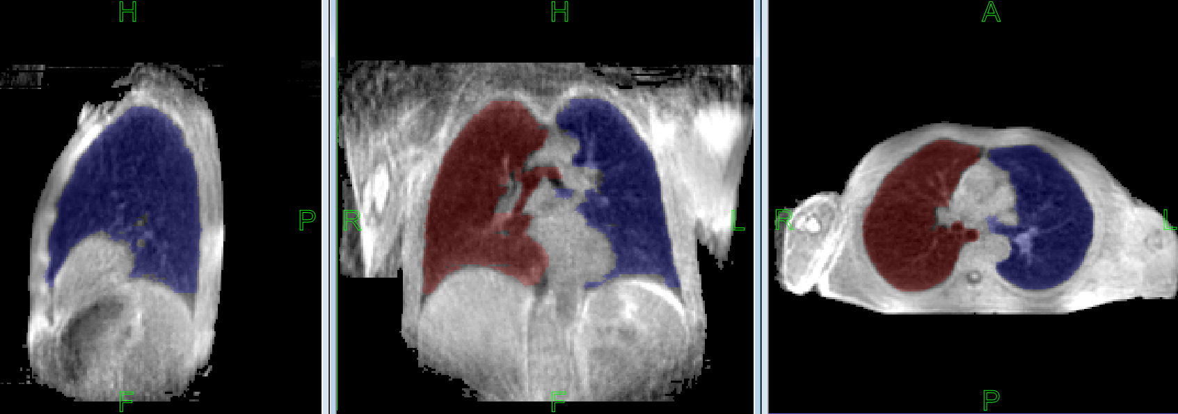
MRI is an attractive modality for monitoring dynamic lung function since it does not expose patients to ionizing radiation. In this study, a method to automatically segment the right and left lungs using XD-GRASP MRI was developed. The accuracy of our segmentation algorithm was assessed by comparing the automated segmentation results to manual segmentations outlined by expert observers. Excellent agreement was seen between the automated technique and the ground truth, suggesting clinical applicability of the method.
INTRODUCTION
MRI is an attractive modality for monitoring dynamic lung function since it does not expose patients to ionizing radiation. However, a well-known limitation of lung MRI is the slow acquisition speed and associated need for breath-holding, which is hard in patients with pulmonary disease. XD-GRASP, a recently developed fast 4D radial imaging method based on compressed sensing may enable clinically useful dynamic lung imaging in free-breathing subjects [1]. This sequence provides a clinically useful temporal (~10 Hz) and spatial resolution. Compared to breath-hold images, XD-GRASP images improve image quality but may present intensity inhomogeneities and artifacts that make it challenging to automate lung segmentation. The present study describes and validates a method to automatically segment the right and left lungs from clinical free-breathing dynamic XD-GRASP acquisitions. The accuracy of our segmentation algorithm is assessed by comparing the automated segmentation results to manual segmentations outlined by expert observers as the ground truth.MATERIALS AND METHODS
This study was IRB approved and all subjects provided written informed consent for dynamic lung MRI. XD-GRASP MR images of the lungs were obtained on a whole body 3T scanner (Tim Trio, Siemens AG, Erlangen, Germany) equipped with the standard 12-element body matrix coil array and with the subject in the supine position [1]. Images from five healthy volunteers and five patients diagnosed with COPD were randomly selected from 50 dynamic imaging studies performed on 24 subjects. XD-GRASP Images were reconstructed into 180 x 180 x 180 x 30 matrix of isotropic 2 mm voxels and 30 respiratory phases.
The automated lung segmentation procedure starts with a data cropping stage. Signal nonuniformity-correction, locally adaptive thresholding (using a large 10 cm radius) and maximum connected component analysis is applied to the initial time-point. The resulting 3D region is presumed to include all soft tissue in the torso. The corresponding bounding box is then constructed and inflated by 3 cm in x, y, and z directions to allow for chest motion. The extended bounding box is then used to crop each time-point volume, i.e., the entire 4D dataset. This preprocessing step leaves a substantially smaller dataset and accelerates further processing. Next, a binary object BodyMask(t) is generated at each 3D time point t separately using the same procedure as above. LungMask(t) is then constructed by applying a hole filling operator to BodyMask(t). Two largest 3D connected components from LungMask(t) are extracted and all other components are removed. Then BodyMask(t) are updated as:
BodyMask(t) := BodyMask(t) AND NOT LungMask(t) .
The average signals, Slung and Sbody, are measured from the two updated binary masks LungMask(t) and BodyMask(t). The lung upper threshold is then calculated as (SLung+SBody)/2, and the auxiliary lung mask Aux(t) is extracted from gray-scale images as voxels with signal intensity below this threshold. The lung masks are augmented using set union LungMask(t) AND Aux(t). Finally, the masks is decomposed into two distinct regions for the left and the right lung using a custom wavefront propagation algorithm that tracks the ancestors of each voxel of the wavefront.
While some steps (including nonuniformity correction) of the overall procedure are controlled by adjustable parameters, these were kept constant for the entire validation project. There were no manual interactions. The segmentation accuracy was measured by comparing to the ground truth obtained by the consensus segmentation of two independent experts.
RESULTS
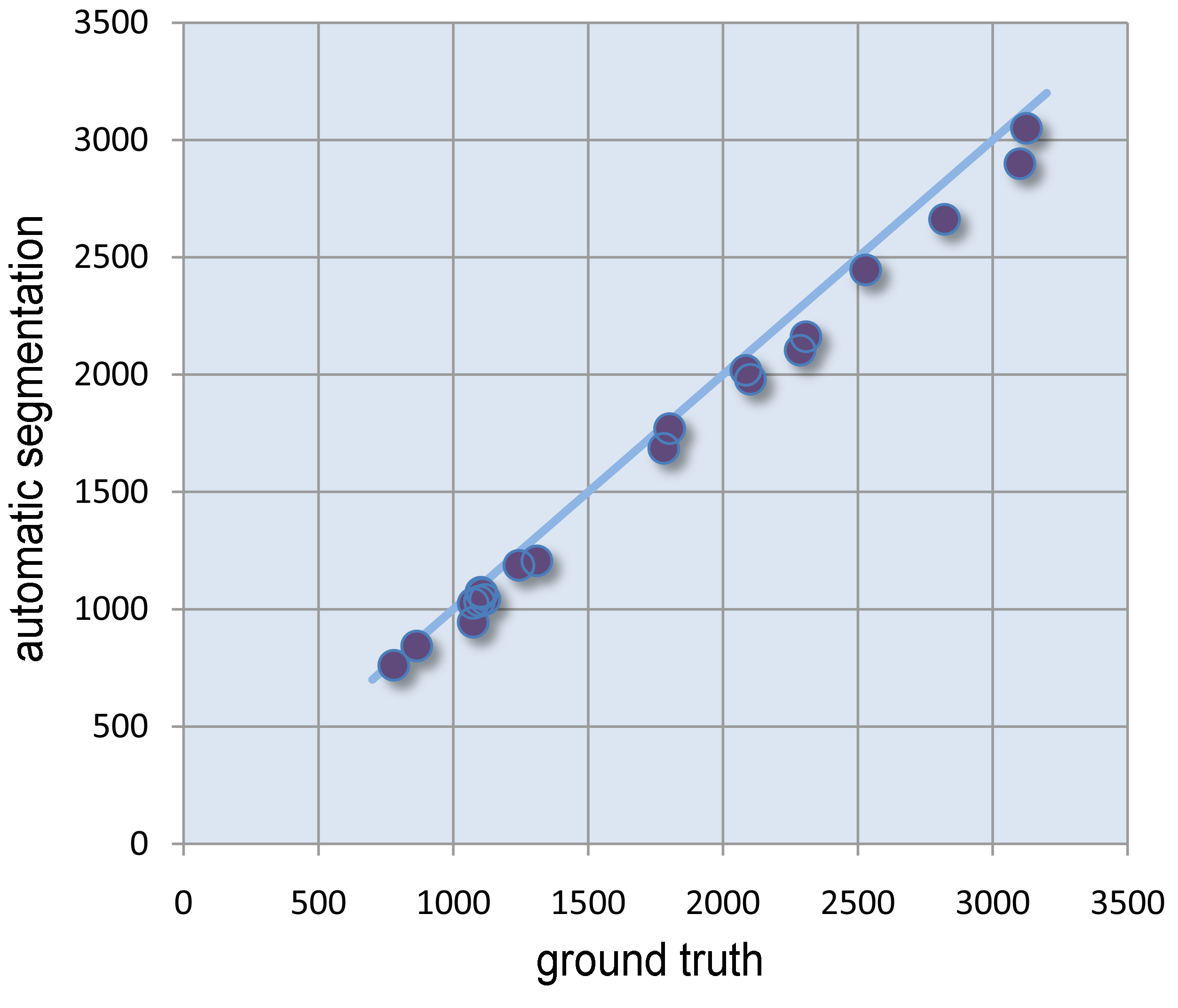
The 20 lungs (10 right and 10 left) selected for validation ranged in volume from 780 to 3125 ml (true volumes). The results of automatic segmentation differed from true values by 4.9%+/-2.5% (average +/- st.dev.). The absolute agreement was excellent, with the intraclass correlation coefficient of 0.991 and its 95% confidence interval [0.973,0.999]. As shown in Figure 2, the segmentation tends to slightly but consistently underestimate the true lung volume.
The average time required for the 4D segmentation of a 30-frame study was 5 min 19 sec on i7-5930K 3.5Ghz processor using all 4 cores\8 threads.
DISCUSSION
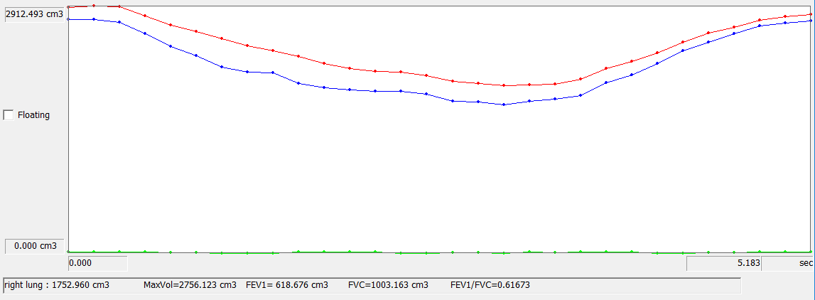
We have developed a fully automatic segmentation method that, when combined with XD-GRASP acquisition, provides structural and functional information of each lung. This method shows an excellent agreement with manual lung volume measurements, which are the current gold-standard. These results suggests that the free-breathing MRI can soon be translated to clinical use and potentially provide spatially resolved respiratory information, which may contribute to better management of patients with lung diseases.Acknowledgements
This work was performed under the rubric of the Center for Advanced Imaging Innovation and Research, an NIBIB Biomedical Technology Resource Center (NIH P41 EB017183).References
1. Feng L, Axel L, Chandarana H, Block KT, Sodickson DK, Otazo R. XD-GRASP: Golden-angle radial MRI with reconstruction of extra motion-state dimensions using compressed sensing. Magn Reson Med 2016 Feb;75(2):775-88.
2. Mikheev A, Nevsky G, Govindan S, Grossman R, Rusinek H. Fully automatic segmentation of the brain from T1-weighted MRI using Bridge Burner algorithm. J MRI. 2008. 27:1235-1241.
3. Mikheev A, Rusinek H, Wiggins G. Non-Uniformity Normalization Using 3D Canny Edges and Legendre Polynomial Approximation of the Bias Field: Validation on 7T T1W Brain Images. ISMRM 2013.
Figures