2428
Robust 3D UTE T2* Mapping in MSK Using Fractional Order Bloch Equation1Radiology and Nuclear Medicine, Erasmus Medical Center, Rotterdam, Netherlands, 2Medical informatics, Erasmus Medical Center, Rotterdam, Netherlands
Synopsis
Determination of the T2* relaxation times with biexponential and monoexponential models has limitations, especially in case of complex, heterogeneous materials1. Using fractional order fitting model we can overcome these limitations8,9. In this model the introduced α parameter is the measure of the deviation from the monoexponential decay, and it accounts for micro-structural complexity. To evaluate the fractional order fitting, it was performed in patella tendon from UTE measurements, and we could demonstrate that compared to biexponential and monoexponential models it is less sensitive to variations to SNR.
Introduction
The $$$T_2^*$$$ values of highly organized tissues such as cartilage or trabecular bone are extremely short (0.5ms-5ms). Hence, there is an increasing interest for sequences with ultrashort echo time (UTE) or with zero echo times (ZTE)2,3,4,5. These short TE techniques in complex, heterogeneous materials reveal stretched exponential $$$T_2^*$$$ decay6,7 (model 1) as opposed to a simple exponential (model 2) or biexponential decay (model 3). Fractional order generalization of the Bloch equation provides and alternative way for the description of the relaxation processes. We compared the performance of all three models on in-vivo data from 3D-UTE-MRI measurements.
Methods
As proposed by Magin et al.8,9 in the fractional order representation the transversal relaxation is described by the following equation:
$$ M_{xy}(TE)=M_{xy}(0)E_{\alpha} \Big[ -\Big( \frac{TE}{T_2} \Big) ^{\alpha} \Big] +M_{xy}(\infty)$$
where $$$M_{xy}(0)$$$ is the transversal magnetization at TE=0, $$$M_{xy}(\infty)$$$ is the transversal magnetization at the steady state and $$$E_{\alpha}$$$ is the stretched Mittag-Leffler function: $$$E_{\alpha}(z)=\sum_{k=0}^{\infty} \frac{z^k}{\Gamma(\alpha k+1)}$$$. In case of α=1 , the function is equivalent to the simple exponential function.
The basis of comparison between the different mathematical models were resulting $$$T_2^*$$$ maps of in-vivo volunteer scans.
Data was retrospectively collected from the Jumper Study (a randomized controlled trial in athletes with patellar tendinopathy who play tendon loading sports at least three times a week, aged 18-35 years). MRI of the symptomatic knee was performed using a 3T MR system (Discovery 750, General Electric, Boston, Massachusetts, USA) using a flexible 3.0 T 16-Channel surface coil (NeoCoil, Pewaukee, Wisconsin, USA). The center of the surface coil was aligned with the patellar apex. Regarding 3D-UTE-MRI, a total of 16 echoes were acquired at TEs of 0.032, 0.49, 0.97, 2.92, 4.87, 6.82, 8.77, 10.72, 13.6, 12.67, 16.57, 18.52, 18.7, 20.47, 22.42, 24.37, 26.32 msec. The 16 echoes were scanned in 4 separate multi-echo sequences with 4 echoes in interleaved order. For each multi-echo acquisition the same TR (83.4 ms) was used.
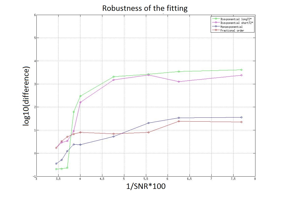
The fitting with all three methods was performed with an in-house developed Matlab script (R2011a; TheMathWorks). In order to test the effect of SNR on the derived $$$T_2^*$$$ values we retrospectively added extra Rician noise to the original magnitude data. The relative $$$T_2^*$$$ difference (difference=$$$\frac{|S_0-S_n|}{S_0}$$$) was evaluated as the function of the SNR ($$$SNR=\frac{mean signal}{\sigma (noise)}$$$).
Results
Figure 1. shows the result of the SNR dependence for all three fitting methods. It is visible that the monoexponential and the fractional order models are less influenced by the noise than the biexponential model.
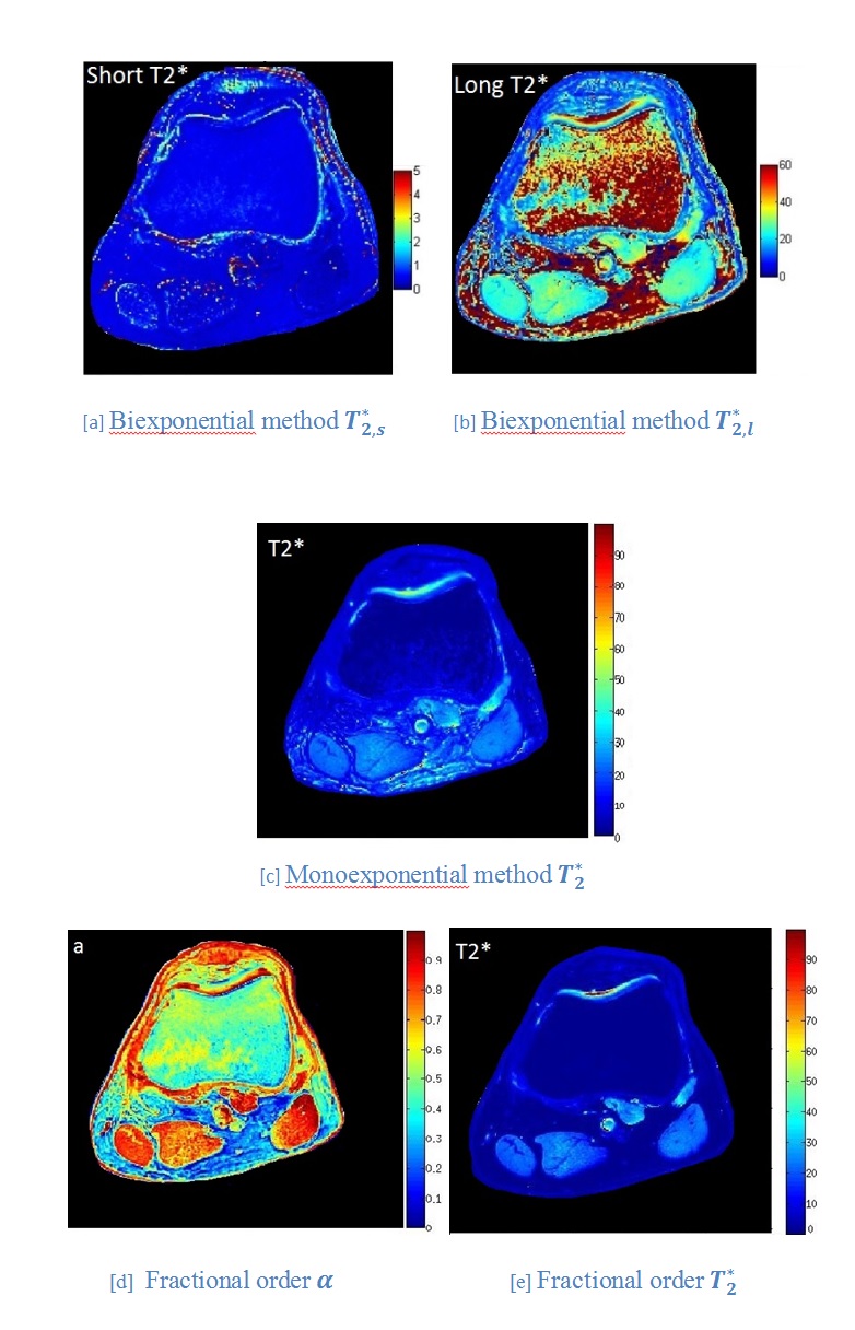
The $$$T_2^*$$$ maps derived by the different mathematical models are shown in figure 2. The background of the images is masked.
Discussion
Testing the robustness of the three different models shows that in case of high SNR the biexponential method performs the best. Above SNR= 25 the mono-exponential and the fractional order methods perform better than the biexponential model, the relative $$$T_2^*$$$ difference is significantly lower. The fractional order $$$T_2^*$$$ maps are smooth, the contrast between different tissues is higher than in case of the monoexponential and biexponential maps. The results depicts the expectation of $$$T_2^*$$$ mapping. The estimated values in the femur (~6ms) are the closest to the ones in the literatue11 in case of the fractional order fitting. The α parameter is different for the different tissues, the parametric maps clearly show the anatomy.Conclusion
The fractional order model is less sensitive to low SNR compared to the biexponential model. The SNR is usually less controlled in longitudinal MR Imaging, the fractional order model outperforms the biexponential model from the perspective of repeatability.
The $$$\alpha$$$ is a novel parameter, potentially a new biomarker characterizing microscopic structure. The fractional order model could be the preferred fitting method in case of low SNR, for tissues which are only visible with UTE or ZTE techniques (e.g.: patella tendon).
Acknowledgements
No acknowledgement found.References
[1] Barbieri, S. , Donati, O. F., Froehlich, J. M. and Thoeny, H. C. (2016), Impact of the calculation algorithm on biexponential fitting of diffusion-weighted MRI in upper abdominal organs. Magn. Reson. Med., 75: 2175-2184. doi:10.1002/mrm.25765
[2] Gatehouse PD, Bydder GM. Magnetic resonance imaging of short T2 components in tissue. Clin Radiol 2003;58:1–19.
[3] Gold GE, Pauly JM, Macovski A, Herfkens RJ. MR spectroscopic imaging of collagen: tendons and knee menisci. Magn Reson Med 1995;34:654–674.
[4] Robson MD, Benjamin M, Gishen P, Bydder GM. Magnetic resonance imaging of the achilles tendon using ultrashort TE (UTE) pulse sequences. Clin Radiol 2004;59:727–735.
[5] Chappell KE, Patel N, Gatehouse PD, et al. Magnetic resonance imaging of the liver with ultrashort TE (UTE) pulse sequences. J Magn Reson Imaging 2003;18:709–13.
[6] Reiter, D. A., Magin, R. L., Li, W. , Trujillo, J. J., Pilar Velasco, M. and Spencer, R. G. (2016), Anomalous T2 relaxation in normal and degraded cartilage. Magn. Reson. Med., 76: 953-962. doi:10.1002/mrm.25913
[7] Johnson, Kevin M. et al. “Optimized 3D Ultrashort Echo Time Pulmonary MRI.” Magnetic resonance in medicine: official journal of the Society of Magnetic Resonance in Medicine / Society of Magnetic Resonance in Medicine 70.5 (2013): 1241–1250. PMC. Web. 12 June 2018.
[8] R. Magin, X. Feng, D. Baleanu, ”Solving the fractional order Bloch equation”, Concepts Magn. Reson.-A, vol. 34, pp. 16-23, 2009.
[9] Magin, R. L., Li, W., Pilar Velasco, M., Trujillo, J., Reiter, D. A., Morgenstern, A., Spencer, R. G. (2011). Anomalous NMR relaxation in cartilage matrix components and native cartilage: Fractional-order models. Journal of Magnetic Resonance, 210(2), 184-191. DOI: 10.1016/j.jmr.2011.03.006 Access to Document 10.1016/j.jmr.2011.03.006
[10] Anastasios Anastasiou, L. D. Hall Optimisation of T2 and M0 measurements of bi-exponential systems Magn Reson Imaging. 2004 Jan; 22(1): 67–80. doi: 10.1016/j.mri.2003.05.005
[11] Tsai PH, Lee HS, Siow TY, et al. Sequential change in T2* values of cartilage, meniscus, and subchondral bone marrow in a rat model of knee osteoarthritis. PLoS One. 2013;8(10):e76658. Published 2013 Oct 18. doi:10.1371/journal.pone.0076658
Figures