2424
90 min in 12 min? Accurate Surface Reconstruction from Short Ultra-High (0.5mm iso) Resolution T1-Weighted Image1McGill Vision Research Unit, Research Institute - Montreal General Hospital, Montreal, QC, Canada, McGill, Montréal, QC, Canada, 2Massachusetts General Hospital, Charlestown, MA, United States
Synopsis
Gray matter (GM) thickness is a marker of injury and is detected using magnetic resonance imaging (MRI). Even though it possible to acquire images that
INTRODUCTION
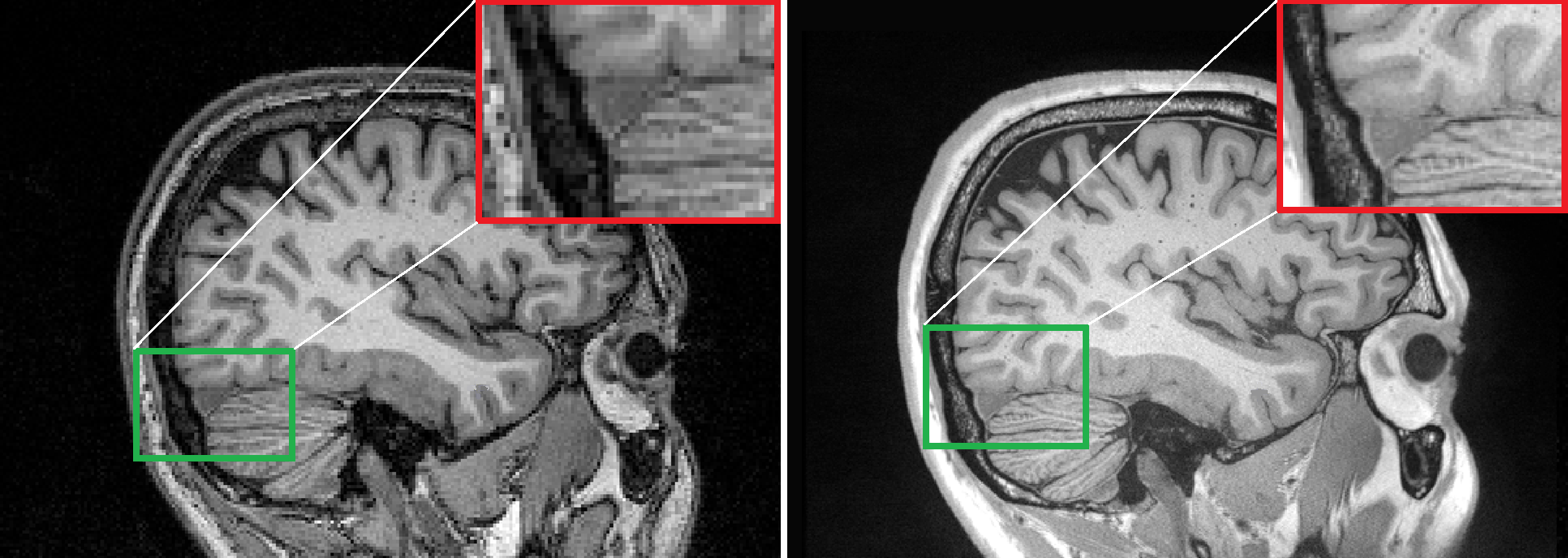
Brain tissue loss has been a hallmark for multiple neurodegenerative diseases association. Expressed as the subtraction of the pial surface and white matter surface of the brain [1]., the gray matter (GM) thickness loss is diagnostic of brain injury and neurodegenerative disease [2]. Higher-resolution images (i.e., smaller than 1mm resolution) are likely to provide more diagnostic information on gray matter loss, but their acquisition suffers from high thermal noise—images at 0.5mm isotropic resolution require multiple acquisitions and averaging in order to yield suitable quality for analysis (Fig. 1). Given that the only relevant information to GM thickness measurement is good contrast at the boundaries of white and gray matter and cerebrospinal fluid, we sought to determine if a single short high-resolution acquisition could be denoised to yield accurate surface reconstructions, comparable to those generated from the average of multiple acquisitions.
METHODS
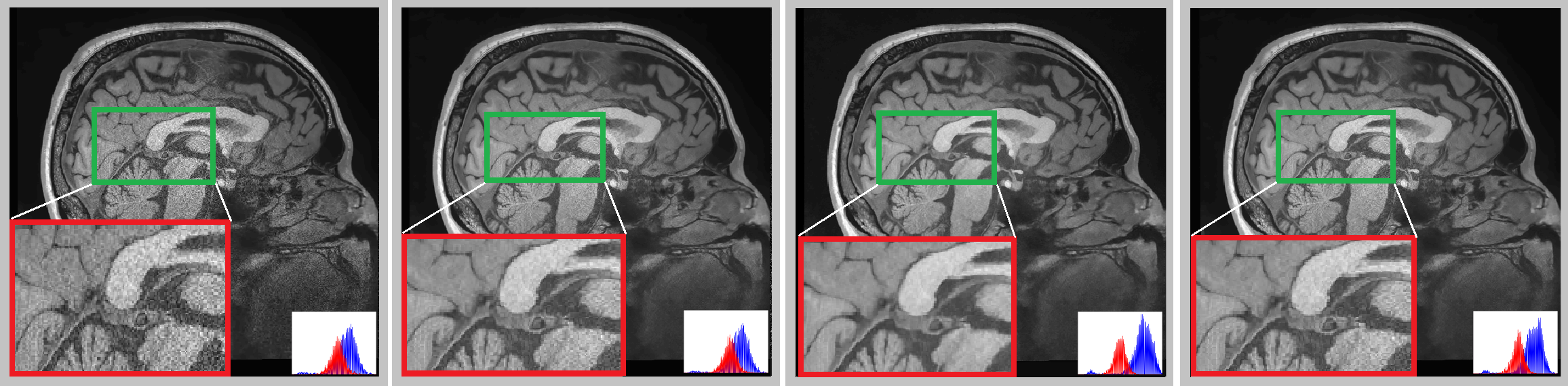
One MEMPRAGE with prospective motion correction was acquired on a Siemens 3T TrioTrim MR scanner, with a 0.5mm isotropic voxel size (TR=2530ms, TE=2.45, BW=548Hz/px, 480x480 matrix, 3D acquisition). Four denoising image filters were evaluated: Oracle-based DCT filter (ODCT) [3], Optimized Nonlocal Means (ONLM) [4], Adaptive Optimized Nonlocal Means (AONLM) [5], and Prefiltered Rotationally Invariant Nonlocal Means (PRINM) filter [3] (Fig. 2). Surfaces were reconstructed using the FreeSurfer package, with manual inspection of each processing step. The GM thickness estimates from surface reconstruction of the gold-standard (GS; mean of 6 0.5mm acquisitions with no denoising) and the resulting filtered image were compared. Later, a parameter optimization was implemented in the image preprocessing step to correct critical regions where the GM thickness differed between the denoised and our gold standard.RESULTS
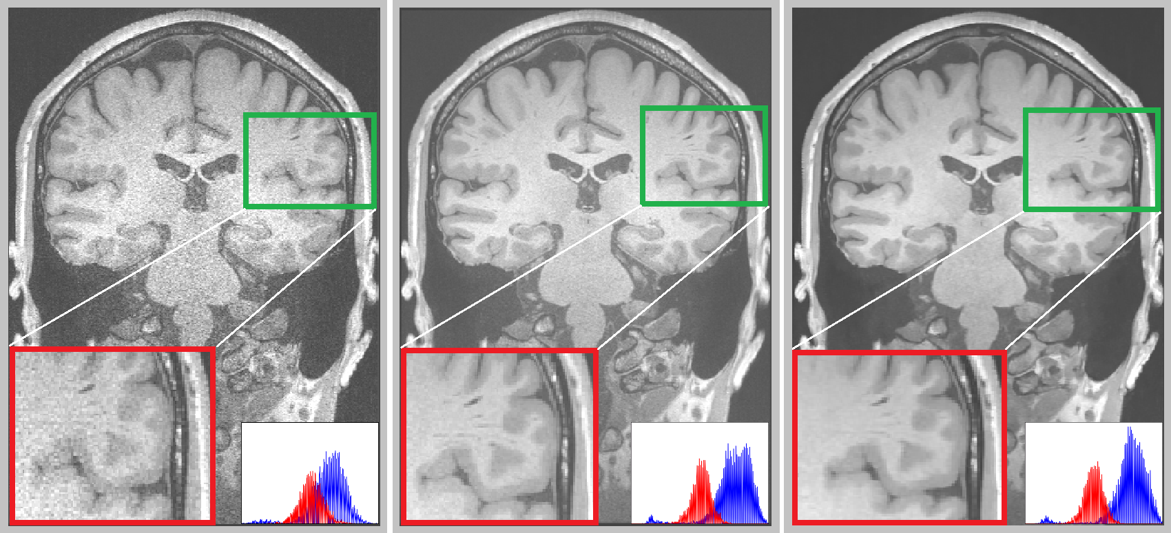
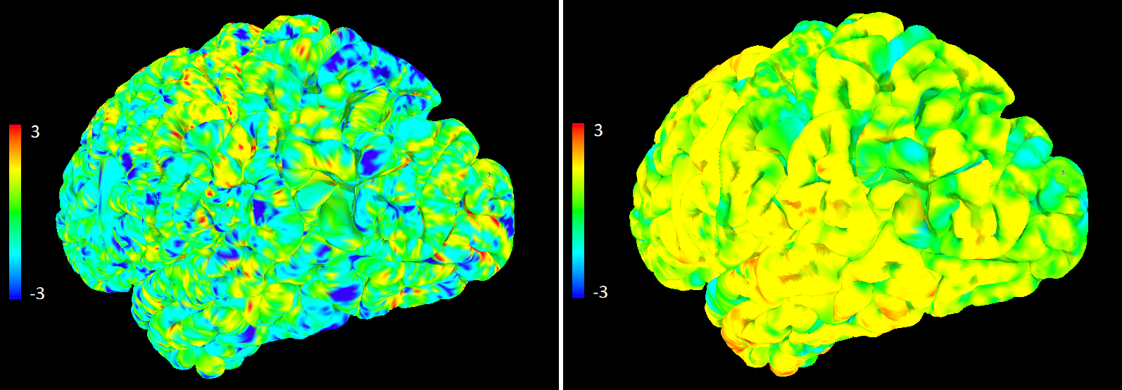
The AONLM filter exhibited the best segregation of white and GM based on an analysis of the voxel histograms (Fig. 2). Overall, GM thickness did not differ between the denoised image (one acquisition, 12 min) and the GS (mean of 6, or 90min acquisition), but there were regions where the denoised image yielded different GM thickness estimates as compared to the "Gold Standard" (Fig. 3). Therefore, further optimization of B1 non-uniformity correction was implemented to correct such regions (Fig. 4). As a bonus, the denoised images were preprocessed faster following this additional optimization.CONCLUSION
This work showed that it is possible to reduce MRI acquisition time and maintain relevant image features for the GM thickness, plus the feasibility of 0.5mm isotropic anatomical imaging in clinically-relevant duration using software denoising to emphasize the crucial image features for extracting GM thickness estimates. Further optimization will be needed, including a repeatability analysis between multiple subjects.Acknowledgements
No acknowledgement found.References
[1] Fischl, Bruce, et al. Measuring the thickness of the human cerebral cortex from magnetic resonance images. Proceedings of the National Academy of Sciences 97.20 (2000): 11050-11055.
[2] Winkler, Anderson M., et al. Cortical thickness or grey matter volume? The importance of selecting the phenotype for imaging genetics studies. Neuroimage 53.3 (2010): 1135-1146.
[3] Mafjón, José V. et al. New methods for MRI denoising based on sparseness and self-similarity. Medical image Analysis. 2012 Jan 1;16(1):18-27.
[4] Coupé, Pierrick, et al. Adaptive multiresolution non-local means filter for three-dimensional magnetic resonance image denoising. IET Image Processing 6.5 (2012): 558-568.
[5] Manjón, José V. et al. Adaptive non-local means denoising of MR images with spatially varying noise levels. Journal of Magnetic Resonance Imaging. 2010 Jan;31(1):192-203.
Figures