2422
THE IMPACT OF MR IMAGES ACQUISITION PROCESS ON RADIOMIC FEATURES: PHANTOM STUDIES TO SUPPORT CLINICAL RESEARCH1Physics Department, Università degli Studi di Milano, Milan, Italy, 2INSTM, Milan, Italy, 3IEO, European Institute of Oncology IRCCS, Milan, Italy
Synopsis
Radiomic analysis of radiological images allows the extraction of quantitative features that can represent a support tool for clinical decision. The investigation of these features stability during the MR image acquisition process represents the aim of this study. The features short- and long-term repeatability was tested on a common MR phantom, imaged with the clinical protocol for gynecological imaging. The non-repeatable features were identified and can be excluded a priori in potential clinical studies. Simultaneously, a dedicated phantom was designed to mimic the pelvis and to investigate the stable features, especially the ones characterizing the texture of the imaged tissue.
Introduction
The term "Radiomics" refers to a process of quantitative analysis of medical images to obtain data useful for pathologies assessment and treatment. "Radiomic features" are extracted from the images with mathematical algorithms starting from the numerical value associated to each voxel in the image and they can be an expression of the phenotype of the imaged tissue. The features could be integrated into predictive models helpful for prognosis and the choice of treatment strategy, especially in clinical oncology. Radiomics analysis of Magnetic Resonance (MR) images is showing promising, although preliminary, results in different oncological diseases1,2. However, due to the recent development of this discipline, a standard and robust methodology for the features extraction is currently missing. This lack leads to the need for an investigation of the features stability during the MR image acquisition process. With this aim, a study was performed with main focus on the pelvis (prostate and ovarian cancer).Methods
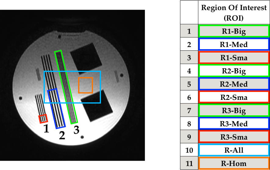
The methodological study was carried out with two phantoms on a 1.5 T MR scanner available at the European Institute of Oncology IRCCS, Milan. The first phantom (PH1) is used for routine quality control on the scanner. PH1 images were acquired with the protocol used for gynecological imaging. Ten consecutive and identical acquisitions were repeated without any change in the MR sequence parameters on different days (t0, t1, t2 and t3). On these images, radiomic features were calculated on selected Regions Of Interest (ROI) with variable volume (Fig. 1), exploiting IBEX software.
In order to test the short-term repeatability of the features, the coefficient r = 1.96σ / |μ| was evaluated. In this equation μ is the mean of the ten values obtained for each feature on each acquisition day ti (i= 0, 1, 2, 3) and σ is the corresponding standard deviation.
In order to test the long-term repeatability, only the features with r < 10% for each ti and for each ROI were considered. At fixed ROI and feature, a paired sample t-test was performed between each couple of μ(ti).
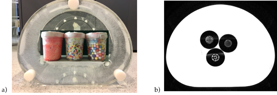
At the same time a second dedicated phantom (PH2) was designed in order to mimic the acquisition conditions of patients. An abdomen-shaped container was filled with a solution of MnCl2 to obtain relaxation times similar to the pelvis organs. The relaxation times of the solution were measured with an NMR spectrometer at the University of Milan. The relaxation times of tissues of interest were measured with dedicated sequences in vivo both on healthy and non-healthy subjects.
With the aim to test radiomics ability to characterize different texture, some test objects were inserted in the phantom solution to mimic various patterns in the image.
Results
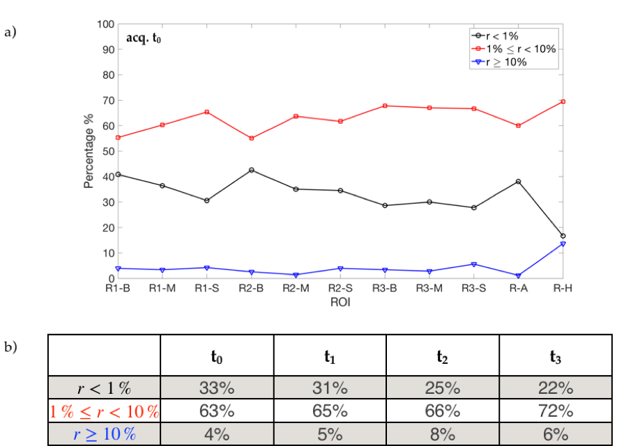
The results for the short-term repeatability test are reported in Fig. 2. On average the percentage of features with r < 1% resulted around 27.8%, while the features with 1% ≤ r < 10% were about 66.5% of the total.
Moreover, the analysis showed that the instability of the features increases by decreasing the volume of the ROI. The long-term analysis showed that only 8% of the radiomic features have r < 10% for all ROI and all the acquisitions.
As regards PH2, the first goal to reproduce tissues with different texture in a phantom compatible with MR scanner was achieved. The phantom is shown in Fig. 3.
The features analyses were handled with in-house scripts developed in MATLAB®.
Discussion
The majority of the radiomic features varies less than 10% in repeated phantom acquisitions. However, this study showed that 6% of the features in hypothetical clinical studies should be excluded a priori as not stable.
It is interesting to notice that all the stable features in long-term acquisitions describe the image texture. This suggests that texture features can actually play a key role in radiomic analyses and deserve further investigation.
The study gave evidence of the link between the ROI volume and the instability of the features extracted. The identification of a threshold volume, below which the features begin to be unstable and thus must be excluded, is strongly suggested, especially when small volumes are investigated (e.g. lymph nodes, dominant intraprostatic lesion, small ovarian lesions).
Conclusion
A protocol to test the stability of radiomic features extracted from MR images -with regard to short- and long-term repeatability in the same experimental condition- was established. The impact of the image acquisition process on the radiomic features variability was characterized and the unstable features identified. This protocol, once optimized and tested on PH2 (that mimic patients), could be used to support the radiomic clinical studies.Acknowledgements
No acknowledgement found.References
1. Yang, Fei, et al. "Magnetic resonance imaging (MRI)-based radiomics for prostate cancer radiotherapy." Translational andrology and urology 7.3 (2018): 445.
2. Tsougos, Ioannis, et al. "Application of Radiomics and Decision Support Systems for Breast MR Differential Diagnosis." Computational and mathematical methods in medicine 2018 (2018).
Figures