2414
Enhanced MR-STAT by a multi-coil reconstruction framework1Center for Image Sciences, UMC Utrecht, Utrecht, Netherlands
Synopsis
MR-STAT is a framework for obtaining multi-parametric quantitative MR maps using data from single short scans. A large-scale optimization problem is solved in which spatial localisation of signal and estimation of tissue parameters are performed simultaneously by directly fitting a Bloch-based volumetric signal model to the time domain data. Previously, only data from a single receive channel could be incorporated into the reconstruction. In this work we extend the MR-STAT framework to allow parameter maps to be reconstructed from multi-coil data resulting in a more robust reconstruction process that has higher scan-efficiency and is less prone to coil shading artefacts.
Introduction
MR-STAT1 is a framework for obtaining multi-parametric quantitative MR maps using data from single short scans. A large-scale optimization problem is solved in which spatial localisation of signal and estimation of tissue parameters are performed simultaneously by directly fitting a Bloch-based volumetric signal model to the time domain data. In previous work2, only data from a single receive channel could be incorporated into the reconstruction. This is suboptimal in terms of scan-efficiency and may result in coil shading artefacts. In this work we aim to solve by extending the MR-STAT framework to allow parameter maps to be reconstructed from data from multiple receive coils simultaneously.
Theory
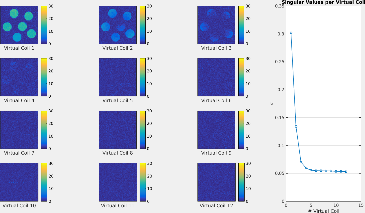
Previously, in (single-coil) MR-STAT, parameter maps were obtained by iteratively solving$$ \hat{\alpha} = \text{argmin}_\alpha \frac{1}{2}\left\lVert d - s(\alpha) \right\rVert^2_2,$$where $$$d$$$ is the measured data from a single channel, $$$s$$$ is a Bloch-equation based volumetric signal model and $$$\alpha$$$ are the quantitative parameter maps concatenated into a single vector. Since data is typically acquired with multiple receive coils simultaneously, the singular value decomposition (SVD) was used to compress coil data to a single virtual coil before reconstruction. In figure 1 it can be seen that SVD compression works well for tube phantoms at 1.5.
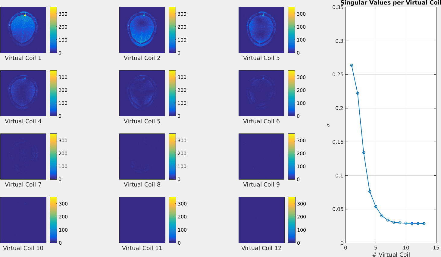
However, as seen in figure 2 for in-vivo brain data at 3T, the compression may result in a dominant virtual coil that displays strongly inhomogeneous receive sensitivity over the field-of-view. This will result in significant image shading artefacts in the reconstructed parameters.
Moreover, as can be seen in both figures 1 and 2, coil data is not perfectly compressible into a single coil and relevant signal is present in the remaining virtual coils. In the single-coil MR-STAT reconstructions, this additional signal was essentially discarded, which is suboptimal from a scan-efficiency point of view.
To overcome these issues (coil shading and discarding data), the MR-STAT reconstruction has been extended to allow reconstruction of parameter maps from multiple (virtual) receive coils simultaneously by solving $$ \hat{\alpha} = \text{argmin}_\alpha \frac{1}{2}\sum_{i=1}^{N_c} \left\lVert d_i - s(\alpha,C_i) \right\rVert^2_2,$$where $$$d_i$$$ and $$$C_i$$$ are the data and coil sensitivity maps from coil $$$i$$$ respectively, and $$$N_c$$$ is the total number of coils.
Methods
The multicoil MR-STAT method has been tested on data from phantom tubes (Philips Ingenia, 1.5T) and from an in-vivo brain scan (Philips Ingenia, 3T). For the data acquisition, a transient-state 2D gradient-balanced sequence with smoothly varying flip angles and Cartesian k-space filling was employed (figure 3). Gel phantoms (Eurospin) and the brain of a healthy volunteer were scanned with scan times of 2.85s (phantoms) and 7.45s (in-vivo) and with a scan resolution of 1x1x4mm3 using a 13 channel receive head-coil. A fully parallelized and matrix-free Gauss-Newton reconstruction algorithm was extended with the multi-coil functionality ($$$N_c=13$$$).
Reconstructions on phantom data were performed using single-coil MR-STAT and also using multi-coil MR-STAT with four virtual coils and thirteen real coils, respectively. For the in-vivo data, reconstructions were performed using single-coil MR-STAT and multi-coil MR-STAT with four virtual coils. Noise-prewhitening was applied before generating virtual coils with the SVD.
Coil sensitivity maps used in the multi-coil reconstructions were generated from the MR-STAT scans directly using the ESPIRiT method.3 The reconstruction algorithm was implemented in the Julia programming language4 and was run on a computing cluster using 64 cores.
Results
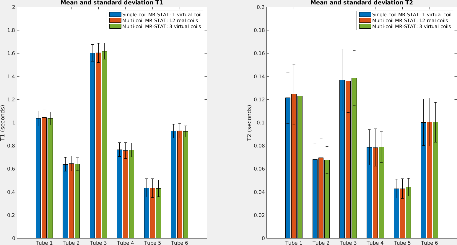
In figure 3, mean $$$T_1$$$ and $$$T_2$$$ values and standard deviations for the reconstructions on phantom tube data are shown. Using three virtual coils for the phantom tubes results in lower standard deviations compared to using all real coils.
In figure 4, reconstructed $$$T_1$$$, $$$T_2$$$ and proton density maps for the in-vivo brain data are shown. For the single coil reconstruction, the coil shading not only affects the proton density map but also the $$$T_1$$$ and $$$T_2$$$ maps.
Discussion
Using one virtual coil can result in severe coil shading problems. Multi-coil MR-STAT addresses this issue by including coil sensitivities into the reconstruction. We hypothesize this is especially beneficial at 3T since coil shading is more pronounced at 3T.
An additional benefit of multi-coil MR-STAT is the higher data-efficiency since no signal is discarded.
The SVD allows us to select only a limited amount of channels to capture all signal while reducing noise (many virtual coils contain mostly noise). This also reduces reconstruction times since less coils need to be simulated. Benchmarks show ~50% computational overhead when simulating twelve coils and ~15% when using four (virtual) coils.
Conclusion
The MR-STAT framework has been extended to allow parameter maps to be reconstructed from multi-coil data, resulting in a more robust reconstruction process that takes into account all the signal and is less prone to coil shading.Acknowledgements
This research is funded by the Netherlands Organisation for Scientific Research, domain Applied and Engineering Sciences, Grant #14125.
References
[1] Sbrizzi A, van der Heide O, Cloos M, van der Toorn A, Hoogduin H, Luijten PR, van den Berg CAT. Fast quantitative MRI as a nonlinear tomography problem. Magnetic Resonance Imaging, 46:56–63, 2018.3.
[2] van der Heide O, Sbrizzi A, Luijten PR, van den Berg CAT. Proc. 26th Sci. Meet. Int. Soc. Magn. Reson. Med. Paris, 2018:0266.
[3] Uecker M, Lai P, Murphy MJ, Virtue P, Elad M, Pauly JM, Vasanawala SS, Lustig M. ESPIRiT - An eigenvalue approach to autocalibrating parallel MRI: Where SENSE meets GRAPPA. Magnetic Resonance in Medicine, 2014.
[4] Bezanson J, Edelman A, Karpinski S, Shah VB. Julia: A fresh approach to numerical computing. CoRR, abs/1411.1607, 2014.
Figures
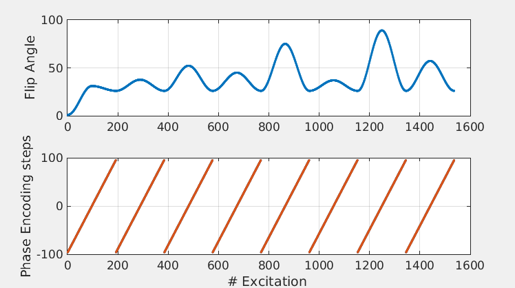
The transient-state pulse sequence with smoothly varying flip angle train and linear, Cartesian k-space filling. For the in-vivo brain scan, a TR and TE of 8.3ms and 4.15ms were used. For the tube phantoms, a TR and TE of 7.4ms and 3.7ms were used. For both scans the equivalent of four k-spaces was acquired with a total scan time of 2.85s(phantoms) and 7.45s (in-vivo).