2411
Full 3D ky-kz-kx GRAPPA reconstruction of SMS MB EPIChan Hong Moon1, Hoby P. Hetherington 1, and Jullie W. Pan2
1Radiology, University of Pittsburgh, Pittsburgh, PA, United States, 2Neurology, University of Pittsburgh, Pittsburgh, PA, United States
Synopsis
EPI with sub-second sampling rates is essential for fMRI to increase tSNR and filter out physiological noise. Simultaneous multiple slice (SMS)-EPI using multi-band (MB) slice excitation has been successfully applied to acquire whole brain fMRI data in < 1s. However, the reconstruction of SMS-EPI remains in separate 2D/1D GRAPPA or partial 3D GRAPPA in ky-kz’-kx domains. To further increase acquisition speed, TR< 500ms, higher k-space dimensional GRAPPA can be used to improve reconstruction performance, e.g. increase SNR and decrease aliasing artifacts. To meet this need we developed a full 3D ky-kz-kx GRAPPA reconstruction for SMS-EPI validated it by simulation and experiment at 7T.
Introduction
SMS-EPI (1, 2) simultaneously acquires aliased 2D images with phase encoding in ky and kz’ (counterpart of MB slices) w/ or w/o blipped-CAIPHIRINA where z blipped gradient is applied to the MB slice for further encoding in kz’ (2-4). Because SMS-EPI encoding is partial 3D FT in kx-ky-kz’ domain (5), it should also be possible to directly apply a GRAPPA reconstruction in ky-kz’ domain. However, given the limited kz’ sampling (e.g., 2) and the discontinuous coil sensitivity, an alternative reconstruction of SMS-EPI is required. In this study, we describe a full 3D ky-kz-kx GRAPPA for SMS-EPI imaging.Theory
A FT of Nyquist-sampled spin-density (e.g., Δz ) is the band-limited spectrum in k-space (black-dotted rectangle in Fig. 1a). When the spectrum is subsampled and acquired by multi-receivers, the original spectrum can be reconstructed by GRAPPA with low frequency ACS (xxx). When the sampling rate is lower than Nyquist condition (e.g., 2Δz ) and the missing data are zero filled (ZF) and the spectrum is aliased in k-space (Fig. 1b). Alternatively, when the subsampled data without ZF (e.g. # of data points, N/2) - same as MB (N/2) slice excitation), are FTed, the spectrum is aliased with narrowed bandwidth (Fig. 1c) – same as the red windowed spectrum of the ZF subsampled spectrum (Fig. 1b). The ACS data can be aliased (Fig. 1c), which means the subsampled spectral signal in kz’ can not be reconstructed by using the ACS in Fig. 1a, Fig 2. The subsampled data w/o ZF shows an aliasing artifact in the spectrum (red vs. black in right panel Fig. 2c). For the utilization of full kz band (i.e., full feature of coil sensitivity map in z), kz’ spectrum needs to be converted to a kz spectrum. MB slice selection and slice-encoding (regardless of z-gradient blipping) is same as a spatio(z’’)-frequency(kz’) transformation for slice profile (right of Fig. 3a). This linear transformation can be easily reversed to spatial domain (z) – inverse FT of kz’ to z’ and then rearranged with the signal in original MB slice at z’’. FT of the rearranged signal in z becomes full spectrum in kz. Subsampling in kz’ causes the aliasing in z’ and the aliasing pattern is projected to z axis following the rule of MB selection (i.e., same aliasing operation). Therefore, subsampling in kz’ is exactly the same as that in the rearranged kz axis. The y or ky axis is orthogonal to z or kz’ (or kz), independent of the sampling scheme applied from that in kz’. So SMS-EPI (w/ or w/o blipped-CAIPHIRINA encoding) data can be reconstructed in the full 3D ky-kz-kx domain. Fig. 3b summarizes the procedure from data acquisition to the proposed reconstruction.Methods
Imaging data from a uniform phantom was acquired at 3T and 7T. The MB acquisition was simulated by FT of selected multiple slices. 2D subsampling in k-space was done in ky and kz'. Applied MB (Ns in Fig. 4) was 4 to 16 and subsampling factors, Ry and Rz were 1 to 8 (e.g., Nz 10, Ry 5 and Rz 2). ACS ky lines were 28. For an experimental study, a subject brain was scanned by conventional 2D EPI and SMS-EPI with MB 2 and no z-blip gradient at 7T with high order shimming (1st – 4th+); TR/TE 3500/25ms, matrix 96x96x58 and Ry 3. The proposed technique was applied to resting state fMRI (~6 min acquisition) using an ICA analysis to determine the feasibility of the method.Results
Simulation results (Fig. 4) shows the superiority of the full 3D ky-kz-kx GRAPPA reconstruction in comparison to partial 3D ky-kz’-kx GRAPPA (1st vs. 2nd column in Fig. 4b). Although AF=9 (3x3) seems feasible, AF=16 (4x4) is too high to get reasonable image quality when using 32 receivers. SMS-EPI w/ MB 2 at 7T reconstructed by the proposed method showed comparable image quality to conventional 2D EPI, except slightly higher background noise in superior brain regions (Fig. 5a,b). In addition, the application of full 3D GRAPPA to rsfMRI study showed a clear default mode network (Fig. 5c).Conclusions
Full 3D ky-kz-kx GRAPPA was successfully established for a SMS-EPI acquisition, and its superiority to conventional GRAPPA was shown and demonstrated for rsfMRI at 7T. The proposed method enables multi-dimensional acceleration, and the combination with CAIPHIRINA will further improve the image quality as well as acquisition speed via higher AFs on ky and kz axes.Acknowledgements
n/aReferences
[1] Feinberg et al,PlosOne,5(12),2010. [2] Setsompop et al,MRM,67(5):1210-24,2012. [3] Breuer et al,MRM,53(3):684-91,2005. [4] Zhu et al,ISMRM,518,2012. [5] Zahneisen et al,MRM,71(6):2071-81,2014.Figures
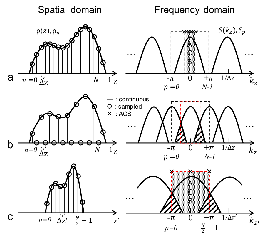
Fig. 1 a, Sampling of data at Nyquist rate
(e.g., Δz) results in the repeated band-limited
spectrum with period of 1/Δz, and the original data can be restored from –π to π. b, Subsampling with zero-filling. When the sampling rate is lower
than the bandwidth of the spectrum, there is aliasing artifact in k-space
(hashed regions). c, Subsampling
without zero-filling (total N/2 points). The spectrum is widened due to the
shrunk spatial profile in z, and the spectrum is aliased. The red-dotted
spectrum is same as that in b.
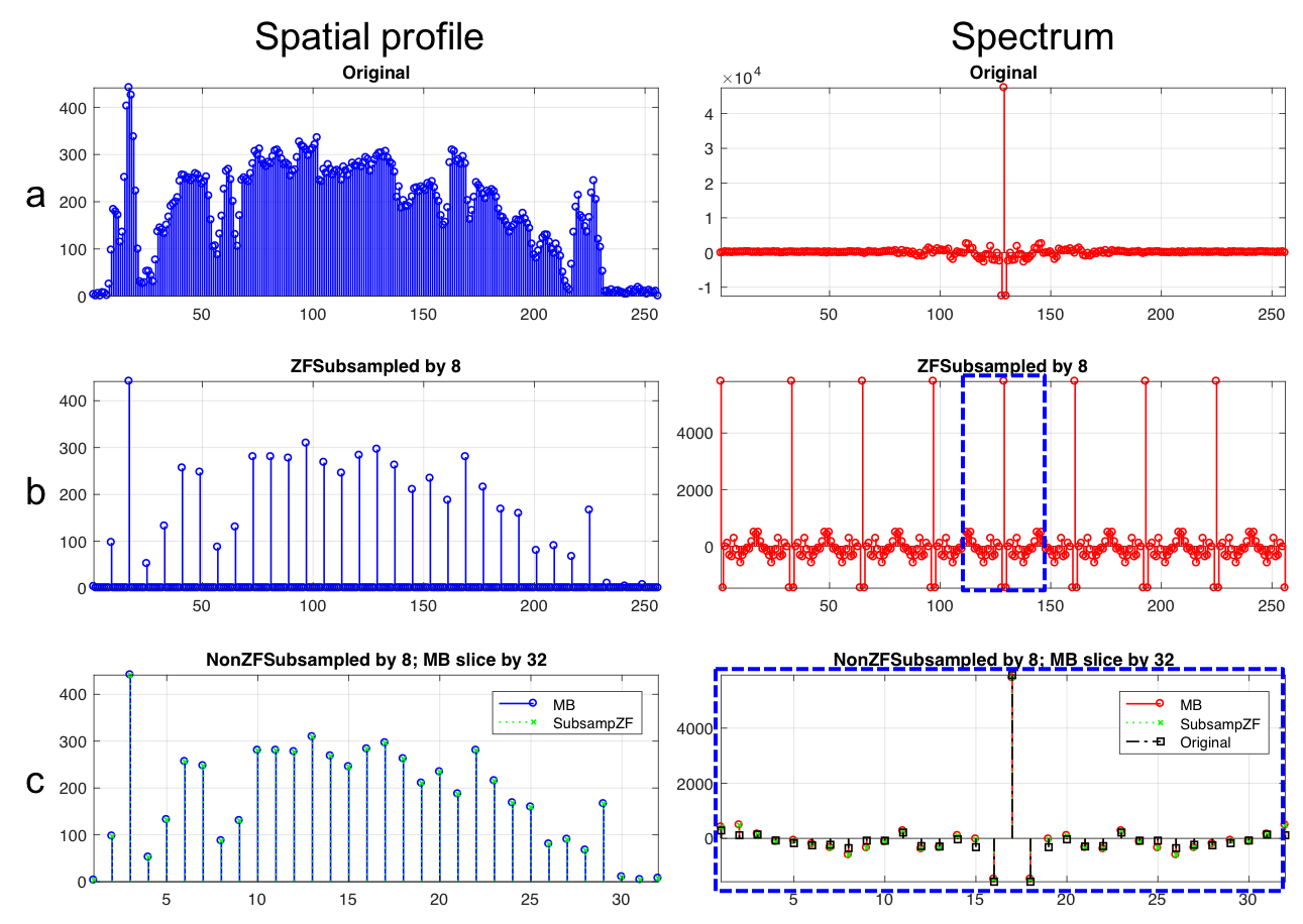
Fig. 2
Simulation of theory in Fig. 1; # of
original samples 256, subsampling 8, MB 32. a, Full sampled data. b,
Subsampled, zero-filled data. c,
Subsampled, non-zero-filled data – same configuration as MB slicing with 32.
(Right) FT of the subsampled data (red color w/ MB legend); Copy of spectrum
for zero-filled subsampled spectrum in blue dotted box in right panel of b (green color w/ SubsampZF legend). The
original spectrum is black colored. (Left panel) Subsampled data w/o zero-filling
(blue color w/ MB legend); Inverse FT of green spectrum in right panel (green
color w/ SubsampZF legend).
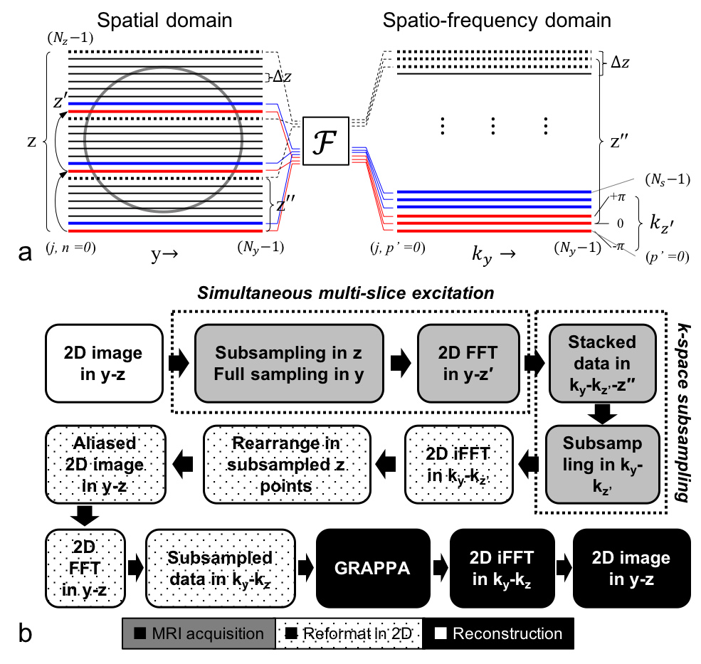
Fig. 3 a, Partial Fourier encoding. Slices in z are sampled by factor of 3
(left panel). Subsampled data is FTed to kz’. Subsampling-FT are repeated to
adjacent slices, and stacked in z’’-kz’ (right panel); final data in spatio(z’’)-frequency(kz’)
domain. Other orthogonal data (e.g., y axis) can be FTed or left as in spatial
space. b, Flow chart of SMS
acquisition (ky-kz’) and full GRAPPA (ky-kz). Slice subsampling and ky-kz’
encoding occurs with MR acquisition. The acquired data are inverse FTed,
rearranged and forward transformed to ky-kz domain. The sub-sampled data in
ky-kz domain can be reconstructed by GRAPPA.
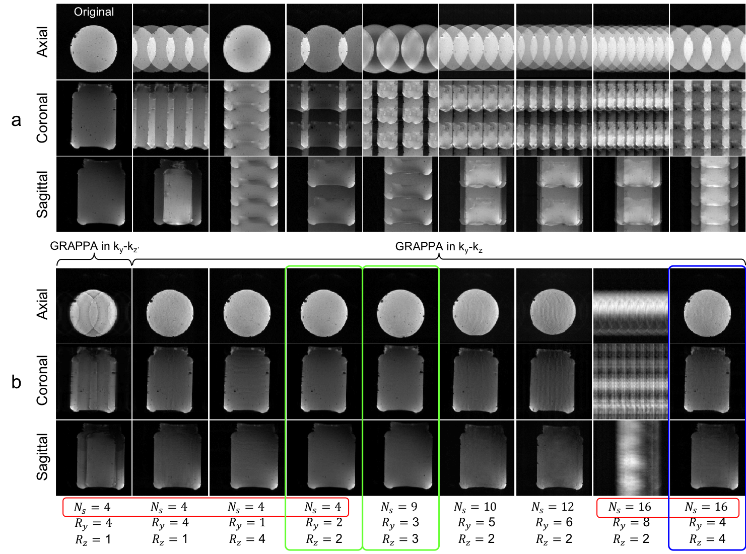
Fig. 4
Comparison of partial vs. full 3D ky-kz-kx GRAPPA. The original full sampled
data are acquired at 3T with 32-ch receiver. a, No GRAPPA. b,
ky-kz’-kx or ky-kz-kx GRAPPA. The simulation results show the superior imaging reconstructed
by ky-kz-kx to ky-kz’-kx GRAPPA (1st vs. 2nd column); in addition,
multi-dimensional PI and GRAPPA can improve the image (2nd vs. 3rd column). By
using full 3D GRAPPA, total AF 9 (3x3) seems feasible, but 16 (4x4) seems not
supported by current 32-ch coil.
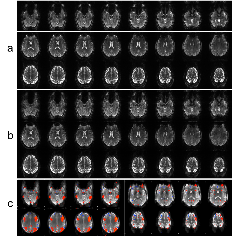
Fig. 5 SMS-EPI data at 7T w/ 16-ch
receivers. Comparison of conventional 2D EPI w/ total AF 3 (a) vs. SMS-EPI w/ MB 2 and total AF 3x2
reconstructed by full 3D GRAPPA (b)
– the data acquired at 7T w/ 16-ch receivers. The EPI image by proposed method
shows comparable image quality to conventional 2D EPI, except lightly higher
background ghost noise in upper region of the brain. c, Example of default mode brain network analyzed by ICA.