2405
Self-Estimated Subspace Reconstruction for Highly-Accelerated Dynamic Golden-Angle Radial MRI1Department of Medical Physics, Memorial Sloan Kettering Cancer Center, New York, NY, United States, 2Department of Radiology, Indiana University School of Medicine, Indianapolis, IN, United States, 3Center for Advanced Imaging Innovation and Research (CAI2R) and Bernard and Irene Schwartz Center for Biomedical Imaging, New York University School of Medicine, New York, NY, United States, 4Department of Radiology, Memorial Sloan Kettering Cancer Center, New York, NY, United States
Synopsis
Subspace-constrained reconstruction is a powerful technique to accelerate dynamic MRI. However, its performance is relatively limited for applications where a robust temporal model is not available. This work proposes to estimate temporal basis from undersampled dynamic golden-angle radial data without the need of a model or additional navigators, and to apply the estimated temporal basis for subspace-constrained reconstruction of undersampled dynamic images. The reconstruction algorithm also enforces an additional low-rank constraint on the resulting low dimensional dynamic images in the subspace. The proposed self-estimated subspace-constrained reconstruction technique was demonstrated for DCE-MRI of the prostate.
INTRODUCTION
Subspace-constrained reconstruction methods have shown encouraging promise to accelerate dynamic MRI acquisitions [1-6]. These methods typically estimate a temporal basis set from a physical signal model, such as T1 or T2 relaxation, which is used by a sparse reconstruction algorithm to represent the undersampled dynamic time-series in a low-dimensional subspace. However, in many dynamic MRI applications, such as dynamic contrast-enhanced MRI (DCE-MRI) , a signal model may not be robust and sufficient enough to accurately represent the temporal signal evolution. A temporal basis can also be obtained from additional navigation data, but this reduces imaging efficiency [7]. In this work, we propose a subspace-constrained image reconstruction approach for undersampled dynamic golden-angled radial MRI, in which the temporal basis set is directly estimated from the acquired undersampled data without a signal model or any navigation data. The proposed method was tested on accelerated DCE-MRI of the prostate.METHODS
Self-estimated subspace reconstruction of dynamic golden-angle radial MRI: Golden-angle radial sampling offers a unique sampling geometry where undersampling is much lower around the central k-space region, as shown in Figure 1. Therefore, even at high acceleration rates, a low-resolution dynamic image-series can still be successfully reconstructed with conventional sparse reconstruction methods, such as the Golden-angle RAdial Sparse Parallel (GRASP) technique [8]. This reconstructed image-series, despite reduced spatial resolution, still provides full temporal information from which a temporal basis set can be estimated to represent the original high spatial resolution image-series in a low-dimensional subspace. Figure 2a shows an example of low spatial resolution DCE-MRI of the prostate (2D+time), where each temporal frame is reconstructed using 13 golden-angle radial spokes with the GRASP method [8]. After converting the image-series to a Casorati matrix (reforming each image as a column), singular value decomposition (SVD) can be performed to obtain a full basis set. The singular values (Figure 2b) of such decomposition show that image information is mainly restricted to the first 4 main components, which suggests that the entire image-series can be represented in a low-dimensional space given by the first 4 principal components (Figure 2c). This observation is further validated in Figure 3, where an undersampled full-resolution image-series is compressed to a low-dimensional subspace with only 4 principal components. More importantly, images in the subspace show dramatically reduced streaking artifacts and improved image quality compared to images in their original space. With this pre-estimated subspace, sparse image reconstruction can be performed by solving the following cost function:
$$d=\underset{d}{\text{argmin}} \frac{1}{2}\parallel EΦd-y\parallel _2^2+R(d) $$
where y is the sorted dynamic radial k-space, E is an encoding operator incorporating coil sensitivities, Φ is the temporal basis of the subspace and d is the coefficients to represent the image-series under the basis set Φ. An additional regularization function R can be applied to the coefficients d to further promote sparsity in the subspace.
Evaluation of reconstruction: The proposed method was tested in one prostate DCE-MRI dataset from a prior study [9]. Relevant imaging parameters included: TR/TE = 4.12/1.96 ms, matrix size=224x224x26, FOV=240x240 mm2, voxel size=1.07x1.07x3.0 mm3. 1755 spokes were acquired for each partition, with a scan time of 221 seconds. Subspace-constrained image reconstruction was performed after grouping every 13 consecutive spokes into one temporal frame, with a temporal resolution of ~1.6 seconds/volume. The temporal basis set was estimated from a low-resolution image-series reconstructed using GRASP with the same temporal resolution but with a reduced image matrix size of 48x48. The first 4 basis components were used to impose subspace constraint with an additional locally low-rank constraint with a block size of 16x16 in the subspace [4,10,11]. This method, referred to as Subspace-GRASP, was compared with standard GRASP reconstruction with a temporal total variation constraint.
RESULTS
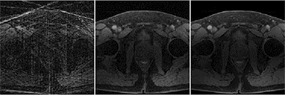
Figure 4 compares gridding, standard GRASP and Subspace-GRASP reconstruction. Although GRASP was able to recover most image features, it suffered from residual artifacts and blurring due to the high acceleration rate. Subspace-GRASP achieved visually improved image quality both in the prostate region (red circle) and in the muscle (green arrows). The signal evaluation curves in an ROI placed on the prostate (red circle) is shown as well. Figure 5 shows a movie for the same comparison as in Figure 4. Subspace-GRASP (right column) shows improved visual image quality over standard GRASP (middle column).DISCUSSION
Golden-angle radial sampling enables estimation of a temporal basis set that represents the subspace directly from acquired data. Self-discovery of the basis functions improves signal modeling compared with handcrafted models and enables the application of subspace-constrained reconstruction to imaging studies where a physical signal model is not available or not reliable.Acknowledgements
No acknowledgement found.References
[1] Doneva M et al. Magn Reson Med 2010;64:1114–1120.
[2] Lam F et al. Magn Reson Med 2014;71:1349–1357.
[3] Zhao B et al. Magn Reson Med 2015;74:489–498.
[4] Tamir JI et al. Magn Reson Med. 2017 Jan;77(1):180-195.
[5] Assländer J et al. Magn Reson Med. 2018 Jan;79(1):83-96.
[6] Zhao B et al. Magn Reson Med. 2018 Feb;79(2):933-942.
[7] Christodoulou AG et al. Nat Biomed Eng. 2018 Apr;2(4):215-226.
[8] Feng L et al. Magn Reson Med. 2014 Sep;72(3):707-17.
[9] Benkert T et al. Magn Reson Med. 2018 Jul;80(1):286-293.
[10] Zhang T et al. Magn Reson Med. 2015 Feb;73(2):655-61.
[11] Zhant T et al. J Magn Reson Imaging. 2015 Feb;41(2):460-73.
Figures