2399
Optimization of a 3-channel gradient waveform for FRONSAC encoding1Radiology and Biomedical Imaging, Yale University, New Haven, CT, United States
Synopsis
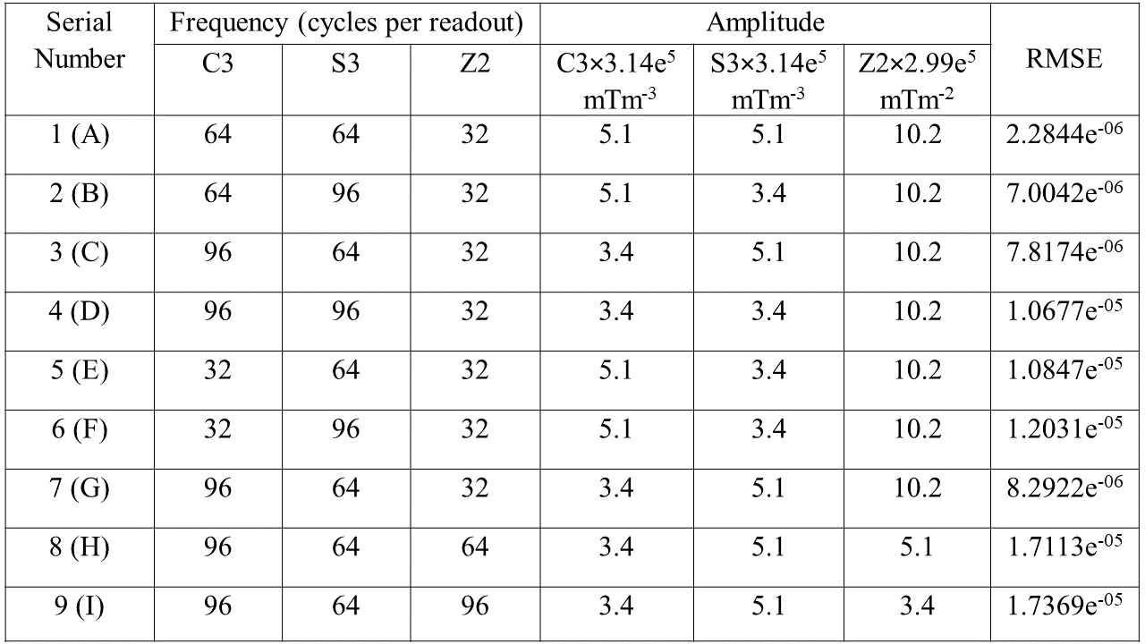
This work reports the performance of various gradient waveforms for Fast Rotary Nonlinear Spatial Acquisition (FRONSAC) encoding, varying the amplitude, frequency and phase of the oscillation on different channels. Waveforms using three NLG channels were used to image an American College of Radiology (ACR) phantom, and root-mean-square error (RMSE) relative to a fully sampled reference was used to evaluate performance. Experimentally observed trends support those reported in previous work, which was based on theory and simulations. For the given hardware, the results suggest that the best combination for C3, S3 and Z2 are 64, 64 and 32 cycles per readout and 1.74×10⁶mTm-3, 1.74×10⁶mTm-3 and 3.05×10⁶mTm-2 respectively.
Background
Parallel imaging can significantly accelerate acquisition and consequently reduce the cost of MR imaging.1-8 While many non-Cartesian sequences show excellent potential for parallel imaging, they generally do so at the cost of complicated contrast and enhanced sensitivity to off-resonance spins and timing errors. However, previous work has suggested that FRONSAC can maintain the contrast and reliability of standard Cartesian acquisitions while improving artifacts due to undersampling.9-17
With FRONSAC encoding, as the sampling function moves across k-space on the trajectory defined by the linear gradients, a rapidly changing nonlinear gradient changes the shape and orientation of the sampling function. These broader and more diverse sampling functions, modulated by coils and aided by oversampling, provide more measurements in the gaps of the linear trajectory in k-space, which subsequently reduces undersampling artifacts. However, the optimal gradient waveform to fill gaps in k-space is an open question with a staggering number of degrees of freedom.16-19 Here we undertake an experimental optimization using three nonlinear gradients available at our site and restricted to sinusoidal waveforms.
Methods
Gradient waveforms were tested at 96, 64 and 32 cycles per readout and two phases (sin and cos). For these high frequency waveforms, maximum amplitude is limited by slew limits over one period of the waveform. To test various amplitudes, waveforms were tested at the maximum achievable amplitude for that frequency and half the maximum amplitude. These tests were performed applying the existing 380mm ID gradient coil which delivers 10.1mT/m3/A, 9.8mT/m3/A and 1.5mT/m2/A, for C3, S3, and Z2, respectively. After collecting field maps and 8-channel coil profiles as previous described, an ACR phantom was imaged. A 3mm slice in the transverse plane was imaged with 250mm FOV, 128 phase encodings, 4x oversampling in the readout (512 points), TE/TR/BW= 16ms/50ms/130Hz/pixel.20-21 Images are reconstructed as previously described with a GPU accelerated CG algorithm.Results
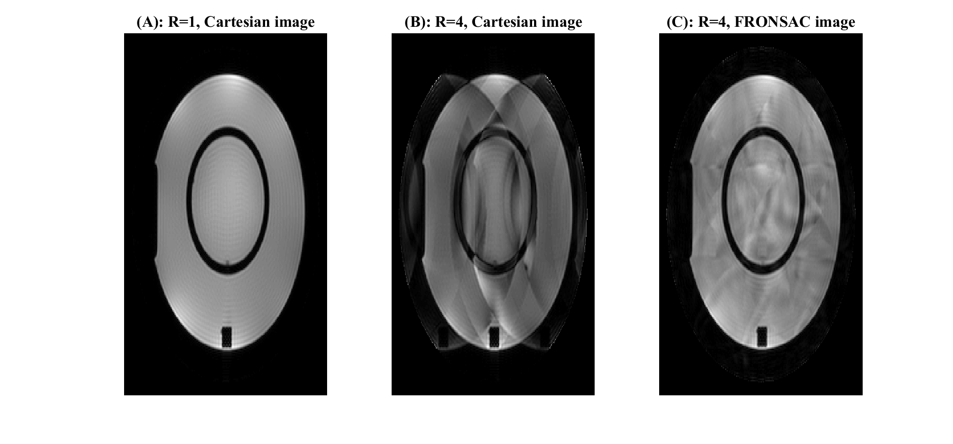
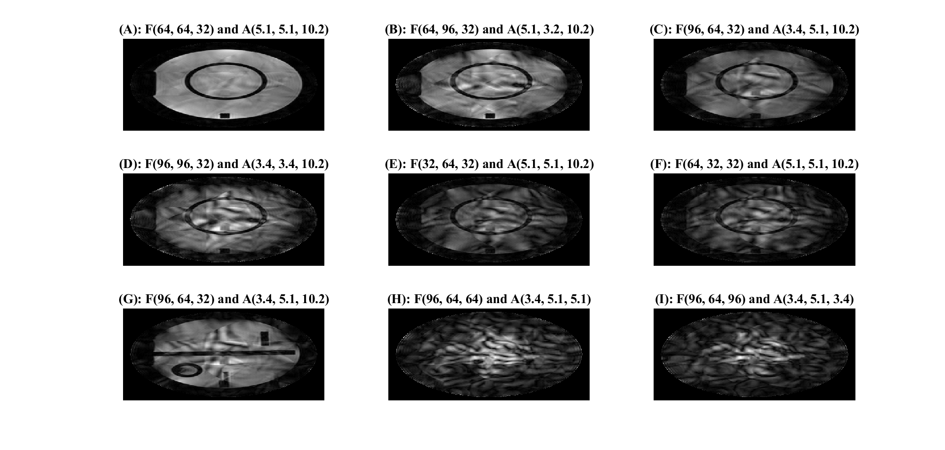
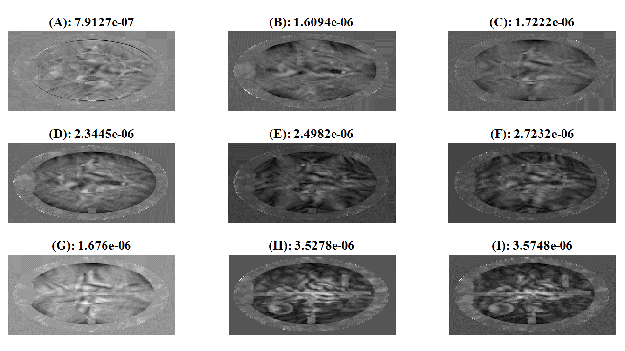
Figure 1 shows a fully sampled Cartesian reference image of the phantom, along with an R=4 undersampled Cartesian image and an R=4 FRONSAC image. While the low number of channels and possible imperfections in the coil profiles limit the image quality in the undersampled images, these results demonstrate that FRONSAC encoding does significantly improve undersampling artifacts. Figure 2 shows a representative set of reconstructions achievable for various FRONSAC gradients, showing dramatic variations in image quality for different candidate waveforms. Difference images and RMSE, compared to a fully sampled Cartesian reference, are shown in Figure 3, and this data is summarized in the table of Figure 4, which tabulates these results ordered by RMSE. These experiments show that dephasing becomes an issue at large moments that are reached when lower frequency waveforms are applied on the strong and steep C3 and S3 gradients, as seen by comparing panels (A)-(C) to (E) and (F). Dephasing is further appreciated in the difference images, which show significant darkening across the image as well as the periphery. However, running these gradients at very high frequency does not allow for sufficient moment to maximally improve image quality, as seen in a comparison of panel (A) with panels (B)-(D). A similar trend is seen in variations of the Z2 gradient field, as shown in panels (G)-(I), for data taken on a different slice of this phantom. In the case of Z2, the gradient is less strong and steep, so a lower frequency and higher moment are favoured.Discussion and Conclusion
Previous simulation studies on the effect of
frequency in the FRONSAC waveform suggested higher frequency always improves
image quality, but that work assumed a fixed gradient moment in each lobe of
the sinusoid. Experimentally, there is a tradeoff between achievable moment and rapid
oscillation in the sampling function.17 In
agreement with previous simulations, these experiments did verify that very
high gradient moments, especially in higher order gradient shapes, cause
dephasing which inhibits image quality. This work tested a large set of possible waveforms
to identify one well suited to the gradient capabilities, coil configuration,
and noise characteristics of the scanner. The results are in agreement with previous generalizations deduced from
theory and simulations. Furthermore, the
results provide a waveform suitable for future studies of clinical sequences
and contrasts in human brain imaging. A
general strength of FRONSAC encoding is that this waveform, optimized for a
particular protocol, will provide significant improvements to scans with
different contrasts, resolutions, and geometries.Acknowledgements
We would like to thank Andrew Dewdney (Siemens) and Terry Nixon for supporting the nonlinear gradient hardware.References
1. Sodickson DK, Manning WJ. Simultaneous acquisition of spatial harmonics (SMASH): fast imaging with radiofrequency coil arrays. Magnetic Resonance in Medicine. 1997;38(4):591‐603.
2. Griswold MA, Jakob PM, Heidemann RM, Nittka M, Jellus V, Wang J, Kiefer B, Haase A. Generalized autocalibrating partially parallel acquisitions (GRAPPA). Magnetic Resonance in Medicine. 2002;47(6):1202‐10.
3. Pruessmann KP, Weiger M, Scheidegger MB, Boesiger P. SENSE: sensitivity encoding for fast MRI. Magnetic Resonance in medicine. 1999;42(5):952‐62.
4. Blaimer M, Breuer F, Mueller M, Heidemann RM, Griswold MA, Jakob PM. SMASH, SENSE, PILS, GRAPPA: how to choose the optimal method. Topics in magnetic resonance imaging: TMRI. 2004;15(4):223‐36. Epub 2004/11/19. PubMed PMID: 15548953.
5. Bydder M, Larkman DJ, Hajnal JV. Generalized SMASH imaging. Magnetic Resonance in Medicine. 2002;47(1):160‐70.
6. Tsao J, Boesiger P, Pruessmann KP. k‐t BLAST and k-t SENSE: Dynamic MRI with high frame rate exploiting spatiotemporal correlations. Magnetic Resonance in Medicine. 2003;50(5):1013-42.
7. Lustig M, Pauly JM. SPIRiT: Iterative self-consistent parallel imaging reconstruction from arbitrary k-space. Magn Reson Med. 2010;64(2): 457-71. Epub 2010/07/29.
8. Wiggins GC, Polimeni JR, Potthast A, Schmitt M, Alagappan V, Wald LL. 96‐Channel receive only head coil for 3 Tesla: design optimisation and evaluation. Magnetic Resonance in Medicine. 2009;62(3):754.
9. Stockmann JP, Ciris PA, Galiana G, Tam L, Constable RT. O‐Space imaging: Highly efficient parallel imaging using second-order nonlinear fields as encoding gradients with no phase encoding. Magnetic Resonance in Medicine. 2010:64(2):447-56.
10. Stockmann JP, Galiana G, Tam LK, Nixon TW, Constable RT. First O‐Space images using a high‐power, actively shielded 12cm Z2 gradient insert on a human 3T scanner. Proceedings of the ISMRM 19th Annual meeting. 2011:717.
11. Stockmann JP, Galiana G, Tam L, Juchem C, Nixon TW, Constable RT. In vivo O‐Space imaging with a dedicated 12 cm Z2 inserted coil on a human 3T scanner using phase map calibration. Mgn Reson Med. 2013;69(2): 444-55.
12. Tam LK, Galiana G, Stockmann JP, Tagare H, Peters DC, Constable RT. Pseudo random center placement O-space imaging for improved incoherence compressed sensing parallel MRI. Magn Reson Med. 2014.
13. Gallichan D, Cocosco C, Dewdney A, Schultz G, Welz A, Hennig J, M. Z. Simultaneously driven linear and nonlinear spatial encoding fields in MRI. Magnetic Resonance in Medicine. 2011;65(3):702‐14.
14. Layton KJ, Gallichan D, Testud F, Cocosco CA, Welz AM, Barmet C, Pruessmann KP, Hennig J, Zaitsev M. Single shot trajectory design for region specific imaging using linear and nonlinear magnetic fields. Magn Reson Med. 2012. Epub 2012/10/09.
15. Schultz G, Weber H, Gallichan D, Witschey WR, Welz AM, Cocosco CA, Hennig J, Zaitsev M. Radial Imaging with Multipolar Magnetic Encoding Fields. IEEE Trans Med Imag. 2011; 16:17.
16. Wang H, Tam L, Kopanoglu E, Peters D, Galiana G, Constable RT. Improving O‐Space Imaging Using High‐Resolution Oversampled Data Acquisitions. Proceedings of the ISMRM 22nd Annual Meeting. 2015: talk.
17. Wang H, Tam L, Constable RT, Galiana G. Fast Rotary Nonlinear Spatial Acquisition (FRONSAC) Imaging. Magn Reson Med. 2017 (March); 75(3) 1154-1165.
18. Galiana G, Stockmann JP, Tam L, Peters D, Tagare H, Constable RT. The role of nonlinear gradients in parallel imaging: A k-space based analysis. Concepts in Magnetic Resonance Part A.40A (5):253‐67.
19. Luedicke N, Tagare H, Galiana G, Constable RT, editors. Trajectory design of optimized repeating linear and nonlinear gradient encoding using a k-space point spread function metric. ISMRM Annual Meeting; 2016; Singapore.
20. Nadine L. Dispenza, Sebastian Littin, Maxim Zaitsev, R. Todd Constable, Gigi Galiana, Clinical Potential of a New Approach to MRI Acceleration, Scientific Report, Nature Publishing (submitted).
21. Nadine L. Dispenza, Maxim Zaitsev, R. Todd Constable, Gigi Galiana, ``Clinical Imaging Potential of FRONSAC”, invited talk for ISMRM Annual Meeting and Exhibition, Paris, France June 18, 2018.
Figures