2395
TrueFISP MRI assay for fast & reliable tumor stage classification in carcinogen induced orthotopic bladder cancer1Radiology & Biomedical Engineering, Northwestern University, Chicago, IL, United States, 2Surgery, Division of Urology, Northshore University Health System, Evanston, IL, United States, 3Urology, Northwestern University, Feinberg School of Medicine, Chicago, IL, United States
Synopsis
Using a TrueFISP sequence we implemented a non-invasive assay for stage classification of bladder tumors using a orthotopic murine bladder cancer model. Because generation of this tumor model is achieved by administration of carcinogenic agents it is naturally heterogeneous. The imaging method proposed allows accurate assessment of tumor burden and provides a tool for randomization to experimental treatments.
Introduction
Recent advances in treatment of bladder cancer have shown that anti-PD-1 and PD-L1-checkpoint inhibitors have durable responses of patients with advanced urothelial carcinoma 1-3 . While exciting, this therapeutic opportunity is limited due to low patient response (~20%). Further investigation is needed to understand the effectiveness of these advanced therapies. A preclinical testing platform4-5 relying on murine bladder cancer models is an essential step in this direction but requires the ability to control for tumor size in order to optimally randomize mice into treatment groups. Several techniques have been used to measure and evaluate tumor burden6-9 but none provide the capabilities of MRI, 3D anatomical visualization of the whole bladder and multiple tissue-contrasts. In this study we validate an MR based assay that combines high throughput screening efficiency and reliability by implementing a fast imaging and simplified post-processing analysis pipeline. To this end we employed fast imaging with steady state precession (True FISP) technique and used the derived images to establish a quantitative assay of bladder tumor burden that correlated well with stage of progression as evaluated through histopathology methods.Methods
All experimental procedures involving animals complied with Northwestern’s IACUC guidelines. Bladder tumors were induced in male C57/B6 mice at least 6 weeks old following administration of N-nitrosobutyl(4-hydroxybutyl)amine (BBN) at a dose of 0.05% in drinking water, provided ad libitum 10 . All mice underwent MRI at 16 weeks post-start of BBN administration and were euthanized at 20 weeks. Histopathology and ex vivo bladder weights (BW) were recorded and compared to MRI. Prior to scanning (~10 minutes) and to improve anatomical evaluation of the bladder (i.e. full and “stretched” bladder) each mouse was injected with a sub-cutaneous dose of 0.1-0.2 ml of warm saline solution. Animals were scanned under anesthesia on a 7T MRI ClinScan (Bruker). A quadrature body coil was used for excitation with a 4-channel phased array for reception placed on the lower abdomen. This coil choice and positioning enabled optimized acquisition of whole bladder images. Rapid multi-directional sets of bladder MR images were acquired in the shortest time possible with high diagnostic quality (i.e. high signal-to-noise) using fast imaging with steady state precession technique (true FISP) (TR=900 msec, TE=2 msec, FA=70, 14 averages) providing T1/T2 weighting in <10 minutes per mouse. Total time from start to end was ~15 minutes/mouse. Images were analyzed using Jim7 (Xinapse). One slice located at the center of the bladder was chosen and two regions of interest (ROIs) were drawn, one around the outer edge of the bladder and one over its lumen (see Figure 1). The bladder lumen area (BLA) was then subtracted from the outer bladder area (OBA) and the resulting surface quantity labelled ΔOBA-BLA was considered directly proportional to bladder wall thickness. The underlying hypothesis being that an increase in bladder wall thickness (i.e. an increase in ΔOBA-BLA ) reflects progression of tumor. 3D-rendering of segmented bladder wall was also generated in some cases for enhanced visualization of bladder abnormalities. Tumor stage was determined for each mouse based on the degree of invasiveness and metastasis and through histopathological evaluation (data not shown). Non-invasive papillary urothelial carcinoma is designated as stage Ta, while stages T1, T2, T3 and T4 refer to invasion into the subepithelial connective tissue, muscle, perivescical tissue and adjacent organs, respectively11
Results & Discussion
Shown in Figure 2 is a representative set of MRI images and the corresponding rendering of
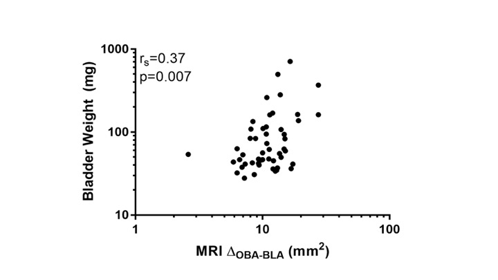
tumor and control mouse as well as the ex vivo sectional images from the corresponding mice. The ΔDOBA-BLA values derived from MRI correlate well with BW
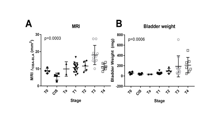
(rs = 0.37, p=0.007) as shown in Figure 2 but more importantly they correlate well with tumor stage classification
as shown in Figure 3 (MRI p=0.0003; BW p=0.0006). The parameter was also shown to
exhibit significant association when
stratifying pathology by non-muscle-invasive bladder cancer and muscle-invasive
bladder cancer (MRI p=0.0002 and BW p<0.0001). The derived area under
the curve (AUC) for ΔDOBA-BLA (AUC=0.81, 95% CI 0.68-093) is statistically similar to AUC for BW
(AUC=0.89, 95% CI 0.80-0.98; p=0.30) reflecting its ability to determine muscle-invasive bladder cancer (Stage >T2) with similar sensitivity and specificity (graphs not shown here). Conclusions
We tested and validated a rapid and reliable MRI based assay of tumor
burden in an orthotopic mouse model of carcinogen induced bladder carcinoma. The surface
parameter ΔDOBA-BLA derived from the MR images correlated significantly with final bladder
weight and histopathology assessments. More importantly we were able to show that this non-invasive MRI parameter reflects and correlates well with tumor stage classification providing us with
an essential tool to conduct high-throughput assays of mice at intermediate stages of tumor development, prior to
randomization for treatment. Acknowledgements
References
1. Rosenberg, J. E., Hoffman-Censits, J., Powles, T. et al.: Atezolizumab in patients with locally advanced and metastatic urothelial carcinoma who have progressed following treatment with platinum-based chemotherapy: a single-arm, multicentre, phase 2 trial. Lancet, 387: 1909, 2016
2 . Sharma, P., Callahan, M. K., Bono, P. et al.: Nivolumab monotherapy in recurrent metastatic urothelial carcinoma (CheckMate 032): a multicentre, open-label, two-stage, multi-arm, phase 1/2 trial. The Lancet Oncology, 17: 1590, 2016
3. Bellmunt, J., de Wit, R., Vaughn, D. J. et al.: Pembrolizumab as Second-Line Therapy for Advanced Urothelial Carcinoma. N Engl J Med, 2017
4. Chan, E., Patel, A., Heston, W. et al.: Mouse orthotopic models for bladder cancer research. BJU Int, 104: 1286, 2009
5. Zhang, N., Li, D., Shao, J. et al.: Animal models for bladder cancer: The model establishment and evaluation (Review). Oncol Lett, 9: 1515, 2015
6. Patel, A. R., Chan, E. S., Hansel, D. E. et al.: Transabdominal micro-ultrasound imaging of bladder cancer in a mouse model: a validation study. Urology, 75: 799, 2010
7. Chin, J., Kadhim, S., Garcia, B. et al.: Magnetic resonance imaging for detecting and treatment monitoring of orthotopic murine bladder tumor implants. J Urol, 145: 1297, 1991
8. Jurczok, A., Fornara, P., Soling, A.: Bioluminescence imaging to monitor bladder cancer cell adhesion in vivo: a new approach to optimize a syngeneic, orthotopic, murine bladder cancer model. BJU Int, 101: 120, 2008
9. Vandeveer, A. J., Fallon, J. K., Tighe, R. et al.: Systemic Immunotherapy of Non-Muscle Invasive Mouse Bladder Cancer with Avelumab, an Anti-PD-L1 Immune Checkpoint Inhibitor. Cancer Immunol Res, 4: 452, 2016
10. Zitvogel, L., Pitt, J. M., Daillere, R. et al.: Mouse models in oncoimmunology. Nat Rev Cancer, 2016
11. Lutzeyer W, Rubben H, Dahm H. Prognostic parameters in superficial bladder cancer: an analysis of 315 cases. J Urol 1982;127:250–2. [PubMed: 7062375]
Figures
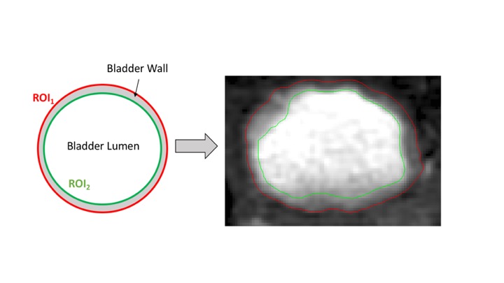
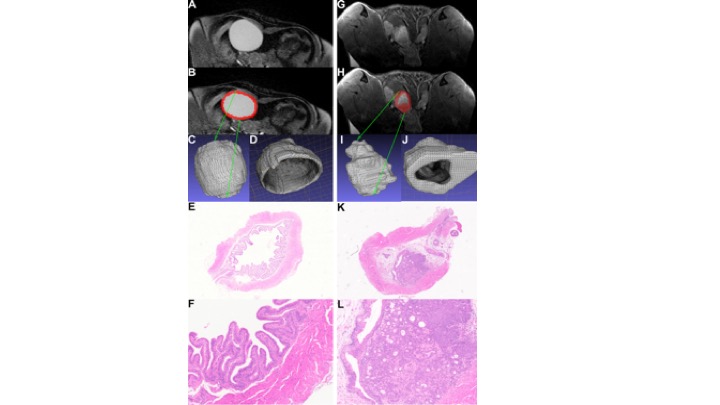
Fig 2.TrueFISP MR images of bladder and the corresponding 3D rendered visualization are shown for (A.B.C.D) control no-tumor mouse and (G.H.I.J.) bladder tumor mouse. Shown in red in (B) and (H) are the outlines used for generating 3D surfaces for each mouse. Low-power (2.5x) and high power (10x) images of bladder sections are shown for mouse with (E.F) no-tumor and for mouse with (K.L.)