2392
MR spectroscopy to assess decreased tumor choline as a marker of response to choline kinase inhibitors1Centre for Preclinical Imaging, University of Liverpool, Liverpool, United Kingdom, 2Department of Radiology, University of Pennsylvania, Philadelphia, PA, United States, 3Biochemistry, Centre for Cell Imaging, Liverpool, United Kingdom
Synopsis
Tumor volume and single voxel in vivo ¹H MRS were used to assess the effects of the choline
kinase inhibitor JAS239 in the F98 rat glioblastoma (GBM) model. Five F344 rats
were inoculated with GBM cells and subsequently treated for 5 consecutive days with
4 mg/kg JAS239 or saline. A reduction in total choline (tCho) in tumors treated
with JAS239, along with tumor growth arrest was noted in comparison to saline
treated rats. JAS239 preferentially inhibited choline metabolism in tumors as
no changes were observed in tCho levels from the contralateral brain.
Introduction
Alterations in lipid metabolism and increased phospholipid metabolism are hallmarks of cancer. Increased total choline (tCho) has been observed in numerous human tumors1,2, manifested by an increase in phosphocholine (PC) and which is typically observed in in vivo MRS. Increased PC in tumors is necessary for cell growth and tumorigenesis1,4 and a key mediator of higher PC is an over-expression of Choline Kinase alpha (ChoKα)4. Inhibiting ChoKα, the first step of PtdCho synthesis, has shown promising results in vivo1,2,3. In recent years, the development of JAS239, a novel ChoKα inhibitor with inherent near infrared fluorescence has enabled it as a theragnostic marker1,5. JAS239 has shown efficacy in a breast cancer xenograft model ¹, however its efficacy in brain tumor models has not been reported. Therefore, our study investigated the effect of JAS239 on tumor growth and tCho using MRS and T2-weighted MRI.Methods
Animal model: Five Fischer F344 (120-130 g) female rats were injected with 5x10⁴ F98 glioblastoma cells in the right cortex.
Treatment: Animals were injected intraperitoneally with either 4 mg/kg JAS239 (n=3) or saline (n=2) for 5 consecutive days.
In vivo single voxel spectroscopy: Anatomical images and spectroscopy data were acquired at baseline (D0) and day 5 (D5) after treatment. T2-weighted images were acquired to localize the tumor and position the MRS voxel. A single voxel of 2x2x2 mm³ was placed within the tumor and a spectrum was acquired using a PRESS sequence with the following parameters: TR = 2500 ms, TE₁ = 8.79 ms and TE₂ = 7.71 ms, number of averages = 256, complex points = 2048 and spectral width = 4401 Hz. Using the same parameters, a second single voxel spectrum was acquired from the contralateral normal brain at each time point from every animal.
Data Quantification: In vivo single voxel 1H MRS spectra from normal brain and tumors were processed and analysed using LC-Model software. Metabolite concentrations were obtained from comparison with the unsuppressed water spectrum. To assess the effect of JAS239 on tumor growth, T2-weighted images from D0 and D5 were used to draw tumor ROIs on multiple slices covering the tumor. Final volume was calculated by summing the number of pixels from all ROIs multiplied by section thickness.
Results
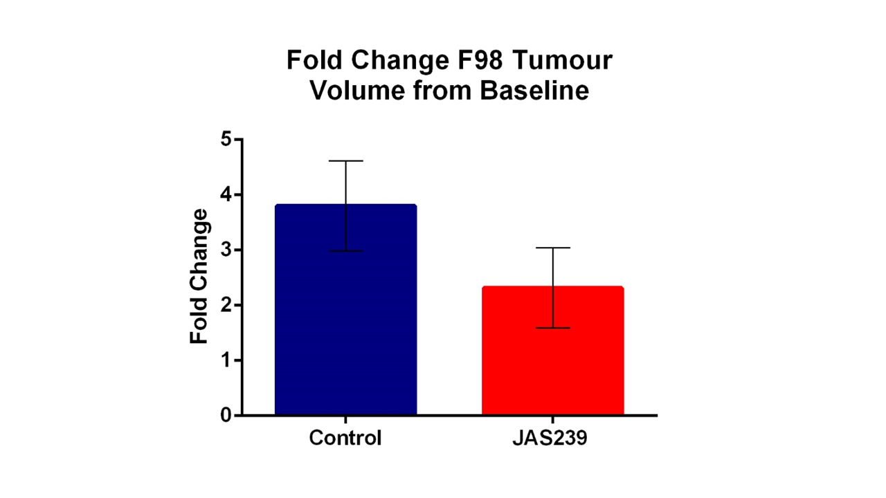
Changes in tumor volumes from day 0 to day 5 after treatment from the three treated and two control rats are shown in Figure 1. Control rats showed a 3.5-fold increase in tumor volume, compared to JAS239 treated animals which only demonstrated a 2.5-fold increase.
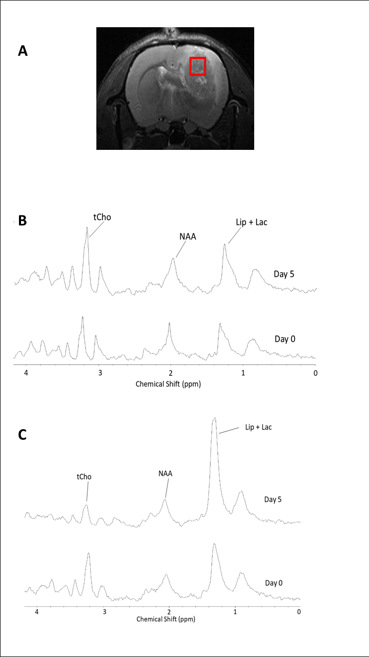
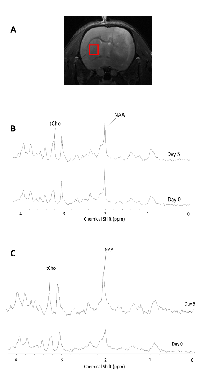
In vivo MR spectra from the tumor and contralateral brain of a representative rat treated with JAS239 and from a control animal treated with saline are shown in Figures 2 and 3. A reduction in tCho peak was observed with JAS239 treatment (Fig 2C) compared to control (Fig 2B) and baseline. Contralateral regions (Figure 3) show minimal change in tCho in control and treated animals before and after treatment.
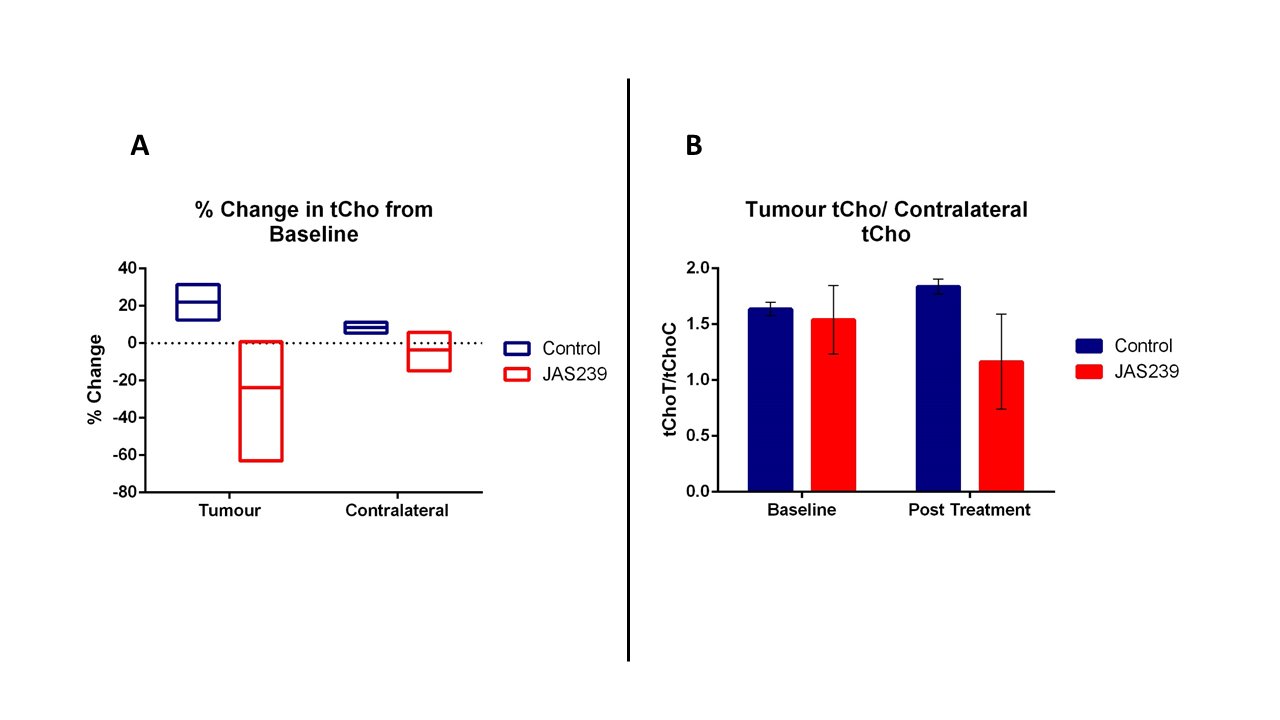
Percentage changes of tCho from baseline to post-treatment are shown in Figure 4A. JAS239 treated animals exhibited a reduction (-24%) in tCho compared to control tumors which showed an increase (+22%). No noticeable changes in the tCho values were noted in the contralateral brain spectra of both JAS239 and saline treated animals (Fig 4A).
A reduction in the tumor tCho to contralateral tCho ratio (0.4) was observed after treatment (Fig 4B) compared to baseline. Control animals however exhibited an increase (0.2) in tumor tCho/contralateral tCho ratio.
Discussion
A delay in tumor growth (indicating growth arrest) and decreases in tCho were observed in JAS239 treated tumors. An earlier study using the first generation ChoKα inhibitor MN58b reported significant tumor growth inhibition and reduction in tCho levels2. However, since JAS239 possesses fluorescent properties5, and has been reported to have similar potency to MN58b in vitro and in vivo1, it may provide an additional non-invasive method of monitoring tumor response using optical imaging methods. In addition, JAS239 has been shown to distinguish cells overexpressing ChoKα1, thus potentially enabling the use of this molecule to stage tumor development, as ChoKα expression has been shown to correlate with tumor grade4,6. The changes in tCho seem to be more pronounced than changes in tumor volume after JAS239, indicating not only the higher sensitivity of MRS in assessing treatment response, but that decreases in tCho may also be used as a pharmacodynamic marker of ChoKα inhibition.Conclusion
Monitoring tCho levels via MRS may provide a non-invasive method to monitor therapy response in glioblastoma models.Acknowledgements
All in vivo experiments were carried out at the Centre for Preclinical Imaging, University of Liverpool.References
1. Arlauckas, S. P., Kumar, M., Popov, A. V, Poptani, H., & Delikatny, E. J. (2017). Near infrared fluorescent imaging of choline kinase alpha expression and inhibition in breast tumors. Oncotarget, 8(10), 16518–16530.
2. Kumar, M., Arlauckas, S. P., Saksena, S., Verma, G., Ittyerah, R., Pickup, S., … Poptani, H. (2015). Magnetic resonance spectroscopy for detection of choline kinase inhibition in the treatment of brain tumors. Molecular Cancer Therapeutics, 14(4), 899–908.
3. Mazarico, J. M., Sanchez-Arevalo Lobo, V. J., Favicchio, R., Greenhalf, W., Costello, E., Carrillo-de Santa Pau, E.,Real, F. X. (2016). Choline kinase alpha (CHK ) as a therapeutic target in pancreatic ductal adenocarcinoma: expression, predictive value, and sensitivity to inhibitors. Molecular Cancer Therapeutics, 15(2), 323–333.
4. Arlauckas, S. P., Popov, A. V., & Delikatny, E. J. (2016). Choline kinase alpha - Putting the ChoK-hold on tumor metabolism. Progress in Lipid Research, 63, 28–40.
5. Arlauckas, S. P., Popov, A. V., & Delikatny, E. J. (2014). Direct inhibition of choline kinase by a near-infrared fluorescent carbocyanine. Molecular Cancer Therapeutics, 13(9), 2149–2158.
6. Al-Saffar, N. M. S., Troy, H., De Molina, A. R., Jackson, L. E., Madhu, B., Griffiths, J. R., … Chung, Y. L. (2006). Noninvasive magnetic resonance spectroscopic pharmacodynamic markers of the choline kinase inhibitor MN58b in human carcinoma models. Cancer Research, 66(1), 427–434.
Figures