2391
Reductive microenvironment responsive gadolinium-based polymers as potential safe MRI contrast agents1Huaxi MR Research Center (HMRRC), Department of Radiology, Sichuan University, Chengdu, Sichuan, China, 2Huaxi MR Research Center (HMRRC), Department of Radiology, Sichuan University, Chengdu,Sichuan, China
Synopsis
The abstract provided an alternative strategy to develop highly efficient and safe gadolinium-based MRI macromolecular contrast agents (Gd-mCAs) via conjugation of small molecular DOTA-Gd to a stimuli-responsive biodegradable and amphiphilic block DHPMA copolymer through a ROX-sensitive biocleavable disulfide bond. Also, its potential as efficient and safe MRI mCAs for cancer diagnosis have been investigated.
Introduction
Traditional clinical used small-molecular gadolinium based MRI contrast agents have some shortcomings for solid tumor diagnosis, such as the lack of specificity, the short blood circulation time and the low imaging efficiency. Gd-based macro-molecular MRI contrast agents (Gd-mCAs) could overcoming many defects of the small ones. However, accumulation of macro-molecular contrast agents in the body tissues potentially induces adverse effects associated with free Gd ion release from the macro-molecular backbone, such as nephrogenic systemic fibrosis and gadolinium deposition in the brain tissue. A novel formulation strategy was proposed herein for Gd-based macromolecular MRI contrast agents (Gd-mCAs), which may significantly reduce Gd retention but maintain sufficient imaging contrast.Methods
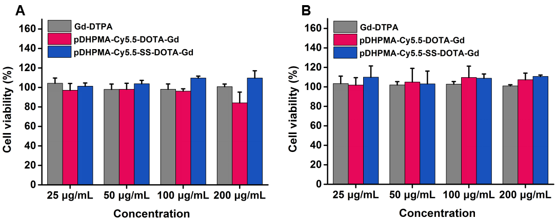
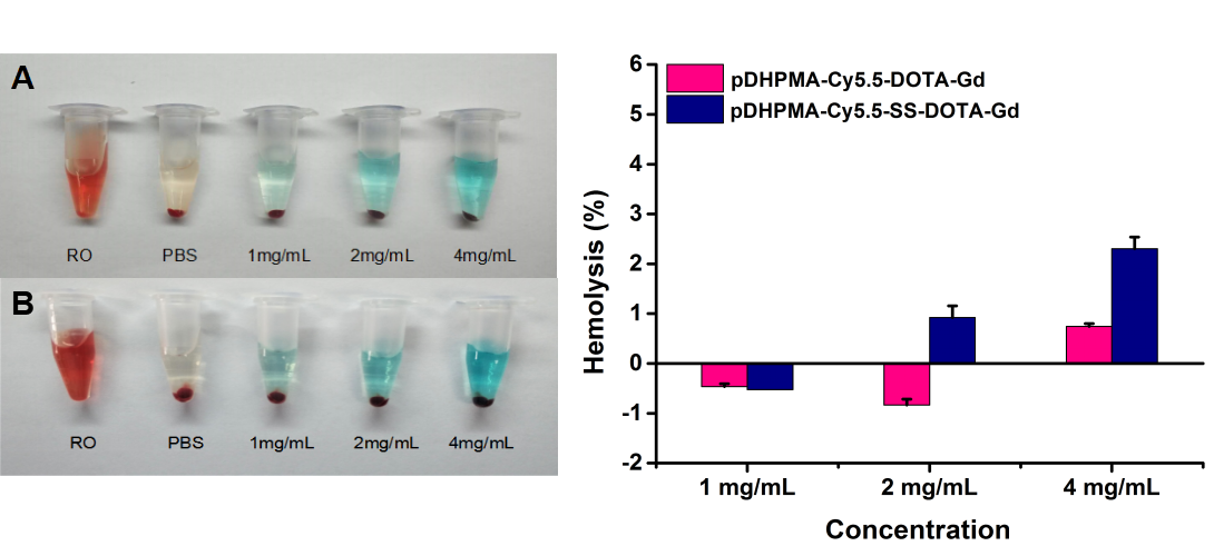
Biodegradable poly[N-(1,3-dihydroxyprpyl) methacrylamide] copolymers (pDHPMA) were synthesized from N-(1,3-dihydroxypropyl) methacrylamide (DHPMA) as a monomer and enzyme-responsive short peptide (GFLG) as a chain transfer agent. Small molecular Gd-chelate (Gd-DOTA) was conjugated onto the copolymer backbone through a sulfide or a GSH-sensitive cleavable disulfide bond to produce two novel Gd-mCAs (pDHPMA-Cy5.5-DOTA-Gd or pDHPMA-Cy5.5-SS-DOTA-Gd) for tumor diagnosis. A clinical MR scanner was used to measure the r1 values of these novel Gd-mCAs before and after incubation of DTT in vitro. An orthotopic tumor model was built by 4T1 cell line on Balb/c mouse. After the tumor size achieved 3-5mm, the tumor efficiency was measured in vivo at different time points after injection (0, 10 min, 30 min, 1, 2, 4, 8, 12 and 24 h), using a clinical MR scanner as well. Furthermore, we explored the gadolinium tissue distribution and pharmacokinetics by ICP-MS. We did tests on bio-safety of the two novel Gd-mCAs in the end. CCK-8 assay was used to measure the cytotoxicity of them while a hemolysis rate was used to test their hematological biocompatibility. And a histological analysis on main organs of healthy Balb/c mice which were injected the new contrast agents and sacrificed 1 or 7 days later. Clinical contrast agent Gd-DTPA was used as a control.Results
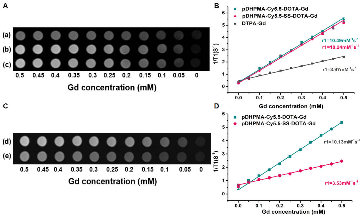
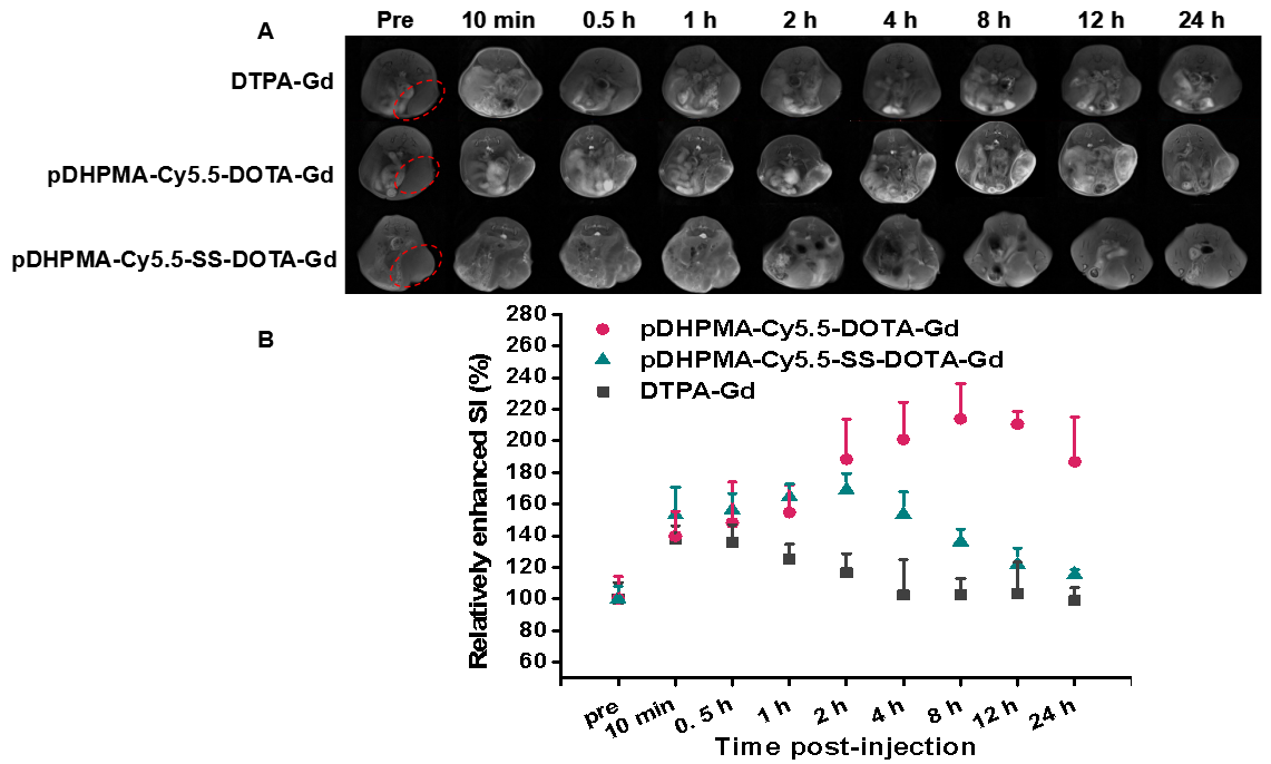
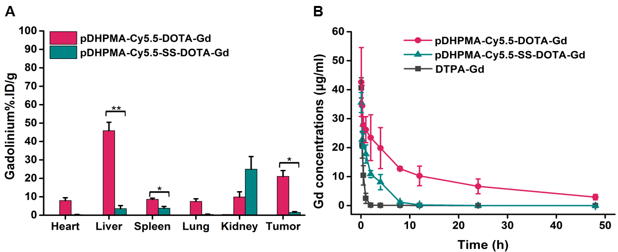
We synthesised pDHPMA-Cy5.5-DOTA-Gd or pDHPMA-Cy5.5-SS-DOTA-Gd successfully. Their relaxivities were 10.49 and 10.24 mM-1s-1 respectively, which were significantly higher than that of DTPA-Gd (3.97 mM-1s-1). After incubation with DTT, the r1 value of pDHPMA-Cy5.5-SS-DOTA-Gd had an obiviously decline (3.53 mM-1s-1) while the r1 value of pDHPMA-Cy5.5-DOTA-Gd stayed stable. In tumor MRI study, most tumors appeared more conspicuous in experimental groups than in control group. Specially, pDHPMA-Cy5.5-SS-DOTA-Gd group tumor showed the brightest signal between 1-2h, while brightest signal of pDHPMA-Cy5.5-DOTA-Gd group appeared around 8h. The results of ICP-MS revealed that compared with pDHPMA-Cy5.5-DOTA-Gd, pDHPMA-Cy5.5-SS-DOTA-Gd had an lower tissue retention and a shorter biological half-life. Relatively bio-safety tests showed our new Gd-mCAs had no obviously bio-toxicity both in vitro and vivo.Discussion
The high relaxivity of the novel Gd-mCAs could be attributed to the macro-molecular weight of the back bone, which would slow down molecular tumbling and increase the rotational correlation time, resulting in a greater relaxivity per unit dose of the paramagnetic ion 1. The obvious r1 value decreasing of pDHPMA-Cy5.5-SS-DOTA-Gd, after incubation with DTT, proved that the disulfide bond was broke in a reductive enviroment and DOTA-Gd was released. The better tumor efficacy in experimental group than in control group could be attributed to EPR effect and their higher r1 value 2-4. Rupture of disulfide bond in reductive tumor microenvironment make the releasing of DOTA-Gd, resulting in a weakening of EPR effect and a decline of r1 value, could explain the earlier time to tumor signal peak in pDHPMA-Cy5.5-SS-DOTA-Gd group. The results of ICP-MS confirmed that using disulfide bond to link DOTA-Gd to copolymer backbone could significantly reduce the body retention of gadolinium.Conclusion
Here, we have successfully synthesized two macromolecular MRI contrast agents. Their macro-molecular weight make they have a higher r1 value, a better tumor imaging efficacy and a longer blood circulation time than clinical MRI contrast agent. Meanwhile, the rupture of disulfide bond of pDHPMA-Cy5.5-SS-DOTA-Gd in reductive tumor microenvironment make the risk of gadolinium retention has a significant decline. Both of the two Gd-mCAs showed a good bio-compatibility. Those results verified that using disulfide bond to link DOTA-Gd to macro-molecular main chain is an good way to maintain sufficient tumor imaging efficacy and reduce gadolinium retention. However, the biological half-life of pDHPMA-Cy5.5-SS-DOTA-Gd still too high when compared with the clinical one, more effective ways should be explored to decrease the retention time of Gd-mCAs.Acknowledgements
This work was supported by National Natural ScienceFoundation of China (51673127, 8162103), InternationalScience and Technology Cooperation Program of China(2015DFE52780), International Science and TechnologyCooperation Program of Sichuan (2018HH0006) andInternational Science and Technology Cooperation Program ofChengdu (2016-GH03-00005-HZ)References
1. Villaraza A J L, Bumb A, Brechbiel M W. Macromolecules, Dendrimers and Nanomaterials in Magnetic Resonance Imaging: The Interplay Between Size, Function and Pharmacokinetics[J]. Chemical Reviews, 2010, 110(5):2921.
2. Matsumura Y, Maeda H. A new concept for macromolecular therapeutics in cancer chemotherapy: mechanism of tumoritropic accumulation of proteins and the antitumor agent smancs.[J]. Cancer Research, 1986, 46(12 Pt 1):6387.
3. Fang J, Nakamura H, Maeda H. The EPR effect: Unique features of tumor blood vessels for drug delivery, factors involved, and limitations and augmentation of the effect.[J]. Advanced Drug Delivery Reviews, 2011, 63(3):136-151.
4. Wei X, Luo Q, Sun L, et al. Enzyme- and pH-sensitive Branched Polymer-Doxorubicin Conjugate-Based Nanoscale Drug Delivery System for Cancer Therapy[J]. Acs Applied Materials & Interfaces, 2016, 8(18):11765.
Figures