2389
Characterization of the arrest and retention of iron-labeled breast cancer cells and the growth and progression of brain metastases in NSG mice1Robarts Research Institute, London, ON, Canada, 2Medical Biophysics, The University of Western Ontario, London, ON, Canada
Synopsis
Patient-derived xenografts in NSG mice provide a novel and more clinically relevant model of studying breast cancer brain metastasis in comparison to traditional cell lines in nude mice. NSG and nude mice both received brain-seeking breast cancer cells and were imaged with MRI to assess cell arrest, retention, and growth. Images revealed significantly more brain metastases and overall whole-body tumour burden in NSG mice than nudes. These results provide characterization of the NSG mouse as a preclinical platform for PDX models and demonstrates the importance of imaging to establish this model for future advancements in drug development and personalized medicine.
Introduction
Metastasis is the primary cause of mortality in breast cancer patients, with brain metastasis occurring in up to 30% of breast cancer cases1-2. Our group developed cellular imaging technologies that allow for tracking metastasis from the initial arrest of cells in the brain through tumour progression with the unique capability of tracking dormant cancer cells by virtue of their retention of iron nanoparticles3. Our previous work has focused on studies using human brain metastatic breast cancer cell lines (such as 231BR) in nude mice3-5. While this is a common type of modeling, there is currently momentum toward use of patient-derived xenografts (PDX) in the more severely immune-compromised NOD/SCID/ILIIrg-/- (NSG) mice6-7. PDX models better represent the tumour heterogeneity which is now recognized to be a critical element for developing personalized treatments8-11. The use of NSG mice is necessary for successful engraftment of PDX models12-13 but studies which characterize the growth of brain metastases in NSG mice are scarce. As we transition from cell lines to PDX models it is important to understand differences that may exist NSG mice, first with human cell lines. Here we demonstrate the use of cellular MRI for the characterization of arrest, retention and metastasis of a human brain metastatic breast cancer cell line in NSG mice, and compare these findings to the same cell line in nude mice.Methods
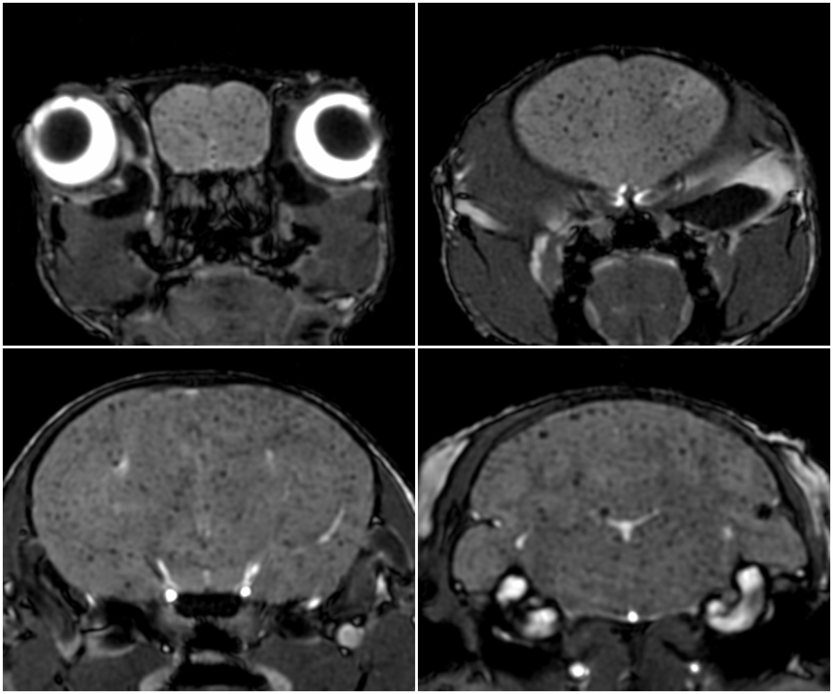
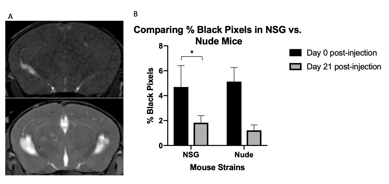
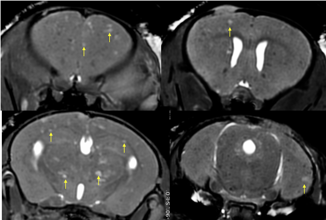
NSG mice (n=8) received an intracardiac injection of 1.5x10⁶ iron-labeled GFP+ brain-seeking breast cancer cells (231BR). Images of the mouse brain and body were acquired at 3T using a high performance gradient coil insert and custom-built mouse RF coils and a 3D balanced steady state free precession (bSSFP) sequence. Images were 100x100x200 microns for brain (28min) and 200 microns isotropic for body (33min). Images obtained on day 0 were assessed for regions of signal void representing iron labeled 231BR cells arrested in the brain; the number of black pixels caused by iron-positive cells was determined. Day 21 images were assessed for retention of signal voids and for brain metastases. Metastases were counted to determine tumour burden. Body images were assessed for metastases in lung, liver, and lymph nodes. Tissues were stained for iron, H&E, and fluorescence. Data was compared to previous studies in our lab which used the 231BR cell line in nude mice.Results
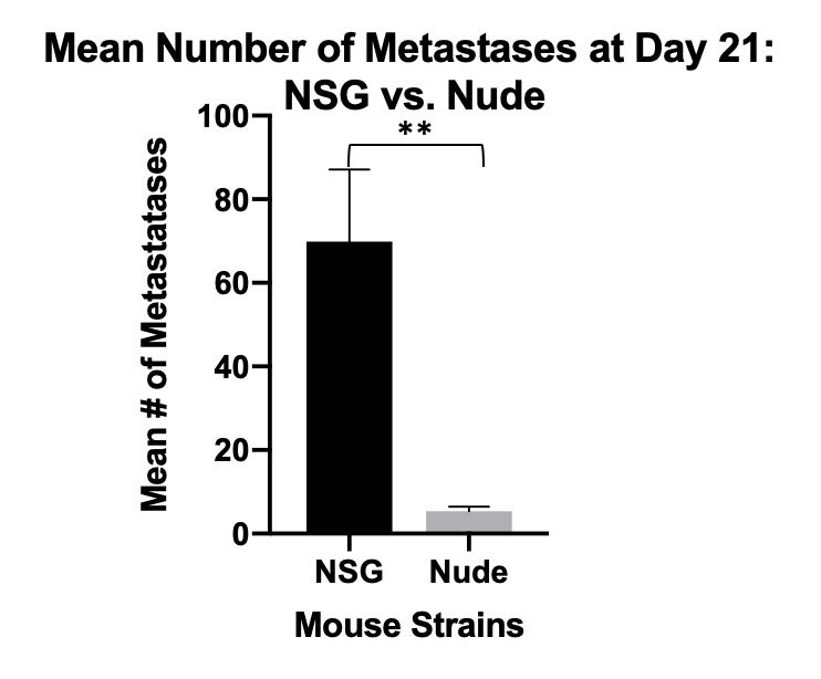
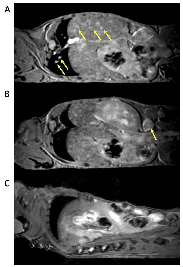
On day 0 iron labeled cells were visualized throughout the brain in all NSG mice (Figure 1) revealing cells that have arrested in the brain. Over time the number of signal voids declined dramatically (Figure 2a) as cancer cells die and are cleared from the brain. Signal voids were quantified by calculating the % of black pixels. This value decreased significantly between day 0 and 21 (Figure 2b). This is consistent with our previous data on nude mice and indicates that the retention of non-proliferating cells is similar in both strains. On day 21 brain metastases were evident in all NSG mice as regions of signal hyperintensity (Figure 3). Significantly more 231BR brain metastases were counted in NSG mice at day 21 compared to nudes (Figure 4). Unlike nude mice, NSG mice had numerous metastases in the body, including in liver, lung, and lymph nodes (Figure 5). The higher brain tumour burden and additional body tumour burden led to an early endpoint for NSG mice based on signs of weakness and weight loss.Discussion
Successful implementation of the PDX models for studying brain metastases due to breast cancer requires that the NSG mouse as a preclinical platform must be fully characterized. Here we showed that the human 231BR cell line grew very differently in NSG mice compared to nude mice. While both are immune-compromised strains, the NSG is more severely deficient; nudes have no T cells, NSG have no T, B, or NK cells14. This may explain the more aggressive growth of brain metastases. Although the 231BR cells were selected to only grow in the brain, which is true in nude mice, in NSG mice we found that liver and lung metastases were frequent and numerous and contributed to a shorter disease model. Additionally, ultrasound revealed that the wall of the heart in NSG mice was substantially thicker than in nude mice, making intracardiac cell injections more challenging. Our work demonstrates the hugely valuable role that imaging can play in the progress toward credentialing these important models.Conclusion
Characterization of immunocompromised mice being used for PDX models of cancer is critical. Cellular MRI allows us to visualize and measure the arrest, retention, and metastasis of cancer cells in vivo.Acknowledgements
This project is supported by the London Regional Cancer Program (LRCP) Translational Breast Cancer Research Unit (TBCRU) and the Western Graduate Research Scholarship (WGRS).References
1. Cheng, X., & Hung, M. C. (2007). Breast cancer brain metastases. Cancer and Metastasis Reviews, 26(3-4), 635-643.
2. Crivellari, D., Pagani, O., Veronesi, A., Lombardi, D., Nole, F., Th¨ rlimann, B., ... & Graffeo, R. (2001). High incidence of central nervous system involvement in patients with metastatic or locally advanced breast cancer treated with epirubicin and docetaxel. Annals of oncology, 12(3), 353-356.
3. Heyn, C., Ronald, J. A., Ramadan, S. S., Snir, J. A., Barry, A. M., MacKenzie, L. T., ... & Yoneda, T. (2006). In vivo MRI of cancer cell fate at the single‐cell level in a mouse model of breast cancer metastasis to the brain. Magnetic Resonance in Medicine: An Official Journal of the International Society for Magnetic Resonance in Medicine, 56(5), 1001-1010.
4. Perera, M., Ribot, E. J., Percy, D. B., McFadden, C., Simedrea, C., Palmieri, D., ... & Foster, P. J. (2012). In vivo magnetic resonance imaging for investigating the development and distribution of experimental brain metastases due to breast cancer. Translational oncology, 5(3), 217-225.
5. Percy, D. B., Ribot, E. J., Chen, Y., McFadden, C., Simedrea, C., Steeg, P. S., ... & Foster, P. J. (2011). In vivo characterization of changing blood-tumor barrier permeability in a mouse model of breast cancer metastasis: a complementary magnetic resonance imaging approach. Investigative radiology, 46(11), 718-725.
6. Hidalgo, M., Amant, F., Biankin, A. V., Budinská, E., Byrne, A. T., Caldas, C., ... & Roman-Roman, S. (2014). Patient-derived xenograft models: an emerging platform for translational cancer research. Cancer discovery, 4(9), 998-1013.
7. Zhang, X., Claerhout, S., Pratt, A., Dobrolecki, L. E., Petrovic, I., Lai, Q., ... & Wong, H. (2013). A renewable tissue resource of phenotypically stable, biologically and ethnically diverse, patient-derived human breast cancer xenograft models. Cancer research.
8. Cassidy, J. W., Caldas, C., & Bruna, A. (2015). Maintaining tumor heterogeneity in patient-derived tumor xenografts. Cancer research, 75(15), 2963-2968.
9. DeRose, Y. S., Wang, G., Lin, Y. C., Bernard, P. S., Buys, S. S., Ebbert, M. T., ... & Neumayer, L. (2011). Tumor grafts derived from women with breast cancer authentically reflect tumor pathology, growth, metastasis and disease outcomes. Nature medicine, 17(11), 1514.
10. Landis, M. D., Lehmann, B. D., Pietenpol, J. A., & Chang, J. C. (2013). Patient-derived breast tumor xenografts facilitating personalized cancer therapy. Breast Cancer Research, 15(1), 1.
11. Reyal, F., Guyader, C., Decraene, C., Lucchesi, C., Auger, N., Assayag, F., ... & De Cremoux, P. (2012). Molecular profiling of patient-derived breast cancer xenografts. Breast cancer research, 14(1), R11.
12. Whittle, J. R., Lewis, M. T., Lindeman, G. J., & Visvader, J. E. (2015). Patient-derived xenograft models of breast cancer and their predictive power. Breast cancer research, 17(1), 17.
13. Zhou, Q., Facciponte, J., Jin, M., Shen, Q., & Lin, Q. (2014). Humanized NOD-SCID IL2rg–/–mice as a preclinical model for cancer research and its potential use for individualized cancer therapies. Cancer letters, 344(1), 13-19.
14. Dobrolecki, L. E., Airhart, S. D., Alferez, D. G., Aparicio, S., Behbod, F., Bentires-Alj, M., ... & Dowst, H. (2016). Patient-derived xenograft (PDX) models in basic and translational breast cancer research. Cancer and Metastasis Reviews, 35(4), 547-573.
Figures