2386
Zero TE based pseudo CT conversion: impact of different HU value assignment methods for bones in the Head.1GE Healthcare, Munich, Germany, 2Umeå University, Umeå, Sweden, 3GE Global Research, Bangalore, India, 4GE Healthcare, Stockholm, Sweden
Synopsis
Patient specific and accurate pseudo CT are needed for the adoption of MR-only in the radiation therapy workflow. Zero TE (ZTE) acquisition has proven to be very robust and reliable for bone segmentation, with the additional advantage of showing a reproducible inverse linear correlation with corresponding CT HU for ZTE intensity values in the bone range. Here we specifically investigate the impact on the dose accuracy of continuous versus single HU value assignment for bones and the strength of the Zero TE inverse linear correlation to CT values for accurate pseudo CT conversion.
Purpose:
Proton density (PD) weighted Zero TE (ZTE) was recently demonstrated to be well suited for bone imaging and pseudo Computed Tomography (CT) conversion in the head1. Different methods have been studied in order to reach an accurate and patient specific pseudo CT conversion. First a threshold-based pixel-wise conversion method1,2 was investigated, showing a reproducible and accurate inverse linear correlation with corresponding CT HU values for bones. In the sinus, the dense presence of air/tissue interfaces could cause an excess of bone false positives. Therefore, the nasal region was identified based on anatomical landmarks and no bone was then allowed in that region. To improve image similarity to the reference CT, a Deep Learning (DL) method based on 3D convolutional neural network of the U-net architecture was explored3. The DL conversion gave superior results when comparing the quantitative accuracy of the two methods to the reference CT in the bone region via Dice coefficient and mean absolute error (MAE)3,4. Here we analyze the impact of the inverse linear scaling and DL conversion on bones, demonstrating the effectiveness and superior dose accuracy of continuous versus single HU value assignment.Methods:
Four patient data-sets from the study evaluated in1,2,3 were analyzed, where a 3.0T GE SIGNA PET/MR scanner (GE Healthcare, Chicago, IL) and a head array coil were used for ZTE data acquisition. A CT scan was also provided for each patient. A CT bone mask was generated for values larger than 150 HU. The DL method was used to generate the pseudo CT bone mask for pseudo CT values larger than 150 HU. The pseudo CT bone mask was rigidly registered to its corresponding reference CT bone mask. The following steps were applied to the original CT data:
1) a single bulk density value of 700 HU was applied to all voxels from the CT bone mask, corresponding to the mean CT bone density value of all patients considered.
2) A single soft tissue value of 42 HU was assigned to all voxels from the CT bone mask.
3) The pseudo CT bone mask was embedded into the data-set generated in step 2) and the inverse linear scaling of (-2000*(ZTEBONE-1) + 42) was applied to the ZTE values of the pseudo CT bone mask voxels.
4) The DL pseudo CT values were applied to the pseudo CT bone mask voxels embedded in the CT data-set from step 2).
The datasets were then imported into a Radiation Therapy Planning software (RayStation, RaySearch, Stockholm, Sweden). Dose distributions for all evaluated plans were analyzed and exported from the treatment planning system into the image analysis software MICE (Medical Interactive Creative Environment)5 to perform a gamma analysis over the full volume as defined by the CT scan.
Results and discussion:
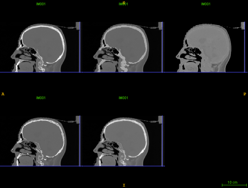
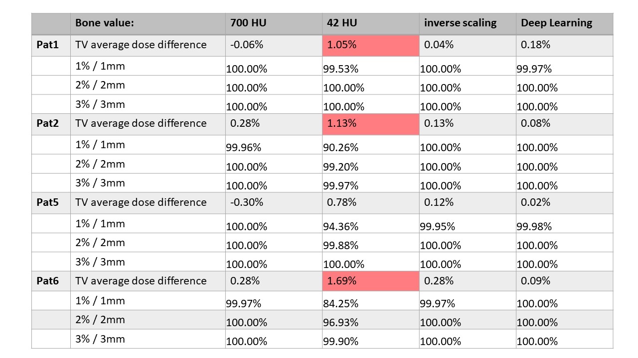
In Figure 1 the images corresponding to the four bone value assignment methods under study are shown and compared to the reference CT data. The Target Volume (TV) average dose difference for all cases is shown in Table 1 together with the gamma pass analysis calculated for the high dose regions, i.e. receiving more than 15% of the prescribed dose. While in step 1) and 2) the pure dose difference originating from different HU value assignment is measured for step 3) and 4) also dice coefficient and registration errors enter in the calculation. The largest dose difference is observed in the case of CT bone values assigned to a soft tissue value of 42 as expected,a milder difference is measured for 700 HU assignment, while an excellent correspondence between dose calculated on the CT with its original bone values and on the CT with pseudo CT bone values and pseudo CT inverse scaling values can be observed, despite registration inaccuracies.Conclusions:
This confirms the strength and the reliability of ZTE based bone value conversion, opening up new possibility for a simpler and faster pseudo CT conversion based on a combination of DL and analytical method explored in6 where a DL classification method is used to identify bones and the inverse linear scaling is applied to convert them into HU values.Acknowledgements
No acknowledgement found.References
1. Wiesinger F, Bylund M, Yang J, et al. “Zero TE-based pseudo-CT image conversion in the head and its application in PET/MR attenuation correction and MR-guided radiation therapy planning”. Magn. Reson. Med. 2018. doi: 10.1002/mrm.27134
2. Cozzini C, Bylund M, Jonsson J H, et al. “Feasibility of Zero TE MR based Radiation Therapy Planning for Head application”, ISMRM 2017
3. Kaushik S, Cozzini C, Bylund M, et al. “Deep Learning based pseudo-CT computation and its application for PET/MR attenuation correction and MR-guided radiation therapy planning”, ISMRM 2018
4. Edmund, Jens M., and Tufve Nyholm. "A review of substitute CT generation for MRI-only radiation therapy." Radiation Oncology12.1 (2017): 28.
5. T. Nyholm, et al, 3rd ESTRO Forum, 2015 [6] Wiesinger F, Kaushik S, Engstrom M, et al. “MR to pseudo CT conversion combining Deep-Learning and Analytical Image Processing” ISMRM 2019, submitted
Figures