2383
Detection and Risk-stratification of Prostate Cancer with MR Molecular Imaging using Extradomain-B Fibronectin as a Biomarker1Biomedical Engineering, Case Western Reserve University, Cleveland, OH, United States
Synopsis
Early detection and differential diagnosis of high-risk prostate cancer is imperative, so as to enable risk-stratification and decision-making in disease management. This research shows that the ECM oncoprotein Extradomain-B Fibronectin (EDB-FN) is strongly associated with high-risk prostate tumors and with low-risk prostate tumors that evolve into high-risk tumors, highlighting the potential of EDB-FN as a promising diagnostic biomarker for prostate cancer imaging. In addition, we have developed EDB-FN-specific peptide targeted MRI contrast agents that facilitate accurate differential detection and risk-stratification of prostate cancers.
Introduction
Prostate cancer (PCa) is the second most common cause of cancer death in men in the US.1 Early detection and differential diagnosis of high-risk prostate cancer is imperative, so as to enable risk-stratification and decision-making in disease management as well as for tailoring the best possible treatment for a diverse spectrum of patients. Unfortunately, current diagnostic tools, including prostate specific antigen (PSA) screening, are unable to distinguish high-risk tumors from indolent ones, resulting in high rates of overdiagnoses and false positives, resulting in unnecessary invasive biopsies and aggressive treatments in most PCa patients.2 Thus, there is an urgent unmet clinical need for the development of molecular imaging technologies based on biomarkers specific to metastatic and invasive properties, to facilitate the non-invasive detection and differential diagnosis of highly aggressive PCa tumors from the indolent ones.
Because tumor heterogeneity plays a major role in the dynamic nature of PCa progression, we have developed a novel MR molecular imaging (MRMI) approach by targeting the tumor extracellular matrix (ECM), which plays a critical role in relaying oncogenic signals to the tumor and supporting its growth, migration, inflammation, and immune evasion.3 The ECM component, Extradomain-B Fibronectin (EDB-FN), is emerging as a biomarker for a plethora of neoplasms4 and is associated with epithelial-to-mesenchymal transition (EMT), cancer cell stemness, proliferation, angiogenesis, and metastasis, all of which reflect tumor aggressiveness.5
Methods
The potential role of EDB-FN as a molecular marker for PCa aggressiveness was determined in multiple prostate cancer lines cultured in Matrigel to facilitate the establishment of a conducive ECM. The expression of EDB-FN secreted into the matrix was analyzed using qRT-PCR and fluorescent-labeled EDB-FN-specific peptide ZD2-Cy5.5.6 For MRMI of EDB-FN, an optimized contrast agent ZD2-N3-Gd(HP-DO3A)7 was synthesized and characterized by MALDI-TOF spectrometry. The efficacy of this agent for MRMI, detection, and risk-stratification was evaluated in athymic male mice subcutaneously implanted with aggressive PC3 and low-risk LNCaP tumor xenografts. T1-weighted MR images were obtained pre- and post- injection of 0.1 mmol/kg ZD2-N3-Gd(HP-DO3A) using 2D spin-echo (TR = 500 ms, TE = 8.1 ms, FA = 90°, FOV = 3.50 cm x 3.50 cm, slice thickness = 1.20 mm, slice number = 16, Nav = 2, matrix = 128 x 128, acquisition time = 128 s) and 3D FLASH coronal sequences (TR = 15 ms, TE = 2.8 ms, FA = 15°, FOV = 10.00 cm x 3.00 cm, slice thickness = 35 mm, Nav = 1, matrix = 128 x 512, resolution = 0.0127 x 0.273 cm/pixel, acquisition time = 61 s) using a 7T Bruker BioSpin small animal scanner. Volume rendering and maximum intensity projections (MIP) were obtained using 3D FLASH sequence and rendered using OsiriX software.Results and Discussion
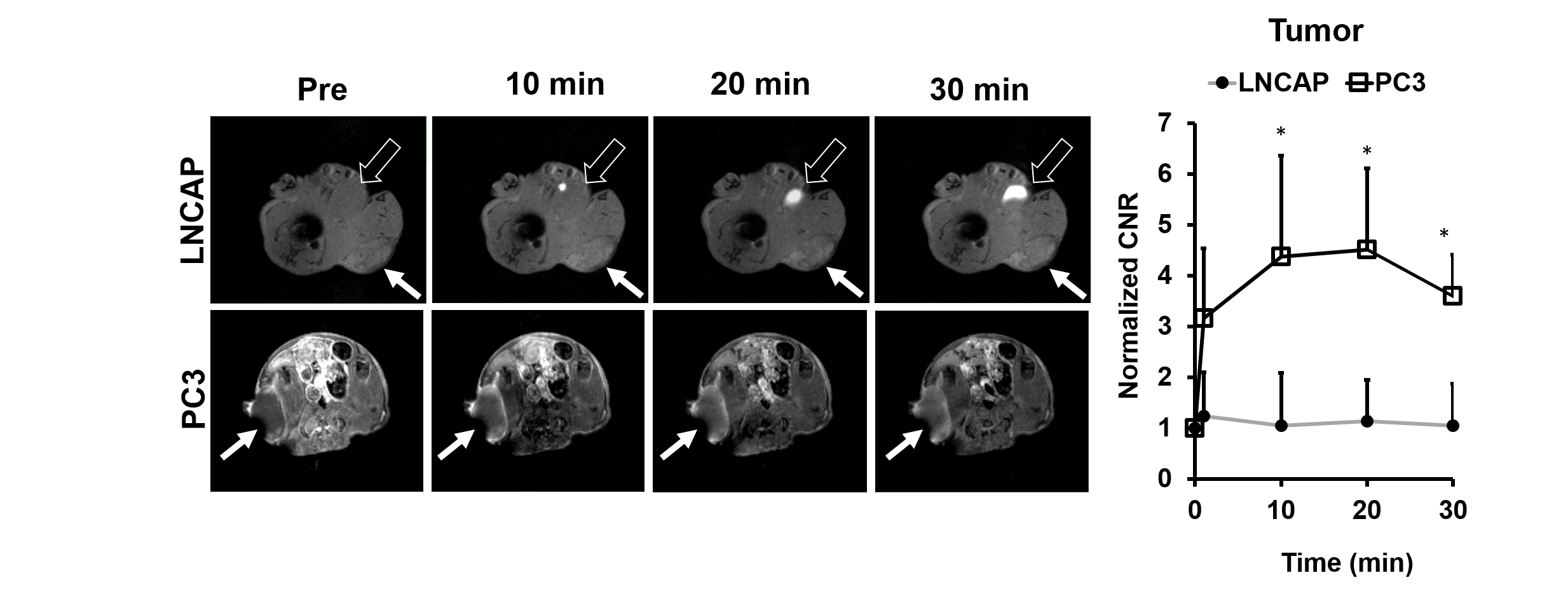
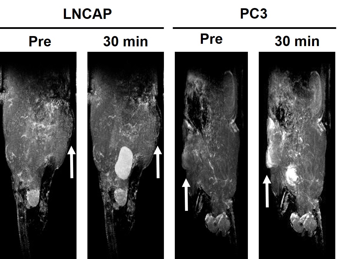
Differential contrast enhanced MRMI of the fast-growing PC3 and slow-growing LNCaP tumor xenografts was performed using the EDB-FN-targeting contrast agent. As shown in Fig 1, ZD2-N3-Gd(HP-DO3A) resulted in strong signal enhancement in the aggressive PC3 tumors, compared to the relatively indolent LNCaP tumors. Quantitative analysis of tumor contrast-noise ratios (CNRs) confirmed 4-fold enhancement for at least 30 min post-injection, while the LNCaP tumors demonstrated negligible increase in the CNR, indicating that contrast enhanced MRI with ZD2-N3-Gd(HP-DO3A) can enable differential detection and diagnosis of high- and low-risk prostate cancers. Strong tumor enhancement was also observed in the MIP images of PC3-bearing mice, with little enhancement in LNCaP tumors (Fig 2).
When grown in 3D culture, the PC3 cells showed proliferative network formation and significantly high levels of EDB-FN secretion, compared to the LNCaP cells, which formed smaller tumor spheroids along with low expression of EDB-FN in the ECM (Fig 3). This EDB-FN expression profile is consistent with the differential enhancement observed in MRMI of the tumor xenografts. When the LNCaP cells were modified by stable expression of the pro-tumorigenic IL8 receptor CXCR2,8 they underwent EMT and gained significant growth and invasive advantages. These pro-survival properties were accompanied by a significant increase in the EDB-FN secretion at the protein and mRNA levels (Fig 3). These results demonstrate that EDB-FN is strongly associated with high-risk PCa tumors and with low-risk tumors that evolve into high-risk tumors.
Conclusions
EDB-FN is a promising diagnostic and prognostic biomarker for aggressive high-risk prostate cancer. Our work highlights the potential of non-invasive MRMI using peptide targeted MR contrast agents specific to EDB-FN for differentiating between high-risk and low-risk PCa tumorsAcknowledgements
This research was supported by the National Institute of Health grants R01CA194518 to ZRL.References
1. Roobol M. Perspective: Enforce the clinical guidelines. Nature. 2015;528(7582):S123.
2. Qaseem A, Barry MJ, Denberg TD, et al. Screening for prostate cancer: a guidance statement from the Clinical Guidelines Committee of the American College of Physicians. Ann. Intern. Med.2013;158: 761-9.
3. Balkwill F, Capasso M, & Hagemann T. The tumor microenvironment at a glance. J. Cell Sci. 2012;125:5591-96.
4. Petrini I, Barachini S, Carnicelli V, et al. ED-B fibronectin expression is a marker of epithelial-mesenchymal transition in translational oncology. Oncotarget. 2017;8:4914-21.
5. White ES, Baralle FE, & Muro AF. New insights into form and function of fibronectin splice variants. J. Pathol. 2008;216:1-14.
6. Han Z, Zhou Z, Shi X, et al. EDB fibronectin specific peptide for prostate cancer targeting. Bioconjug. Chem. 2015;26(5):830-838.
7. Ayat N, Qin J, Han C, et al. Optimization of ZD2 Peptide Targeted Gd(HP-DO3A) for Detection and Risk-Stratification of Prostate Cancer with MRI. ACS. Med. Chem. Lett. 2018;9(7):730-735.
8. Chen H, Sun Y, Wu C, et al. Pathogenesis of prostatic small cell carcinoma involves the inactivation of the P53 pathway. Endocr. Relat. Cancer. 2012; June 19(3): 321-331.
Figures
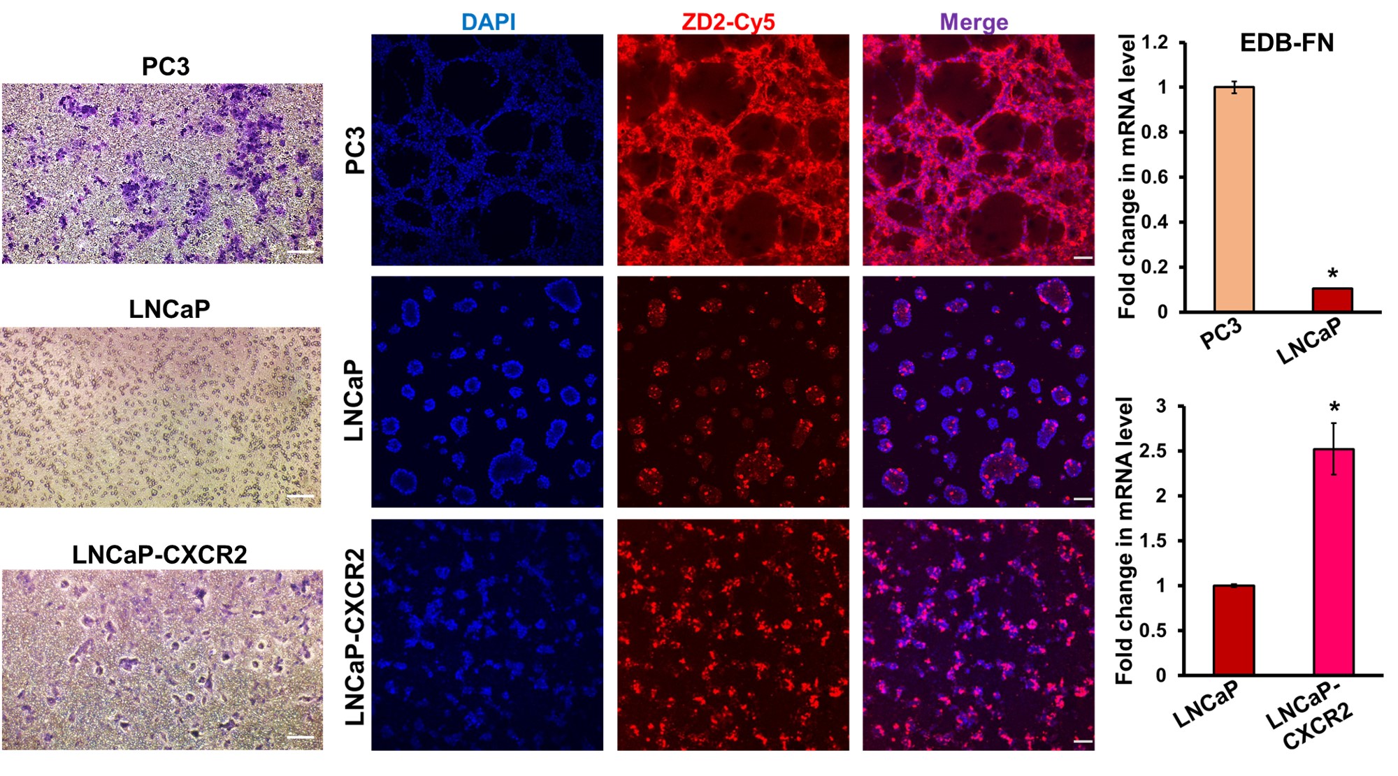
Transwell assays show invasive potential of the prostate cancer lines. ZD2-Cy5.5-binding in 3D cultures and RT-PCR analysis demonstrates that EDB-FN is significantly overexpressed in aggressive PC3 cells, compared to the relatively indolent LNCaP cells. The modified aggressive LNCaP-CXCR2 cells show elevated EDB-FN expression compared to their low-risk LNCaP counterparts.