2379
Understanding the Biomechanical Signature of Pressurised Tumour on the Surrounding Tissue: a Modelling Study1School of Biomedical Engineering and Imaging Sciences, King's College London, London, United Kingdom, 2Biomedical Engineering, University of Michigan, Ann Arbor, MI, United States, 3U1148, INSERM, Paris, France
Synopsis
Solid tumour growth is often associated with the accumulation of mechanical stresses acting on the surrounding host tissue. These forces alter the biomechanics of the adjacent soft tissue, generating a variation in stiffness resulting in a signature pattern that can be probed through MR-Elastography. The probed stiffness, however, is strongly dependent on the direction of propagation of the employed shear waves, leading to the reconstruction of anisotropic mechanical properties of the peri-tumoural tissue. Here we present, using theoretical and experimental means, a closed theoretical understanding of the observed alteration of tangent stiffness of soft tissue generated by pressurised tumour expansion.
Introduction
Solid tumour growth is often associated with the accumulation of mechanical stresses acting on the surrounding host tissue[1]. These forces alter the biomechanics of the adjacent soft tissue, which can be probed with propagating shear waves and quantified through MR-Elastography (MRE)[2]. The reconstructed tangent stiffness in the peri-tumoural habitat inherits a signature from the tumour expansion which depends on the stress-strain response in the tumour and the tissue environment and the wave propagation k-vector. Here we used theoretical and experimental means based on a simplified setup of the tissue-tumour ensemble to understand the shift in tangent stiffness probed with MRE, as a way to bridge such analytical considerations to in vivo tissue.Methods
Due to nonlinear mechanical properties of tissue, the shear modulus of the tumour environment will change according to the local deformation created by the pressurised tumour, increasing when undergoing stretch and decreasing when compressed[3]. This renders the mechanical properties of the peri-tumoral tissue anisotropic. Shear waves can sense these apparent changes in stiffness associated to their direction of propagation, hence ideally probing a softening at the leading edge of the inflated object and a stiffening along the lateral area. This pattern was previously shown experimentally, employing an inflated balloon-catheter inserted inside a PVC-based hyperelastic isotropic phantom[4].
Analytically, tumour expansion can be idealised with a thick-walled sphere subjected to an axisymmetric macro-deformation (Fig.1-A). The pressure generated by the inflation of the inner sphere is analytically given by
$$p_i=\int_a^b\frac{d\sigma_{rr}}{dr}\,dr$$
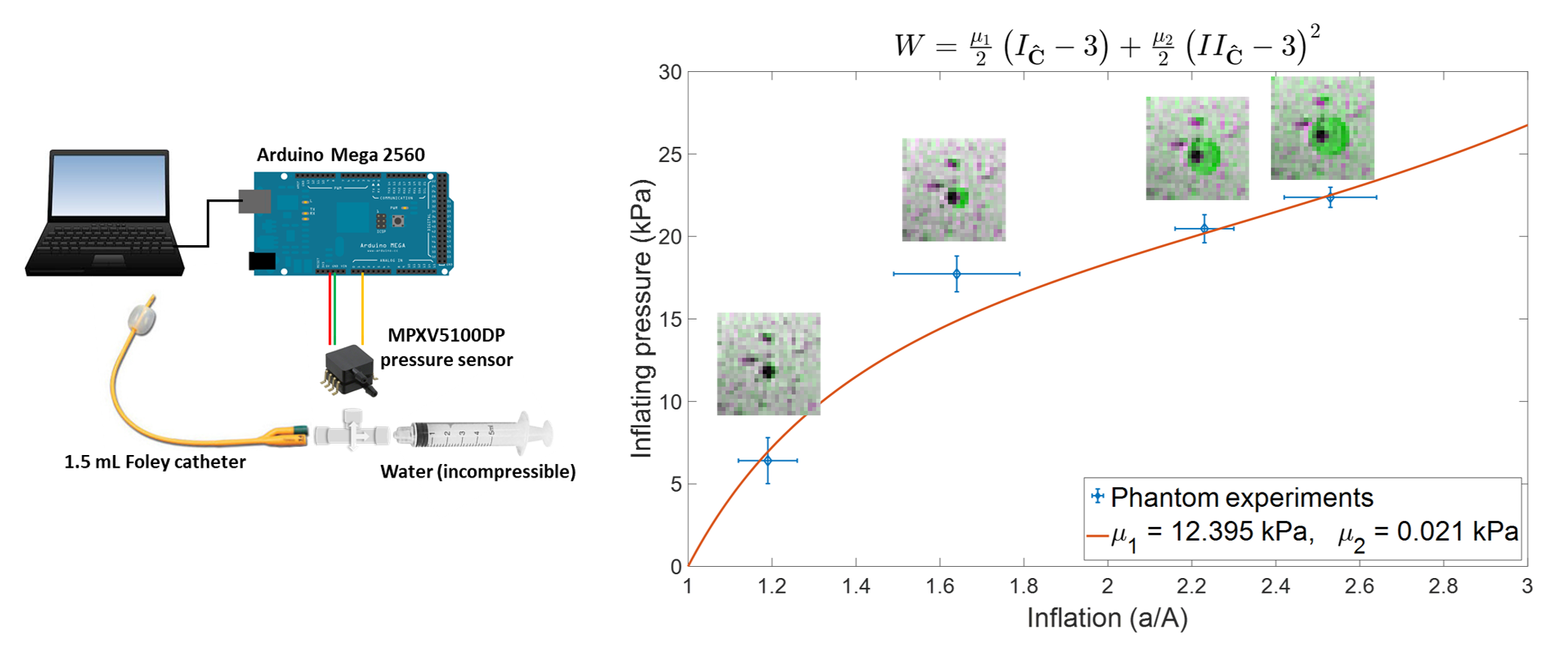
where the radial stress $$$\sigma_{rr}$$$ depends on the chosen material law. Through a pure-stretch rheological investigation we identified a modified Mooney-Rivlin law in the form $$$W=\frac{\mu_1}{2}\left(I_{\hat{\mathbf{C}}}-3\right)+\frac{\mu_2}{2}\left(II_{\hat{\mathbf{C}}}-3\right)^2$$$ that describes the viscoelastic behaviour of the above-mentioned tissue-mimicking phantom (Fig.1-B), yielding an Nth-order polynomial expression of $$$p_i$$$. Fig.1-C shows that, while $$$\mu_1$$$ purely scales the curve produced by the linear term of the constitutive equation, $$$\mu_2$$$ is responsible for the more dramatic variations at higher inflations. Once combined, the two terms result in a characteristic S-shaped pressure curve.
Superimposing the perturbation generated by the low amplitude waves used in MRE on top of the existing macro-deformation, a solution to the wave equation can be found after a linearization process, yielding[3]
$${\rho}J\partial_{t}^{2}\mathbf{u}_{\varepsilon}-\nabla_{\mathbf{x}}\cdot\left[\boldsymbol{\mathcal{C}}:\nabla_{\mathbf{x}}\mathbf{u}_{\varepsilon}+p_{\varepsilon}\mathbf{I}\right]=0$$
with the stiffness tensor being
$$\mathcal{C}_{ijkl}=\frac{1}{J}\frac{{\partial}P_{is}}{{\partial}F_{mn}}F_{ln}F_{js}$$
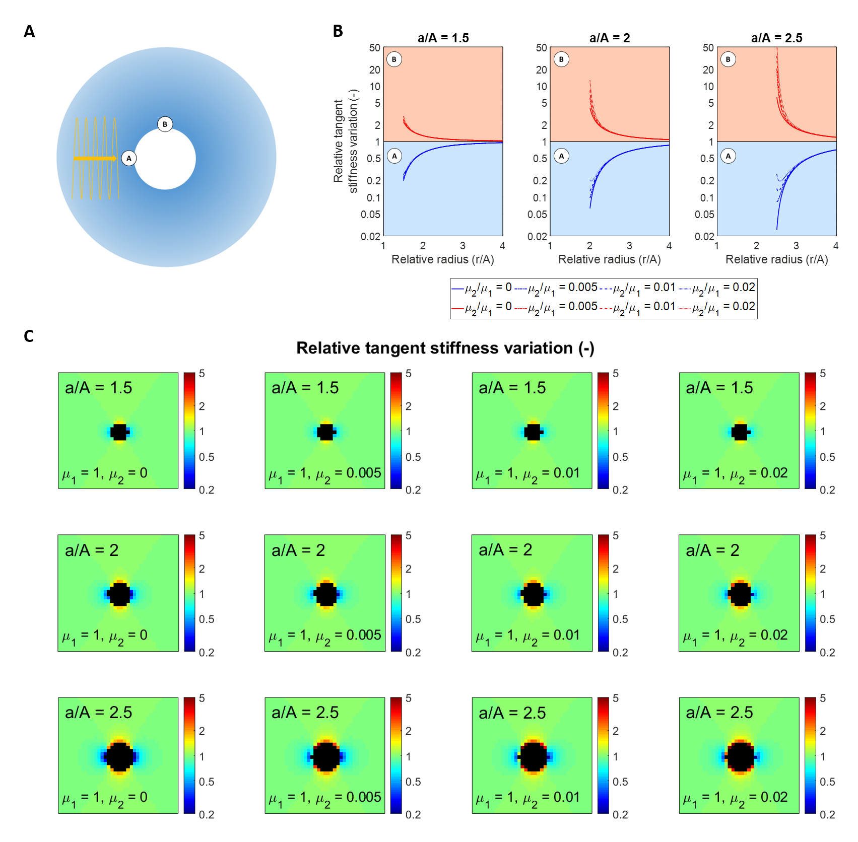
where the first Piola-Kirchhoff stress tensor $$$\mathbf{P}$$$ will depend on the deformation gradient $$$\mathbf{F}$$$ and the constitutive law describing the phantom. In the simple case of a plane shear wave $$$\mathbf{u}_{\varepsilon}=u_{\varepsilon}(x)\mathbf{\hat{e}_y}$$$ (Fig.2-A), the sensed stiffness $$$\boldsymbol{\mathcal{C}}:\nabla_{\mathbf{x}}\mathbf{u}_\varepsilon$$$ will change according to the applied deformation, producing the expected decrease in stiffness when the waves approach the inclusion head-on (Fig.2-B). In contrast, along the peripheral interface of the inner sphere, a stiffening is detected. This matches our expectations from simple considerations of waves in anisotropic media and our previous phantom experiments. The numerically calculated associated signature pattern is shown in Fig.2-C.
We experimentally validated this theory by fitting the model parameters to pressure measurements obtained at different inflation states and comparing the numerically generated apparent stiffness patterns in the implemented model with those acquired from MRE data.
Results & Discussion
Fig.3 shows how three out of four experimental pressure measures, obtained at different inflations through a pressure sensor (left), fit the curve produced using the modified Mooney-Rivlin model (right). Furthermore, the linearised shear modulus of the undeformed material, calculated from the model using
$$\mu=\lim_{a{\longrightarrow}0}2\left(\frac{{\partial}W}{{\partial}I_{\hat{\mathbf{C}}}}+\frac{{\partial}W}{{\partial}II_{\hat{\mathbf{C}}}}\right)=\mu_1$$
matches the background stiffness reconstructed from the MRE data ($$$\mu=11.9\pm2.9$$$ kPa).
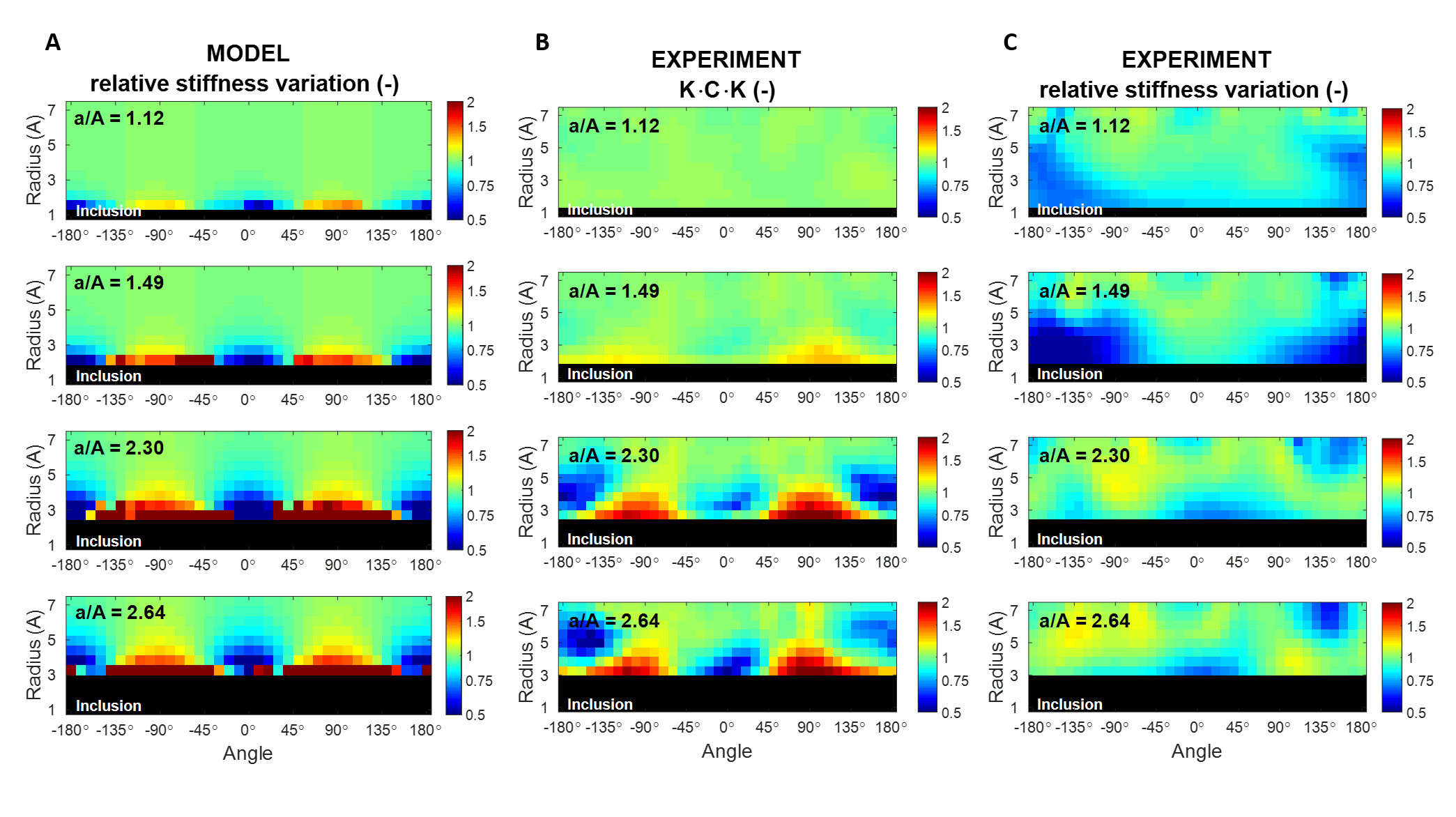
The material parameters were used to numerically calculate the relative
stiffness variation around the inclusion. The patterns presented in Fig.4 were radially
unravelled following the perimeter of the balloon, with the 0° angle associated
to the mean k-vector of the waves. The modelled pattern in Fig.4-A, obtained
simulating simple uni-directional shear waves, shows a qualitative and
quantitative agreement with the voxel-wise projection of the right Cauchy-Green
tensor associated to the real deformation along the local direction of the
k-vector, presented in Fig.4-B. Given the more complex wave behaviour and
deformation field in the experimental case, $$$\mathbf{K}\cdot\mathbf{C}\cdot\mathbf{K}$$$ gives an understanding of the type and intensity of deformation probed
by the waves. Similar patterns are found in the relative stiffness variation
experimentally measured; however the model does not predict the shift generated
in the region immediately adjacent to the inflated balloon, possibly due to the
impact of the phantom/balloon discontinuity on the reconstructed stiffness.
Conclusions
We demonstrated that MRE, in combination with non-linear mechanics, is capable to predict the alteration of tangent stiffness of soft tissue generated by tumour expansion. More experiments are currently underway to further extend this investigation to real tissue and tumour cases.Acknowledgements
This project has received funding from the European Union’s Horizon 2020 research and innovation program under grant agreement No 668039 and by the King’s College London & Imperial College London EPSRC Centre for Doctoral Training in Medical Imaging (EP/L015226/1).References
[1] T. Stylianopoulos, “The Solid Mechanics of Cancer and Strategies for Improved Therapy,” J. Biomech. Eng., vol. 139, no. 2, p. 021004, Jan. 2017.
[2] R. Sinkus, M. Tanter, T. Xydeas, S. Catheline, J. Bercoff, and M. Fink, “Viscoelastic shear properties of in vivo breast lesions measured by MR elastography.,” Magn. Reson. Imaging, vol. 23, no. 2, pp. 159–65, Feb. 2005.
[3] A. Capilnasiu et al., “Magnetic resonance elastography in nonlinear viscoelastic materials under load,” Biomech. Model. Mechanobiol., pp. 1–25, Aug. 2018.
[4] D. Fovargue et al., “Non-linear Mechanics Allows Non-invasive Quantification of Interstitial Fluid Pressure,” ISMRM #3832, 2018.
Figures
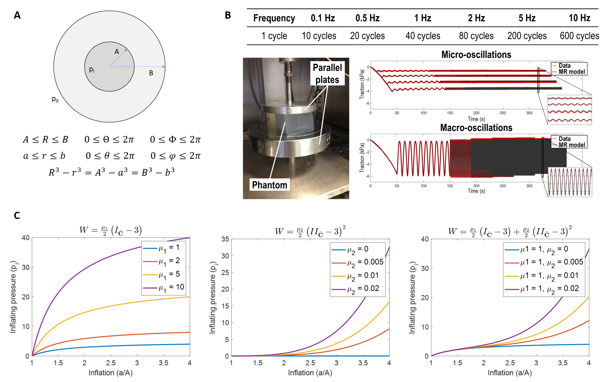
Figure 1. Section of the idealised thick-shelled sphere used to model balloon inflation is soft PVC-phantom (A). The phantom was subjected to uniaxial-sinusoidal micro- and macro-oscillations at different frequencies and compression levels to identify a suitable material law. The proposed modified Mooney-Rivlin model produced a good fit of the experimental data (B). The generated family of pressure curves reveals the scaling effect of μ1 on the linear term of the constitutive equation (C-left), as well as the impact of μ2 at higher inflations on the quadratic term (C-middle). Once combined, the two terms produce a characteristic S-shaped curve (C-right).