2374
Correlation of breast tumor grade and lymphovascular invasion with biomechanical properties: first results from a breast cancer trial1Guy's and St.Thomas' NHS Foundation Trust, London, United Kingdom, 2Division of Cancer Studies, King's College London, London, United Kingdom, 3Division of Imaging Sciences & Biomedical Engineering, King's College London, London, United Kingdom, 4King's College Hospital, London, United Kingdom
Synopsis
Magnetic Resonance Elastography (MRE) has been considered a promising novel imaging modality in the quantification of viscoelastic properties of breast tumours. The purpose of this study was to evaluate reproducibility and repeatability of a newly developed MRE breast system and investigate whether aberrant biomechanical properties correlate with tumour histopathology. MRE was conducted on 20 healthy volunteers and 15 breast cancer patients. Malignant lesions demonstrated an increase in viscoelasticity when compared to adipose or fibroglandular tissue. While lesions with lymphovascular invasion demonstrated a tendency towards more elevated viscoelasticity than those without lymphovascular invasion, histological grades clearly did not correlate with biomechanics.
Introduction
Despite access to an armamentarium of imaging techniques for detecting and diagnosing breast cancer, current imaging modalities are limited in their ability to accurately diagnose breast cancer in dense breasts or in mammographically occult lesions1,2. Furthermore, while predicting tumor grade or metastatic propensity non-invasively may be most valuable for patient stratification prior to surgical excision, clinical reality is far from providing such imaging biomarkers. Magnetic Resonance Elastography (MRE) has in recent years been considered as a promising novel imaging modality for the quantification of viscoelastic properties of breast tumors. Here we aim firstly to evaluate the reproducibility/repeatability of a new developed breast MRE system allowing for bilateral high resolution elasticity imaging, and secondly to test the hypothesis whether aberrant biomechanical properties correlate with tumor histopathological and biological factors.Methods
The first cohort consisted of healthy female participants recruited via King’s College London Advertisement. The second cohort consisted of patients with histologically confirmed invasive breast cancer on core biopsy, due to undergo primary surgery with curative intent, who were recruited from the breast clinic at Guy’s and St. Thomas’ NHS Foundation Trust, London. High resolution T1 and T2 weighted images were conducted in both cohorts with the addition of a dynamic contrast-enhanced T1 weighted scan in patients. MR-Elastography was performed after the anatomical images with a mechanical excitation frequency of 36Hz using a novel MRE setup3 and a GRE-based MRE sequence4 on a 3T Achieva MR scanner (Philips Healthcare, The Netherlands). Total acquisition time was 6.5mins providing entire breast coverage (FOV=340-400mm) at 2mm isotropic resolution covering 15 slices in FH-direction. Parallel imaging was not used. All 3-motion directions plus a reference scan were encoded sampling eight wave phases per oscillatory cycle. Repeatability of the MRE protocol was assessed in a cohort of participants by taking the participant out of the scanner with subsequent repositioning using the identical scan protocol. All cancer patients subsequently underwent resection of their primary tumor providing detailed histopathology of each lesion.Results
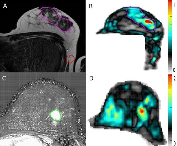
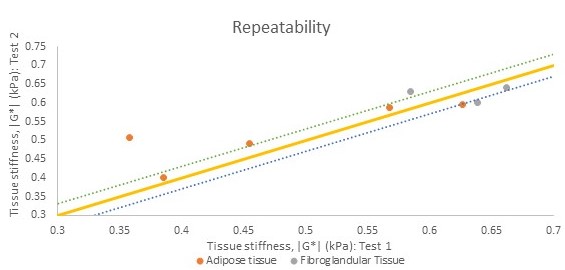
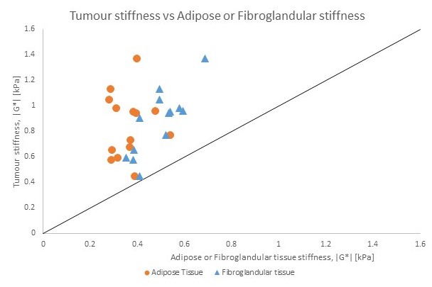
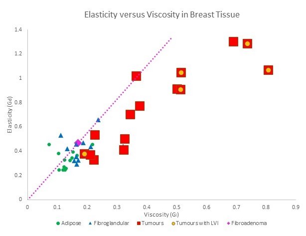
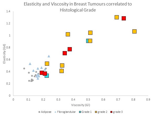
Figure 1 shows excellent correlation between anatomy (tumor/adipose/fibroglandular tissue) and magnitude of the shear modulus. Mean stiffness values between both breasts showed significant correlation (p-value <0.001) and no asymmetry between the left and the right breast. In a cohort of participants, reproducibility was tested and repeatability was around 10% as demonstrated in Figure 2. A paired t-test demonstrated a significant difference between tumor (M=0.89, SD=0.24) and fibroglandular tissue stiffness (M=0.5, SD=0.09); t(13) =6.28, p<0.001. Similarly, a significant difference was established between tumor stiffness (M=0.86, SD=0.24) and adipose tissue stiffness (M=0.38, SD=0.6); t(15) =6.83, p<0.001 with tumors demonstrating significantly increased stiffness in |G*| as seen in Figure 3. Furthermore a significant increase in elasticity (Gd) and viscosity (Gl) in tumors has been noted when compared to adipose and fibroglandular tissue tissue (Gd p<0.001; Gl p<0.01). Interestingly, tumor cases which were confirmed to have lymphovascular invasion (red squares with yellow dot in Figure 4) account for the highest increase in viscoelasticity with the exception of one case which demonstrated lower viscoelasticity. Simultaneously, we observed one case without lymphovascular invasion to account for the third highest increase in viscoelasticity. Despite these two exceptions, there seems to be a general tendency towards more elevated viscoelasticity in tumors with lymphovascular invasion. This corresponds to the results of another study that we are currently conducting which investigates the correlation between tumor pressure and lymphovascular invasion as potential biomarker for metastatic potential. Additionally, we observed a benign lesion in form of a fibroadenoma (pink diamond shape in Figure 4) which demonstrated a higher elasticity to viscosity ratio than tumors. This is coherent with the literature where it has been noted that malignant breast lesions were shown to be more viscous than benign breast lesion5,6. Interestingly, histological tumor grade did not seem to be correlated to any significant increase in viscoelasticity as seen in Figure 5. This is contradictory to what has been found in ultrasound elastography7.Discussion
This study demonstrates the high performance of the newly developed breast MRE system within the clinical breast cancer environment. The significant increase in viscoelasticity in tumors when compared to adipose or fibroglandular tissue that we observed demonstrates the sensitivity of the technique whilst further studies are required to increase specificity. Further investigations in breast cancer patients are required to understand whether different breast tumor types influence the biomechanics of the tumor. However, the increased tumor viscoelasticity when the lymphovascular space has been invaded in most cases is especially interesting when considering the metastatic potential of breast tumors and the lack of current imaging techniques to quantify the aforementioned.Acknowledgements
This project has received funding from the European Union’s Horizon 2020 research and innovation programme under grant agreement No 668039.References
1. Smith, J. A. & Andreopoulou, E. An overview of the status of imaging screening technology for breast cancer. doi:10.1093/annonc/mdh653
2. Bosch, A. M. et al. Interexamination variation of whole breast ultrasound. Br. J. Radiol. 76, 328–331 (2003).
3. Runge JH, Hoelzl SH, Sudakova J, Dokumaci AS, Nelissen JL, Lee J, Stoker J, Nederveen AJ, Nordsletten D, S. R. A novel MR Elastography transducer concept based on a rotational eccentric mass: the gravitational transducer.
4. Garteiser, P. et al. Rapid acquisition of multifrequency, multislice and multidirectional MR elastography data with a fractionally encoded gradient echo sequence. NMR Biomed. 26, 1326–1335 (2013).
5. Sinkus, R. et al. Viscoelastic shear properties of in vivo breast lesions measured by MR elastography. Magn. Reson. Imaging 23, 159–165 (2005).
6. Sinkus, R. et al. Imaging anisotropic and viscous properties of breast tissue by magnetic resonance-elastography. Magn Reson Med 53, 372–387 (2005).
7. Evans, A. et al. Invasive Breast Cancer: Relationship between Shear-wave Elastographic Findings and Histologic Prognostic Factors. Radiology 263, 673–677 (2012).
Figures