2350
Bolus arrival time estimation for DCE-MRI signals without fast up-slope1Department of Medical Physics in Radiology, German Cancer Research Center (DKFZ), Heidelberg, Germany, 2Faculty of Biosciences, University of Heidelberg, Heidelberg, Germany, 3Translational Radiation Oncology, National Center for Tumor Diseases (NCT), German Cancer Research Center (DKFZ), Heidelberg, Germany, 4Heidelberg Institute for Radiation Oncology (HIRO) and National Center for Radiation Research in Oncology (NCRO), Heidelberg, Germany, 5Department of Radiation Oncology and Radiotherapy, University Hospital Heidelberg, Heidelberg, Germany, 6Department of Medical Physics in Radiation Oncology, German Cancer Research Center (DKFZ), Heidelberg, Germany, 7Natural Sciences and Humanities, University of Applied Sciences Würzburg-Schweinfurt, Schweinfurt, Germany
Synopsis
Accuracy in pharmacokinetic modelling of DCE-MRI data can be impaired due to a delay between the contrast agent arrival in the tissue of interest and an artery further upstream. To correct the delay, bolus arrival times (BATs) are estimated from the concentration curves. However, the state-of-the-art method for estimating BATs may give unsatisfactory results if the curves do not exhibit a fast up-slope. We propose a spline-based method for BAT estimation for concentration curves without fast up-slopes which are often observed in small animal data. The proposed method gives accurate results on simulated and in vivo acquired rat data.
Objectives
Accuracy in pharmacokinetic modelling of dynamic contrast-enhanced MRI (DCE-MRI) data can be impaired due to a delay between the arrival of contrast agent (CA) bolus in the artery, selected to extract the arterial input function (AIF), and the arrival of CA in the tissue of interest. When the concentration curves exhibit a fast up-slope in CA concentration, this delay can be corrected for by estimating the difference between both bolus arrival times (BATs) using the state-of-the-art method proposed by Cheong et al.1. We introduce a method for continuous BAT estimation of concentration curves that do not have fast up-slopes. Such data is particularly observed in small animal data.Proposed method
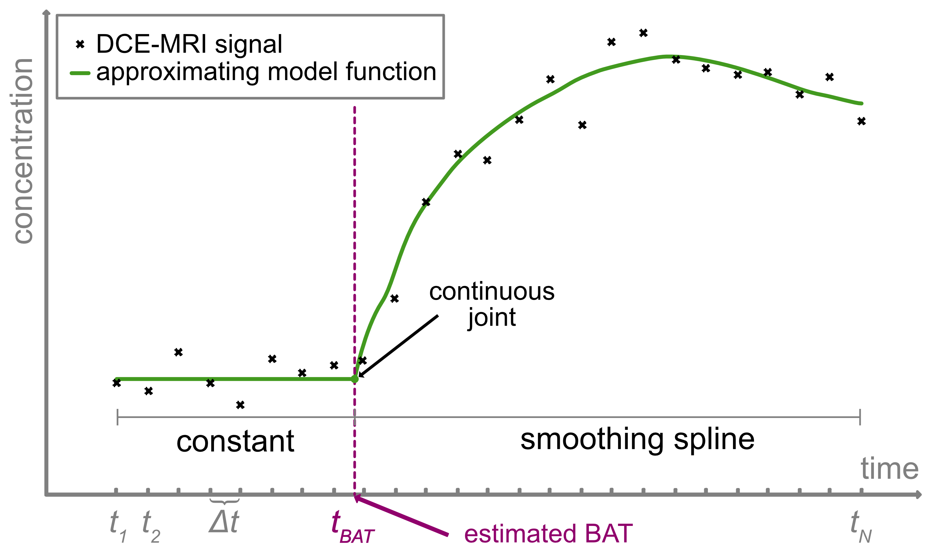
The MR-signal intensities cn are assumed to be recorded at N uniformly sampled time points tn, and the CA to be injected as a bolus after starting the DCE-MRI sequence. The proposed model approximates the data points cn by a continuous piecewise defined function $$$u^*$$$ which is a constant function before the BAT tBAT and a smoothing spline afterwards. Hence, this approach is able to adapt to the shape of the curve (Fig. 1, equ. 1). The BAT tBAT is estimated along with finding the most accurate fit for the sample points cn. The model is described by:
$$\tilde{u}^* = \text{argmin}_{\tilde{u}}\,\sum_{n=1}^{N'} (\tilde u(t_{\text{BAT}}) -c_n)^2 + \sum_{n=N'+1}^{N} (\tilde u(t_n) -c_n)^2 + \alpha\int_{t_{\text{BAT}}}^{t_N} (\tilde u^{(k)}(\tau))^2 \,d\tau \qquad\qquad(1) $$
where $$$\tilde u^{(k)}$$$ denotes the k-th derivative of the function $$$\tilde{u}$$$, α > 0 adjusts the relative weight of data fidelity and smoothness, and N’ is the index of the last sample point before tBAT. The principal interest is to estimate tBAT from the concentration curve. Alongside, the spline parameters α and k are determined, too, as they adapt the model to the measured data points cn. tBAT is estimated on a continuous time scale and therefore is not restricted to the discrete sample points tn. All three parameters are automatically determined by generalized cross validation.
Model validation
The proposed model was validated
using simulated and in vivo acquired
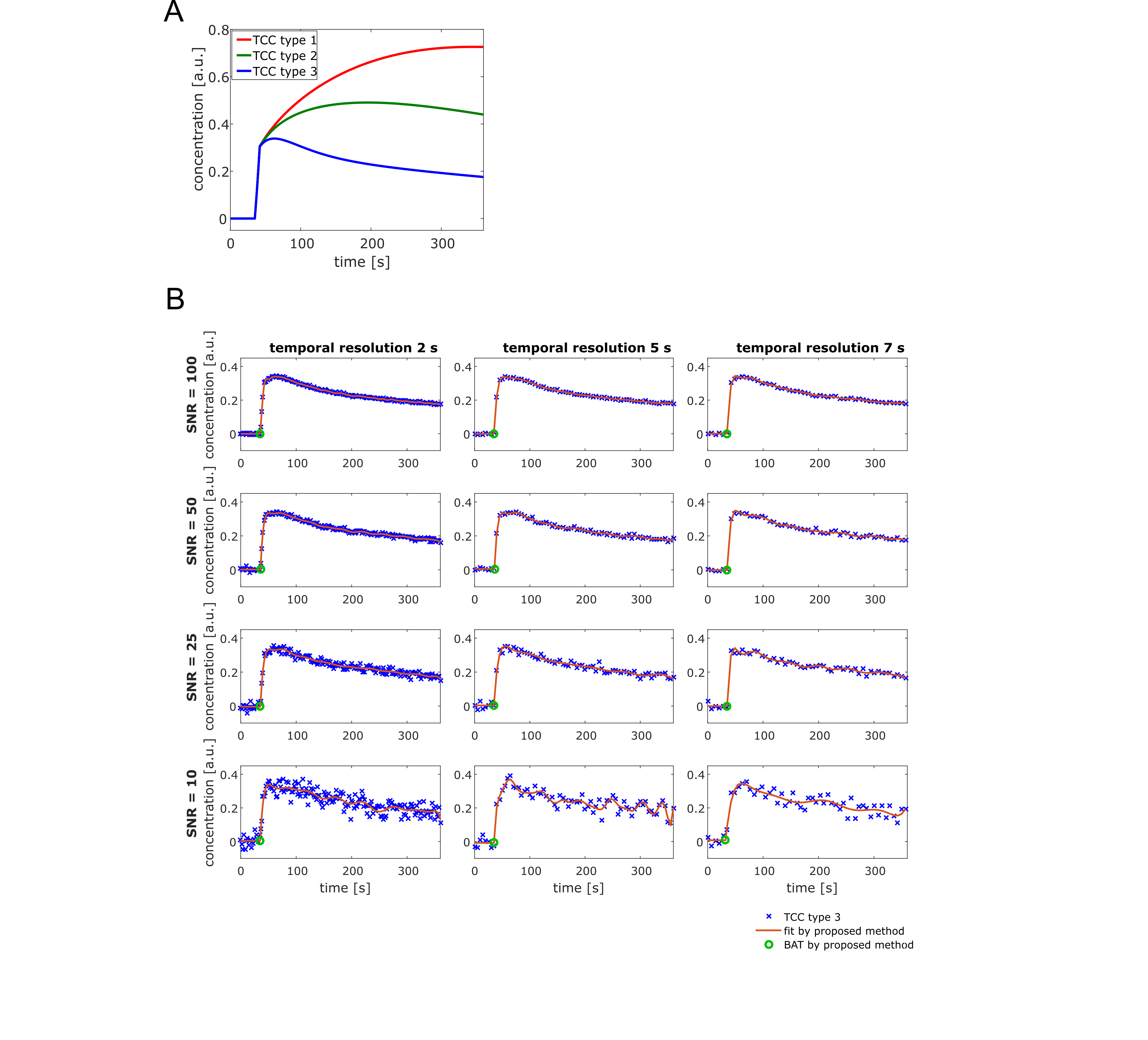
rat data. Three tissue concentration curves (TCCs) were simulated using an
in-house developed software 2 by forward convolution of a
model-based rat AIF 3 with the tissue response
function of the extended Tofts model 4 (Ktrans = 15 ml/min/100ml, vp = 0.05, ve
= 0.3, 0.6, or 0.1, Fig. 2 A). Data were adapted for three temporal resolutions (2 s, 5 s, and
7 s) and four noise levels (SNR = 100, 50, 25, 10). BATs were estimated by the
proposed method for 500 noise realizations for each configuration. For in vivo validation, DCE-MRI data of five
male Copenhagen rats bearing anaplastic Dunning R3327-AT1-tumors transplanted
onto both thighs were acquired. Animals were imaged when tumors reached a size
of 10×10 mm.
DCE-MRI (TURBO-FLASH sequence, temporal resolution: 0.75 s, 380 s total
acquisition time) was acquired per animal for single slices through the largest
diameter of the tumor and through the heart to acquire the AIF. Image signals
were converted to concentrations using absolute signal enhancement.Results
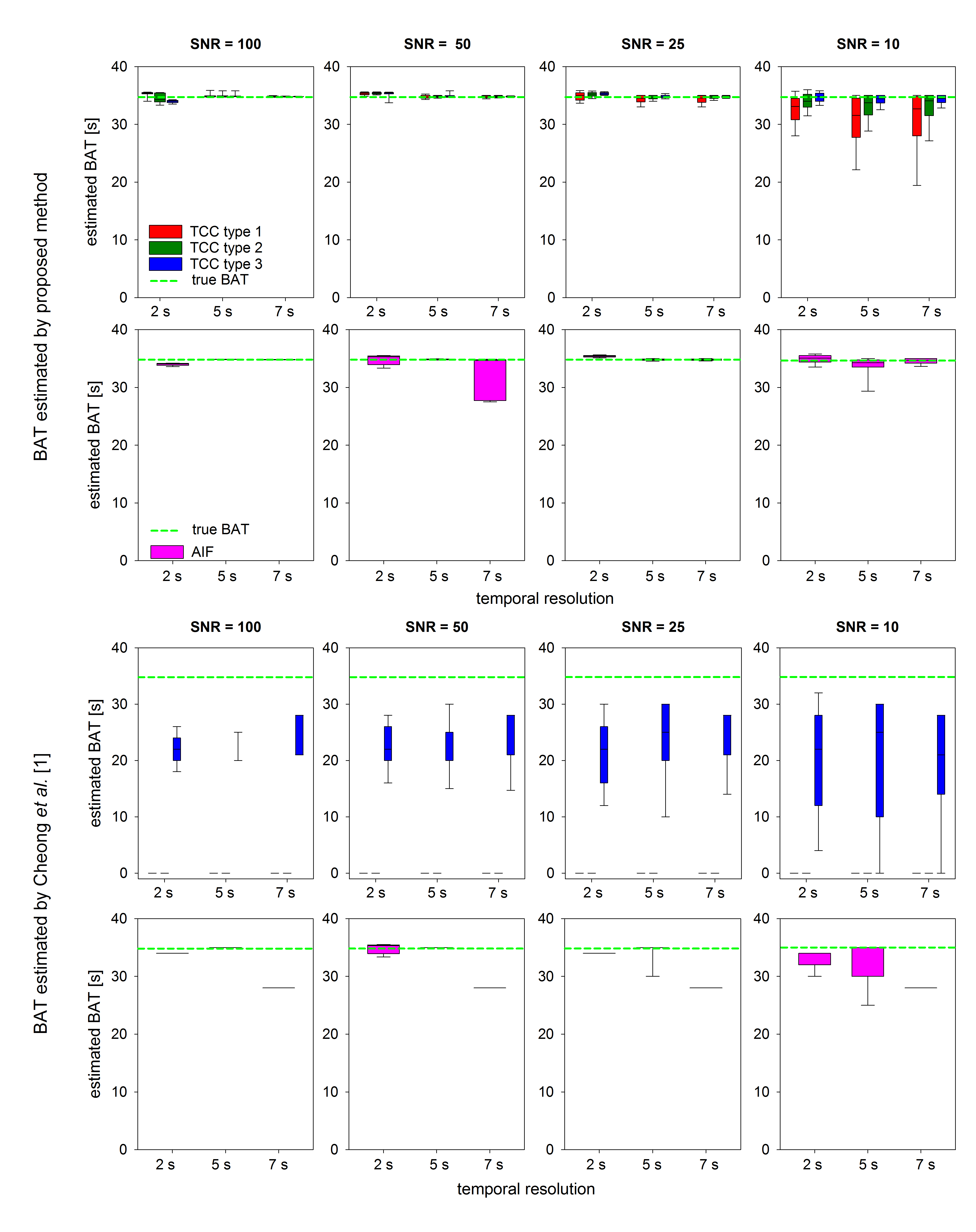
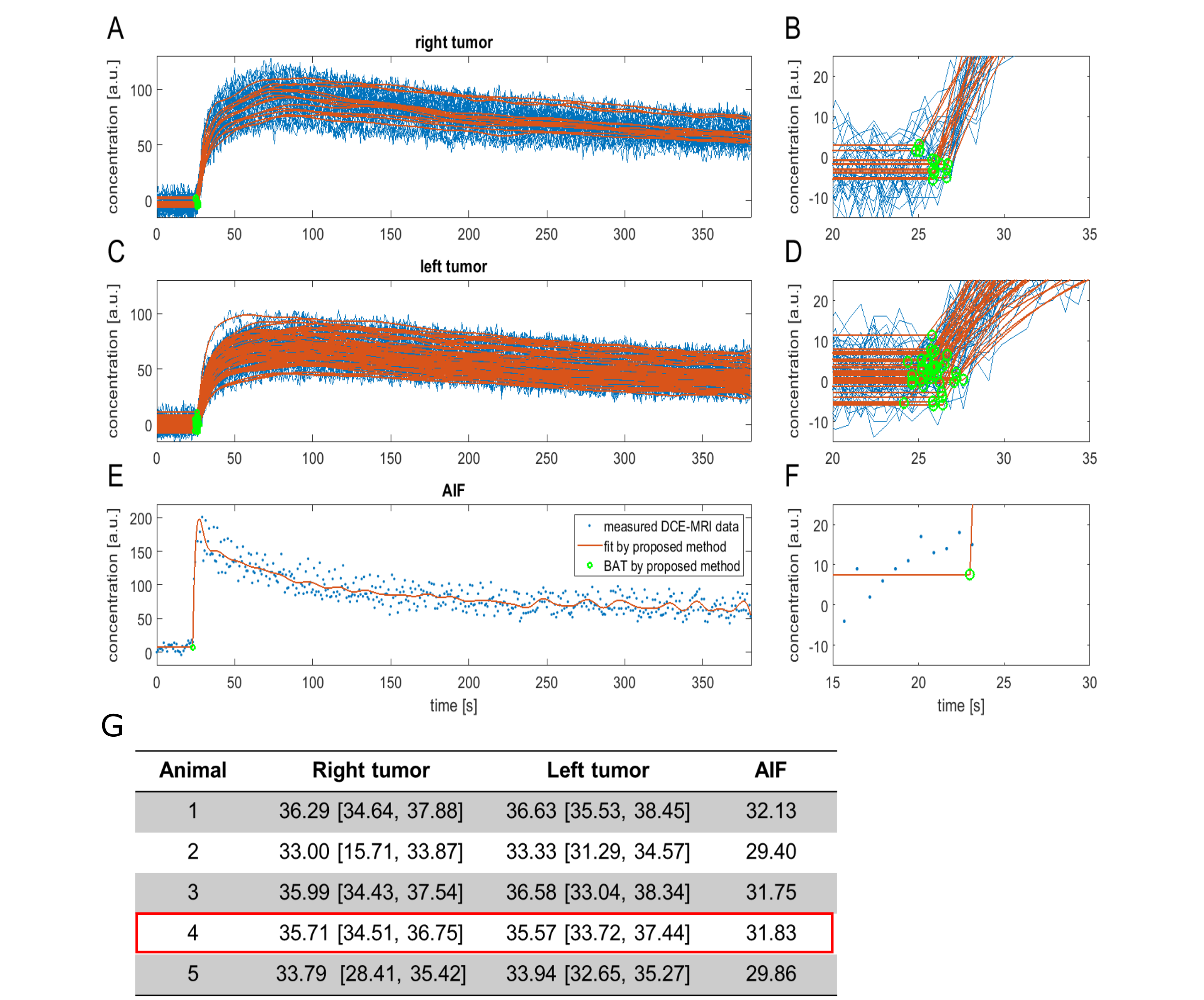
Fig. 2 B displays representative fits of the proposed model to the simulated TCC type 3 and the corresponding estimated BATs of all configurations. The proposed method could accurately estimate the BAT of the simulated TCCs even at low SNR and low temporal resolution (Fig. 3). Accuracy was only compromised for TCC type 1 at low SNR. The BAT of the AIF was accurately estimated for all configurations. BAT estimation by the state-of-the-art gave unsatisfactory results for most configurations (Fig. 3). Qualitative analysis of in vivo acquired data reveals that BATs were estimated at reasonable time points (exemplary fitting results and BAT estimations for animal 4 are displayed in Fig. 4 A-F) and within small margins per animal (Fig. 4 G).Discussion and conclusion
A
new method for BAT estimation on a continuous time scale of DCE-MRI is proposed
for dynamic data lacking a fast up-slope, as often found in small animal data. A
simulation study with known ground truth showed that the accuracy of BAT estimation
was superior to other methods even at low SNR and low temporal resolution. Results
of in vivo acquired data were found
to be at reasonable time points.Acknowledgements
This work was supported by the German Research Foundation (DFG STO1126/2-1, GL893/1-1, KA2679/3-1, and KFO 214).References
1. Cheong LH, Koh TS, Hou Z. An
automatic approach for estimating bolus arrival time in dynamic contrast MRI using
piecewise continuous regression models. Physics in medicine and biology.
2003;48(5):N83-N88.
2. Nolden M, Zelzer S, Seitel A, et al. The Medical Imaging Interaction Toolkit: challenges and advances. Int J CARS. 2013;8(4):607-620.
3. McGrath DM, Bradley DP, Tessier JL, et al. Comparison of model-based arterial input functions for dynamic contrast-enhanced MRI in tumor bearing rats. Magn Reson Med. 2009;61(5):1173-84.
4. Sourbron SP, Buckley DL. Classic models for dynamic contrast-enhanced MRI. NMR Biomed. 2013;26(8):1004-1027.
Figures