2348
Application of 3D_NerveVIEW to neurography of nasopharyngeal carcinoma patients in radiotherapy treatment planning1Sun Yat-sen University Cancer Center, Guangzhou, Guangdong Province, China, 2Philips Healthcare, Guangzhou, Guangdong Province, China, 3Philips Healthcare, Hongkong, China
Synopsis
In radiotherapy, CT/MR images are used to delineate the region of targets and normal structures. In this study, 3D_NerveVIEW sequence was performed on a volunteer, and then the image of cranial nerve was rigidly registered and fused to CT image. In the fused image, the cranial nerve had better visualization than CT and T1w images, and the contour of nerve was easily identified. Which could improve the accuracy of nerve contour and reduce the radiotherapy-induced nerve palsy.
Introduction
Radiotherapy (RT) is regarded as the standard treatment for patients with nasopharyngeal carcinoma (NPC)1, however patients treated with radiotherapy may suffer from some side effects, such as hearing loss, cranial nerve palsy, etc. A previous study has revealed that the most frequent nerve palsy was hypoglossal nerve, followed by vagus nerve2. Currently, some researchers believe that tumor delineation is the weakest link in the delivery of accurate and precise RT3,4. Hence we aimed to use 3D_NerveVIEW sequence to identify the location and contour of cranial nerve in RT planning for nerve preservation of NPC patients to improve the quality of life.Methods
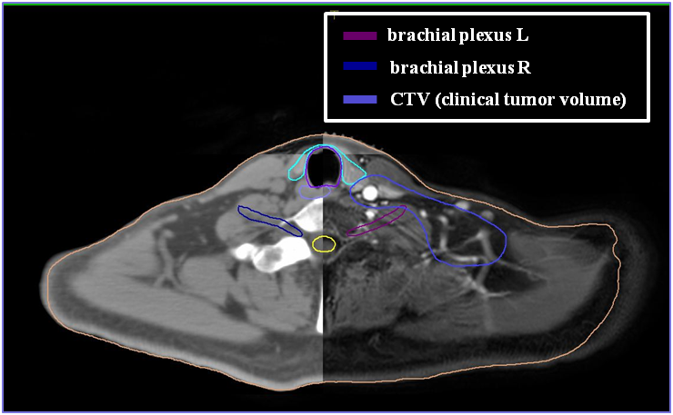
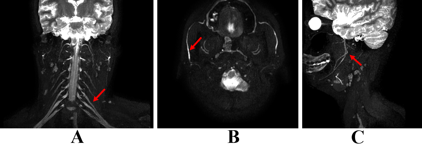
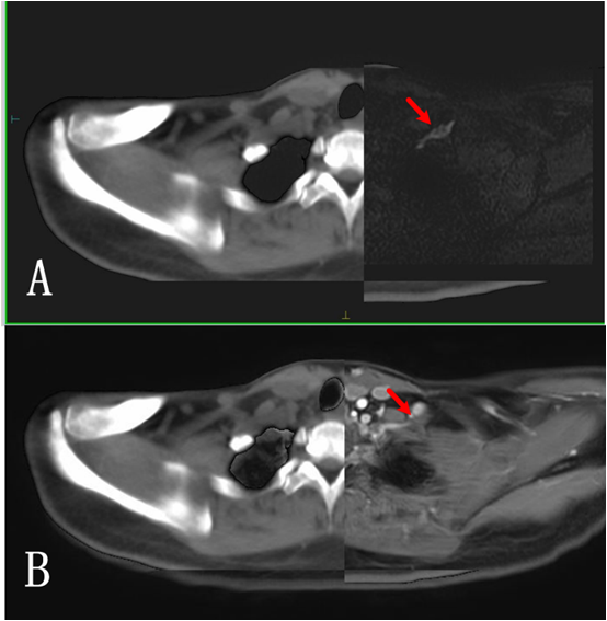
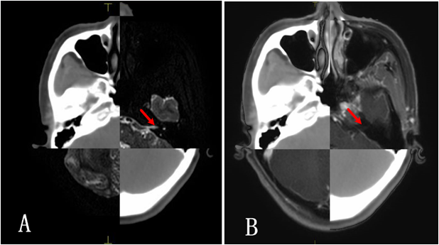
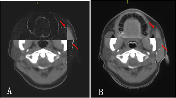
MR scan was carried out on a dedicated 3T MR-RT MR system (Ingenia, Philips) with flat tabletop overlay, coil bridge and external laser positioning system. Routine sequences such as T1W_3D and mDIXON, and 3D_NerveVIEW sequence for neurography were performed after contrast enhancement. The imaging parameters of 3D_NerveVIEW were: TR/TE = 2200/200ms; IR delay = 280ms; acquired voxel-size = 1×1×1mm3; FOV (FH, RL, AP) = 220×249×165; acceleration factor = 3; scan duration = 9:25. The mDIXON images are usually preferred for contour delineation due to excellent fat-suppression performance. The mDIXON images, nerve images and CT images were transferred to Monaco, then the MR images were rigidly registered and fused with CT image at radiotherapy Treatment Planning System (TPS). The fused image was used to delineate the contour of nerves. Then the visibility of brachial plexus, accessory nerve, vagus, parotid duct, optic nerve and hypoglossal nerve were compared between mDIXON and 3D_NerveVIEW.Results
The fused image of mDIXON with CT has superior soft tissue definition, however the nerve contour was not seen (figure 1). The coronal and sagittal reconstruction of 3D_NerveVIEW images (figure 2). The location and contour of the nerves, e.g. brachial plexus (figure 3), hypoglossal nerve (figure 4) and parotid duct (figure 5), was shown on the fused images of 3D_NerveVIEW with CT. Compared to the fused images using mDIXON alone, the nerve contours were more accurately delineated with fused images using 3D_NerveVIEW. The accurate contour of OAR can protect the nerve when patients receive radiotherapy.Conclusion
In this study, 3D_NerveVIEW imaging can provide excellent visualization of cranial nerve, which may improve the precision of OAR delineation, suggesting it will be a potential and effective tool for nerve preservation in radiotherapy planning and treatment.Acknowledgements
No acknowledgement found.References
1. S zhang, B Zhang, J Tian, et al. Radiomics features of Multiparametric MRI as Novel Prognostic Factors in Advanced Nasopharyngeal Carcinoma. Clin Cancer Res 2017;23(15):4259-4269
2.Yao-shiang LIN, Yee-Min Jen, Jiann-Chyun Lin. Radiation-related cranial nerve palsy in patients with nasopharyngeal carcinoma. Cancer 2010, 95(2):404-409
3. Njeh CF. Tumor delineation: the weakest link in the search for accuracy in radiotherapy. J Med Phys 2008; 33: 136–40
4. Pollard JM, Wen Z, Sadagopan R, Wang J, et al. The future of image-guided radiotherapy will be MR guided. Br J Radiol 2017; 90(1073): 20160667
Figures