2336
4DMRI-based abdominal corset study for radiotherapy purposes1Medical Physics in Radiation Oncology, German Cancer Research Center (DKFZ), Heidelberg, Germany, 2Heidelberg Institute for Radiooncology (HIRO), National Center for Radiation Research in Oncology (NCRO), Heidelberg, Germany, 3Department of Physics and Astronomy, Heidelberg University, Heidelberg, Germany, 4X-Ray Imaging and Computed Tomography, German Cancer Research Center (DKFZ), Heidelberg, Germany, 5Medical Image Computing, German Cancer Research Center (DKFZ), Heidelberg, Germany, 6Department of Medicine, Heidelberg University, Heidelberg, Germany, 7Department of Radiation Oncology, Heidelberg University Hospital, Heidelberg, Germany, 8Center for Orthopedic and Trauma Surgery, Heidelberg University Hospital, Heidelberg, Germany
Synopsis
Abdominal organ motion provides challenges for radiotherapy treatments, leading to inhomogeneous dose distributions with over- and underdosage regions in the target volume. Repeated 4D-MRI acquisitions, allow to analyze inter- and intrafractional spatial motion. The aim of this study was to investigate the impact of abdominal corsets for motion reduction purposes, based on repeated 4D-MRI data sets. We found pronounced reductions in cranio-caudal and anterior-posterior direction using corsets, which additionally lead to more reproducible motion amplitudes. Lower amplitudes and better reproducibility are beneficial for radiotherapy and could lead to smaller irradiation margins and dose reductions to healthy tissue.
Introduction
In radiotherapy (RT) treatments of abdominal tumors, organ and tumor motion provide challenges to the treatment delivery. This is especially the case for particle therapy (PT) treatments, where due to the inverse dose profile and the limited range of protons or ions, over- or undershooting effects at the distal edge of the tumor may occur1,2. Moreover, in PT of the pancreas, interplay effects between the scanning pencil beam and intrafractional abdominal organ motion occur3,4. The resulting over- and underdosage depends on the underlying abdominal motion amplitudes. Therefore, motion mitigation techniques, like the usage of abdominal corsets, are a required reduce abdominal motion during delivery. This study investigates the motion and deformation reduction of the pancreas by means of abdominal corsets, based on 10 repeated 4D-MRI measurements of 5 volunteers. Moreover, the important questions of the impact of corsets on the reproducibility of pancreatic motion is investigated. 4D-MRI is a suitable imaging modality for this purpose, since it provides high soft-tissue contrast in the abdomen without exposing the subjects to any imaging dose.Methods
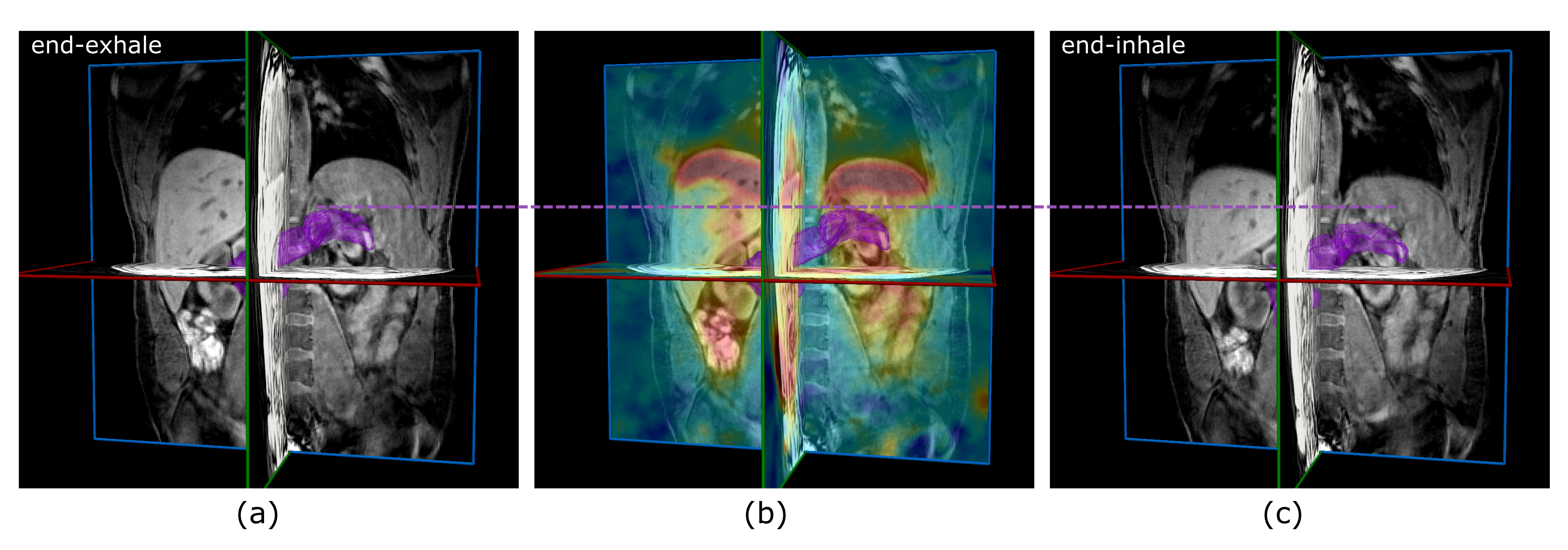
For 5 healthy volunteers each, 10 4D-MRI imaging sessions were acquired each within 2-4 weeks to simulate repeated imaging during fractionated RT. In each session, 4D-MR images were taken with and without a personalized MR-compatible abdominal corset, made of homogeneous polyethylene, which was fitted to the abdominal region of the respective volunteer. The MR images were acquired by means of a T1-weighted gradient echo MR sequence with radial sampling and golden angle spacing (field of view = 400x400 mm2 voxel size 1.5x1.5x3 mm3, spokes per partition = 2100, bandwidth = 610 Hz, TE = 1.5 ms, TR = 3.3 ms, α=12°). The measurements were performed under free breathing on a 1.5 T MR scanner (MAGNETOM Aera, Siemens Healthineers, Erlangen, Germany) with a measurement time of 8 minutes. The data were offline reconstructed by means of an iterative motion-compensated reconstruction algorithm, using a k-space-center-based self-gating signal5. For each 4DMRI data set, 20 overlapping breathing phases were obtained. The pancreas was manually delineated on each end-exhale image. Based on deformable image registration with a Demons algorithm, the motion amplitudes of all voxels within the pancreas delineation were extracted between the breathing phases, see figure 1. The mean motion amplitudes of the resulting motion-volume-histograms (MVHs) in cranio-caudal (CC) and anterior-posterior (AP) direction were used to quantify the motion reduction by the abdominal corset. The standard deviations of the mean motion amplitudes among different imaging sessions were used as a surrogate to quantify the reproducibility of motion amplitudes. Similarly, pancreatic deformations were characterized by the width (standard deviations) of the respective MVHs.Results
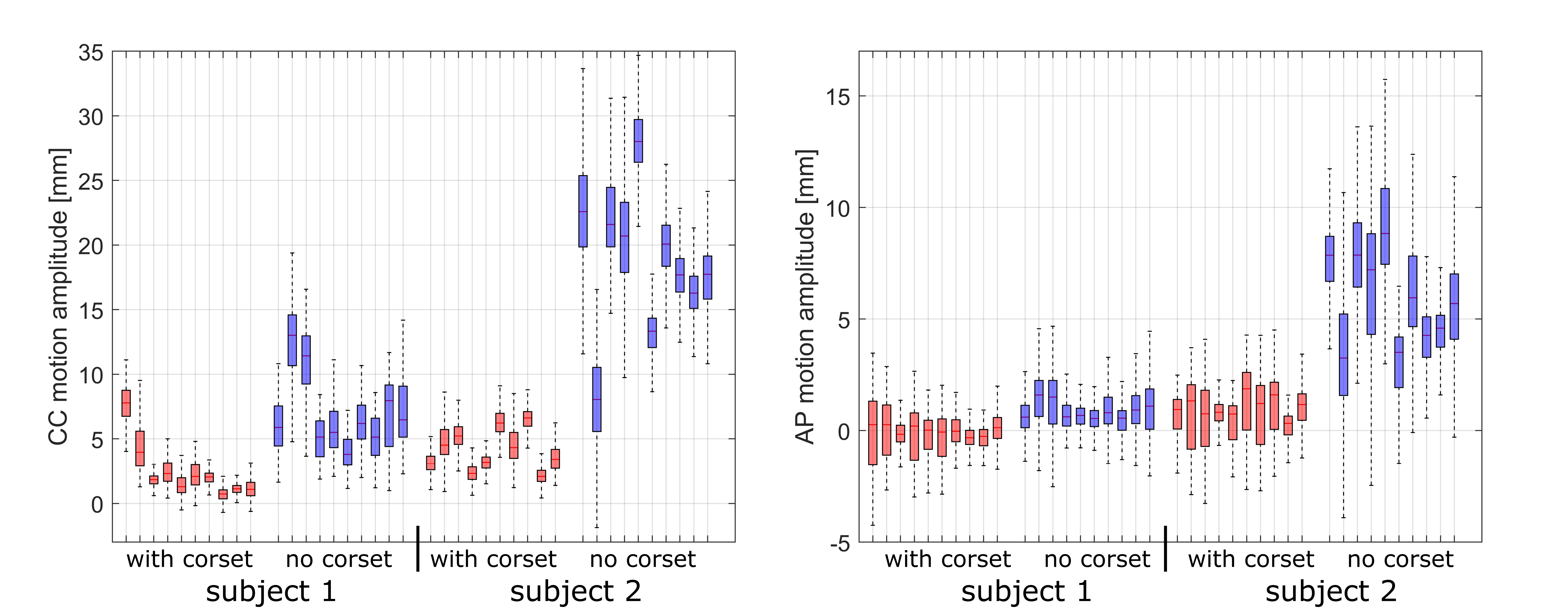
Highly variable pancreatic motion amplitudes were observed with mean cranio-caudal (CC) motion amplitudes of up to 28.5/9.3 mm without/with abdominal corset, respectively, resulting in an averaged CC motion reduction of 48% by the corset. Similarly, a mean anterior-posterior (AP) corset-based motion reduction by 69% was observed among the subjects, as illustrated in figure 2. No differences were found in left-right direction.
The corsets further enabled more reproducible motion amplitudes with smaller day-to-day fluctuations, reduced by 62%/130% in CC/AP direction. With respect to pancreatic deformations, for 4 out of 5 subjects, the corset measurements revealed a more rigid pancreatic motion compared to the pronounced observed deformations without corset.
Discussion
In particle therapy treatments of the pancreas, abdominal organ motion is a major challenge. However, repeated 4D-MRI measurements offer a viable opportunity to investigate intra- and interfractional organ motion and deformation without applying any imaging dose to the patients and provides high abdominal contrast. 4D-MRI can be further used as a viable basis of 4D dose evaluation studies6. This corset study indicates the advantage of using abdominal corsets when aiming at low motion amplitudes and small day-to-day motion variations. The motion reduction in CC/AP direction could contribute to avoid overshooting of the target, which may lead to a reduced dose deposition in adjacent organs at risk. Moreover, smaller safety margins for irradiation may be applicable by means of smaller motion amplitudes and better reproducibility of the motion patterns. Nevertheless, more subject data is necessary to conclude on the significance of the corset benefits, and patient comfort and fitness need to be investigated in further studies.Conclusion
4D-MRI was applied for the investigations of intra- and interfractional motion for radiotherapy purposes. This 4D-MRI-based study showed that the usage of corsets leads to smaller pancreatic motion and deformation and ensures less day-to-day motion variations.Acknowledgements
The authors would like to thank Joao Seco for fruitful discussions.References
1Kumagai M et al. Impact of intrafractional bowel gas movement on carbon ion beam dose distribution in pancreatic radiotherapy. Int. J. Radiat. Oncol. Biol. Phys. 2009; 15:1276–81.
2 Durante M et al. Charged particles in radiation oncology Nat. Rev. Clin. Oncol. 2010; 37:37–43.
3 Dolde K et al. 4D dose calculation for pencil beam scanning proton therapy of pancreatic cancer using repeated 4DMRI datasets. Phys. Med. Biol. 2018; 63:165005 (10pp)
4 Batista V et al. Significance of intra-fractional motion for pancreatic patients treated with charged particles. Radiat. Oncol. 2018; 13:120.
5 Rank C et al. 4D respiratory motion-compensated image reconstruction of free-breathing radial MR data with very high undersampling. Magn. Res. Med. 2017; 77(3): 1170-1183.
Figures