2333
Reproducible radiomic features from post-chemoradiation T2-weighted MRIs can more accurately discriminate pathologic T stage in rectal cancer patients1Department of Biomedical Engineering, Case Western Reserve University, Cleveland, OH, United States, 2Department of General Surgery, University Hospitals Cleveland Medical Center, Cleveland, OH, United States, 3Department of Pathology, University Hospitals Cleveland Medical Center, Cleveland, OH, United States, 4Department of Radiology, University Hospitals Cleveland Medical Center, Cleveland, OH, United States
Synopsis
We present initial results for identifying
Introduction
Expert assessment of neoadjuvant chemoradiation (NAC) and follow-up surgical planning in rectal cancer is impacted by inaccurate tumor re-staging on T2-weighted (T2w) MRI (agreement with pathology ~52%). To overcome this, computer-extracted radiographic imaging (radiomic) features have been evaluated for identifying pathologic response to NAC via rectal MRIs, by quantifying sub-visual imaging characteristics of chemoradiation-induced effects and residual disease. However, radiomic features within chemoradiated regions are likely affected by image acquisition variability between patients. To account for this, we will attempt to exploit radiomic feature reproducibility within reference “non-tumor” regions of the rectal wall. Our hypothesis was that identifying a set of radiomic features that are reproducible across patients can enable construction of a more discriminatory model for characterizing pathologic NAC response within the rectal wall on MRI.Materials and Methods
45 patients diagnosed with rectal cancer and who underwent NAC were retrospectively curated into training and hold-out validation cohorts, based on availability of post-NAC, pre-operative T2w MRI and pathologic tumor stage (TNM) information. An expert radiologist annotated two separate regions of interest (ROI) on T2w MRIs based on information in clinical reports: (1) the “in-plane” ROI, comprising 3 consecutive sections of rectal wall where NAC had been targeted; and (2) the “out-of-plane” ROI, comprising 3 consecutive sections of rectal wall spatially distant from where NAC was targeted. All annotations comprised the entire rectal wall on post-NAC MRIs, to minimize confounding factors for experts. 191 radiomic feature maps were extracted from both ROIs on a per-pixel basis, including 1st order, Gradient, Haralick, Gabor, Laws, and CoLIAGe. Each feature map was then described using basic histogram descriptors (median, standard deviation, skewness, and kurtosis). Feature reproducibility was assessed based on out-of-plane radiomic descriptors, using a previously presented measure by Leo et al with the assumption that reference “non-tumor” regions will be most consistent across all patients. The in-plane radiomic feature set was then pruned to those descriptors that were at least 96% reproducible in out-of-plane evaluation. Top radiomic descriptors were then identified via Wilcoxon rank-sum testing of in-plane descriptors that best discriminated patients by pathologic T stage (ypT0-2 vs ypT3-4). Feature performance was assessed by averaging the area-under-the-ROC (AUC) across 50 runs of 3-fold cross validation in the training set, as well as accuracy on the hold-out validation set. The performance of the reproducibility-informed radiomics model was also compared to a basic radiomics model (where no pruning was performed prior to feature selection).Results
The training set (n=32) comprised 18 patients with ypT0-2 (regressed tumor after NAC) and 14 with ypT3-4 (minimal regression after NAC); all with no nodal (N0) or distant metastatic (M0) disease. Hold-out validation (n=13) comprised patients with nodal or metastatic disease, with 4 ypT0-2N+M+ and 9 ypT3-4N+M+ (see Table 1). The basic radiomics model comprised a mixture of Gradient operators and Laws kernels; which achieved a cross-validated training AUC of 0.683 ± 0.095 and hold-out validation accuracy of 76.9%. By comparison, the reproducibility-informed radiomics model comprised only Gradient operators and resulted in a statistically significantly higher (p<0.001) AUC of 0.747 ± 0.072 as well as significantly higher hold-out validation accuracy of 84.6%. The latter also had no false negative predictions compared to the basic radiomics model (see Table 2).Discussion
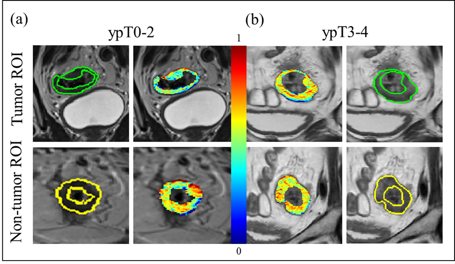
Top-ranked operators in the reproducibility-informed radiomics model were primarily gradient descriptors, indicating their resilience to inherent T2w acquisition differences by not being dependent on absolute intensity values (see Figure 1). By contrast, the basic radiomics model utilized Laws features (capturing sub-visual spottiness or wave patterns in the image), which appear to be more sensitive to these artifacts and less generalizable in hold-out validation (see Table 3). Our relatively high accuracies across both training and validation cohorts suggests that radiomic features are able to accurately characterize post-NAC pathologic T stage independent of disease metastasis in the surrounding nodes or environment.Conclusion
Incorporating feature reproducibility appears to enhance the ability of radiomic features to characterize pathologic response to chemoradiation in rectal cancers via re-staging T2w MRIs. Utilizing reference “non-tumor” portions of the rectal wall to benchmark feature reproducibility enabled us to identify highly discriminative gradient features that capture microscale heterogeneity associated with pathologic response within chemoradiated regions; independent of local or distant metastasis. Further testing in a larger, multi-site study will enable us to develop reproducible radiomics scores for evaluating treatment response via imaging.Acknowledgements
Research reported in this publication was supported by the National Cancer Institute of the National Institutes of Health under award numbers 1U24CA199374-01, R01CA202752-01A1 R01CA208236-01A1 R21CA179327-01; R21CA195152-01; the National Institute of Biomedical Imaging and Bioengineering of the National Institutes of Health under award number T32EB007509; the National Institute of Diabetes and Digestive and Kidney Diseases under award number R01DK098503-02, National Center for Research Resources under award number 1 C06 RR12463-01 the DOD Prostate Cancer Synergistic Idea Development Award (PC120857); the DOD Lung Cancer Idea Development New Investigator Award (LC130463), the DOD Prostate Cancer Idea Development Award; the DOD Peer Reviewed Cancer Research Program W81XWH-16-1-0329 the Ohio Third Frontier Technology Validation Fund the Wallace H. Coulter Foundation Program in the Department of Biomedical Engineering and the Clinical and Translational Science Award Program (CTSA) at Case Western Reserve University. Opinions, interpretations, conclusions and recommendations are those of the authors and are not necessarily endorsed by the Department of Defense and do not necessarily represent the official views of the National Institutes of Health.References
Chen CC, (2005). Dis Colon Rectum. 48(8): 722-8
AJCC Staging Manual, 8th Edition, (2018).
Braman, N et al. (2016). Breast Cancer Research 19: 57
Leo, P et al. (2018). Scientific Reports 8: 14918
Figures