2327
Investigation of Abdominal Organ Respiratory Motion Probability Distribution Function and its Inter-Fractional Reproducibility Assessment Using Fast Volumetric 4D-MRI for Probability-Based Radiotherapy Planning1Medical Physics and Research Department, Hong Kong Sanatorium & Hospital, Hong Kong, China
Synopsis
Respiratory motion is a major concern in radiotherapy (RT) in liver cancer patients. Probability-based treatment planning is an evolving approach for tumor motion management. A major hurdle of this approach is that the dosimetric error is tightly linked to the reproducibility of the tumor motion probability density function (PDF). Previous studies in lung used single-slice dynamic MRI for PDF reproducibility evaluation that could only captured the 2D respiratory motion restricted by the MRI acquisition speed. Moreover, inter-subject and inter-fractional variability of time evolved PDF might be underestimated by using just two fractions. In this study, we aim to investigate the inter-fractional and inter-subject abdominal motion PDF and its reproducibility using an ultrafast volumetric 4D- MRI.
Purpose:
Respiratory motion is a major concern in abdominal radiation therapy (RT) if the planning target volume (PTV) inadequately covers the tumor motion trajectory, resulting in a geographic miss of the radiation dose. Probability-based treatment planning has been proposed in which the dose to a location is weighted by tumor probability being at that location. The major hurdles are the determination of the target tumor motion probability density function (PDF) and its reproducibility assessment for each treatment fraction. Previous studies1-3 in lung used single-slice dynamic MRI for PDF reproducibility evaluation that could only captured the 2D respiratory motion of a representative slice for the entire tumor volume restricted by the MRI acquisition speed. In addition, inter-subject and inter-fractional variability of time evolved PDF might be much underestimated by using just two fractions. In this study, we aim to investigate the inter-fractional and inter-subject abdominal motion PDF and its reproducibility using an ultrafast volumetric 4D- MRI4.Material and Methods:
8 healthy volunteers (34.33±5.77years) were recruited. Each underwent 5 scans of free-breathing 4D-MRI at 1.5T using 3D spoiled-gradient-echo sequence, which (transversal, FOV=350(FE)x262.5(PE)mm, thickness=4mm, matrix size=128x128x56, TE/TR=0.6/1.7ms, flip-angle=6o, RBW=1250Hz/voxel, CAIPIRINHA factor=4, partial Fourier factor=6/8) yielded a temporal resolution of 1s/volume (56 slices) and a reconstructed voxel-size of 2.2x2.2x4mm. 144 frames (totally 8,064 images) were obtained within ~144s. Organ masks, including liver, left and right kidneys, and spleen were manually delineated based on the 2nd frame images and served as reference. Following frames were rigidly registered to the reference masks to calculate the mean position and displacement from the mean position for each organ. The PDF(δ, t, f), a time-evolved probabilistic organ position distribution, was estimated from fitting the organ respiratory displacement (δ) histogram within the scan duration (t) in a session (f) to a Gaussian function. The time evolved inter-fractional PDF reproducibility function R(t), defined as:$$R(t) = 2{{PD{F_1} \cap PDF(\delta ,t,f)} \over {PD{F_1} \oplus PDF(\delta ,t,f)}} \tag{1}$$,was calculated at every 3-second. To simulate the real RT treatment, the PDF calculated from the first scan was considered as the treatment planning reference.The mean PDF reproducibility was fitted to an exponential function
$$\bar R(t) = {A_0}+{A_1}{e^{{\rm{ - }}{A_2}t}} \tag{2}$$ From the fitted curve, the mean initial PDF reproducibility, $$$[{\bar R_0} = \bar R(t=3s)]$$$, the mean ending PDF reproducibility,$$$[{\bar R_e} = \bar R(t=144s)]$$$, and the PDF reproducibility equilibrium time constant $$$T({R_{0.63}})$$$ (the time when PDF reproducibility curve reaches 63% of $$$({R_e}-{R_0})$$$ were determined. Inter-subject and intra-subject coefficients of variance (CVs) were determined. A Mann-Whitney U test was used to assess the differences between organs.
Results
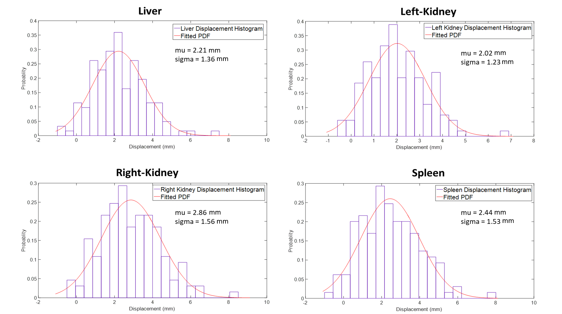
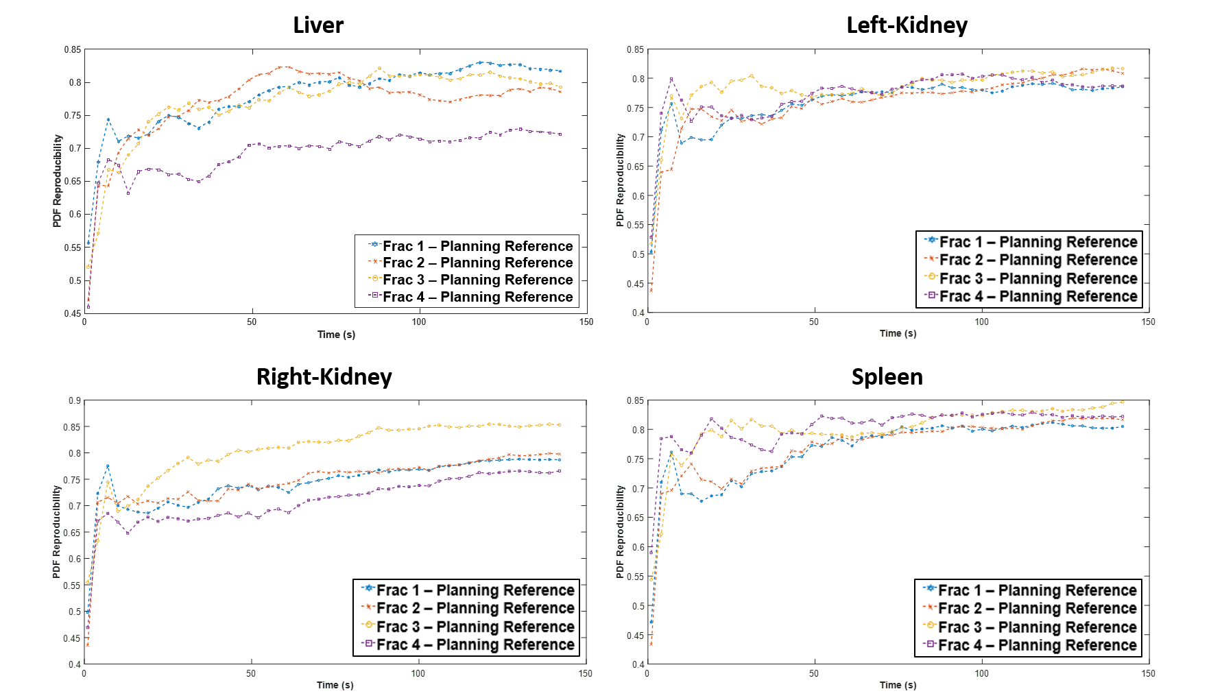
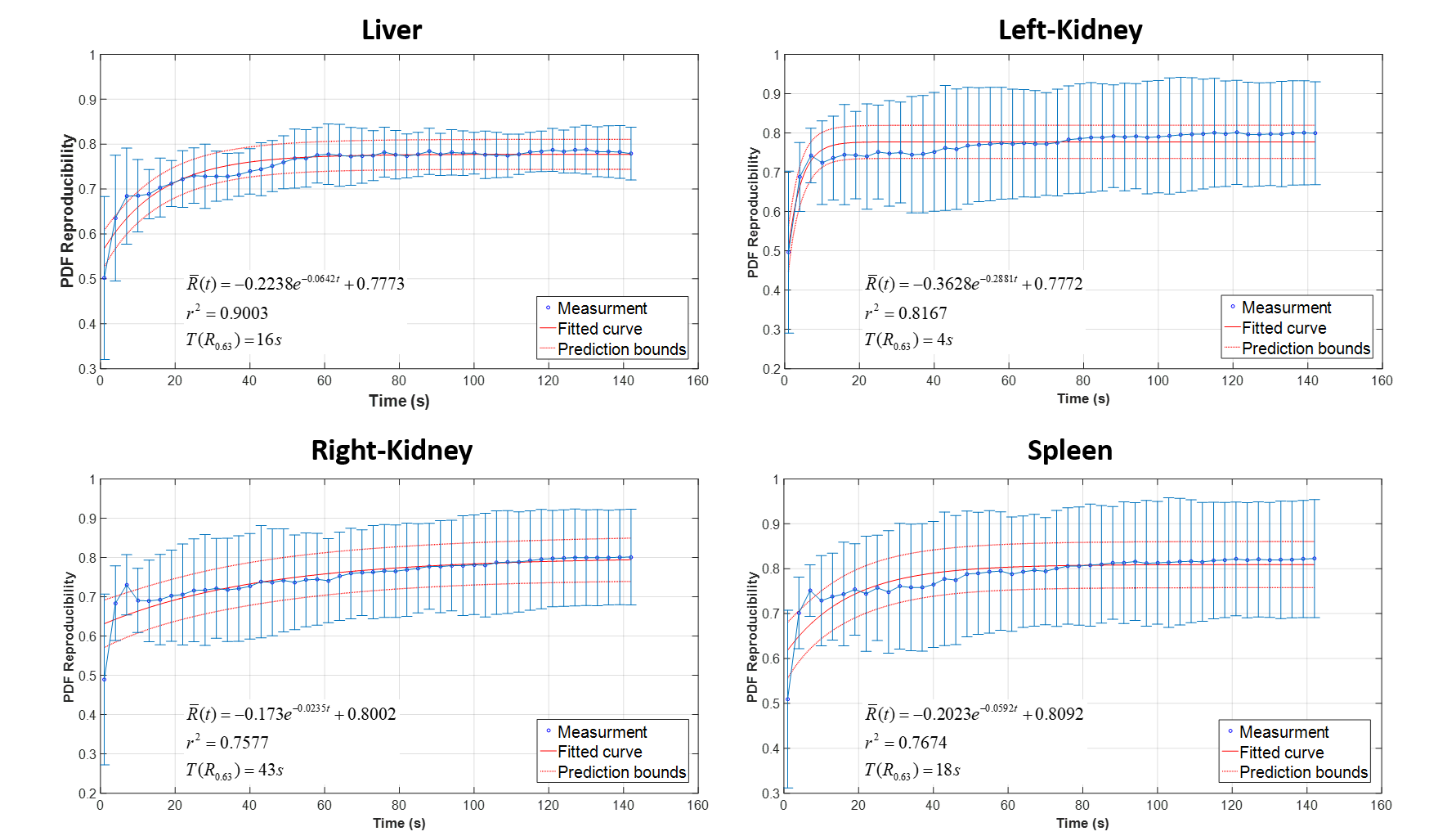
Figure 1 demonstrated the reformatted 4D-MRI images of a volunteer in three orthogonal views acquired by the ultra-fast volumetric 4D MRI. Figure 2. demonstrates the fitted PDFs from the respiratory displacement histograms in a subject. Figure 3 shows time evolved PDF R(t) curves of the simulated treatment fractions relative to the reference in the four organs. All subjects showed an increased PDF reproducibility with longer scan time in all organs. Figure 4 shows the averaged PDF reproducibility curve of all subjects in all fractions. The initial PDF reproducibility of all organs ($$${R_0} = 0.4849-0.5097$$$) increased significantly (p<0.001) at the end of 144-s scan ($$${R_e}{\rm{ }} = {\rm{ }}0.7788-0.8225$$$). The PDF reproducibility equilibrium time constant $$$T({R_{0.63}})$$$ varied largely (4sec–43sec) in organs, but much shorter than the total scan time of 144s, indicating the stable and reproducible PDF could be established in a short period for reliable probability-based treatment planning. Table 1 summarizes the inter-subject and inter-fractional result of R0 and Re for each organ. Significant differences were found between different organs in R0 and Re (p<0.01). The mean variation in R(t) reduced with time from R0 (CV>33.84% all organs) to Re (CV<14.96% all organs).Discussion and Conclusion
In this study, we evaluated the inter-fractional abdominal organ motion PDF reproducibility using a fast volumetric 4D MRI. The PDF reproducibility was observed with a prolonged scan time in a total of 8 healthy subjects. Although the PDF stabilizing time constants varied among organs, they were generally much shorter than the total 4D-MRI acquisition time of 144s. Comparing with previous studies, the volumetric MRI used in our study was fast enough to simultaneously capture the respiratory-induced abdominal organ motion in three-dimension, provides a more reliable reference for MR guided radiotherapy (MRgRT), particularly for large and irregular-shaped tumors. Furthermore, the time evolved PDF reproducibility was assessed in multiple fractions to test the applicability and reliability of the probability-based respiratory motion determined in RT treatment planning for multi-fractional treatment guidance.The main limitation of this study is the recruitment of only healthy volunteers.The respiratory PDFs of tumors in real patients and their reproducibility might be considerably different from healthy subjects. These need to be further verified.Acknowledgements
No acknowledgement found.References
1. Cai J, Read PW, Altes TA, Molloy JA, Brookeman JR, Sheng K. Evaluation of the reproducibility of lung motion probability distribution function (PDF) using dynamic MRI. Phys Med Biol 2007;52(2):365-373.
2. Cai J, Read PW, Larner JM, Jones DR, Benedict SH, Sheng K. Reproducibility of interfraction lung motion probability distribution function using dynamic MRI: statistical analysis. Int J Radiat Oncol Biol Phys 2008;72(4):1228-1235.
3. Zhang F, Hu J, Kelsey CR, Yoo D, Yin FF, Cai J. Reproducibility of tumor motion probability distribution function in stereotactic body radiation therapy of lung cancer. Int J Radiat Oncol Biol Phys 2012;84(3):861-866.
4. Yuan J, Zhou YH, Wong OL, Cheung KY,Yu SK. “Development of a fast 4D-MRI with sub-second volumetric frame rate for respiratory motion tracking in abdominal radiotherapy”, ISMRM, Paris, Jun 2018.
Figures