2301
Choline kinase-α downregulation decreases prostate cancer associated fibroblast viability1Division of Cancer Imaging Research, The Russell H. Morgan Department of Radiology and Radiological Science. The Johns Hopkins University School of Medicine, Baltimore, MD, United States, 2Sidney Kimmel Comprehensive Cancer Center. The Johns Hopkins University School of Medicine, Baltimore, MD, United States, 3Radiation Oncology and Molecular Radiation Sciences, The Johns Hopkins University School of Medicine, Baltimore, MD, United States
Synopsis
Cancer associated fibroblasts (CAFs) significantly influence the proliferation, invasion, and metastasis of cancer cells. CAFs are detected in most tumors and provide a ubiquitous target that is being actively investigated in cancer treatment. In prostate cancer, fibroblasts have been shown to induce growth, confer castration-resistance, and increase metastatic potential. Choline kinase (Chk)-α downregulation has been previously shown to significantly decrease cancer cell viability but its effect on CAFs has not been investigated before. Here we found, for the first time, a significant decrease of prostate cancer fibroblast (PCAF) viability with Chk-α downregulation.
Introduction
Cancer associated fibroblasts (CAFs) present an important target in modifying cancer aggressiveness because of the roles they play in proliferation, invasion, and metastasis of cancer cells 1, 2. We previously observed an increase of Prostate CAF (PCAFs) in prostate cancers that metastasize 3, consistent with the known role of fibroblasts in inducing growth, confer castration-resistance, and increasing metastatic potential 4. While aberrant choline metabolism has been identified in prostate cancer cells 5, choline metabolism in fibroblasts and PCAFs is unexplored and may identify a treatment strategy through downregulation of enzymes such as choline kinase alpha (Chk-α), the enzyme that converts choline to phosphocholine (PC). Chk-α is typically increased in cancer cells. Here we characterized the metabolic profile of normal prostate fibroblasts and PCAFs using 1H MRS and determined the effect of Chk-α downregulation with small interfering RNA (siRNA) on prostate fibroblast and PCAF viability.Materials and Methods
Experiments were performed using the human prostate myofibroblasts (WPMY-1, ATCC, Manassas, VA) and PCAFs (Asterand Bioscience, Detroit, MI). WPMY-1 were derived from stromal cells from the peripheral zone of the histologically normal adult prostate 6. PCAFs were obtained from an adenocarcinoma of the prostate gland. For high resolution 1H MRS, cell extracts were obtained using a dual-phase extraction method as previously described 7. Water-soluble samples were dissolved in 0.6 mL of buffered D2O (Sigma). High-resolution 1H MR spectra were recorded on a Bruker Biospin Avance-III 750 MHz NMR (Bruker Biospin) spectrometer operating at a proton frequency of 750.21 MHz using a 5-mm broad band inverse (BBI) probe head equipped with z-gradient accessories. 1H MR spectra were manually phased and automated baseline corrected using TOPSPIN 3.2 software. Integrals of the metabolites of interest were determined and normalized to the TSP reference and the number of cells. Metabolites were estimated from at least three experimental samples.Statistical significance was evaluated using the Student t test. To target Chk-α, cells were transiently transfected with small interfering RNA (siRNA) against Chk-α at a concentration of 100 nM for 48h using standard protocol. Total RNA was isolated, complementary cDNA synthesised and quantitative real-time PCR (q-RT-PCR) performed using IQ SYBR Green supermix and gene specific primers following an established protocol 7. Viability studies were performed using CCK8 assay as previously reported 8.
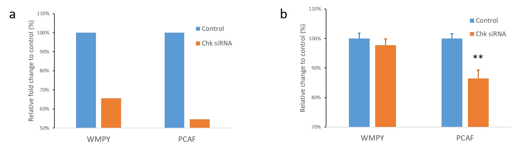
Results and Discussion
A summary of the metabolites detected in 1H MR spectra of WPMY-1 and PCAFs are shown in Figure 1a. Metabolites that were significantly different between WPMY-1 and PCAFs are displayed as a heat map in Figure 1b. As shown in Figure 2a, siRNA directed against Chk-α reduced mRNA levels in both WPMY-1 and PCAFs. We observed a significant decrease of cell viability in the PCAFs but not in normal prostate fibroblasts as shown in Figure 2b. These data support investigating Chk-α as a target to eliminate CAFs in tumors. Increased glutamate observed in PCAFs also support targeting enzymes and transporters in glutamine/glutamate metabolism as potential therapeutic strategies against PCAFs. Future studies with CAFs from different cancers should further validate the metabolic differences between normal fibroblasts and CAFS identified in this study.Acknowledgements
Supported by NIH R35 CACA209960. JPT was supported by Alonso Martin Escudero foundation.References
1. Kalluri R and Zeisberg M. Fibroblasts in cancer. Nat Rev Cancer. 2006; 6(5):392-401.
2. Josson S, Matsuoka Y, Chung LW, et al. Tumor-stroma co-evolution in prostate cancer progression and metastasis. Semin Cell Dev Biol. 2010; 21(1):26-32.
3. Penet MF, Kakkad S, Pathak AP, et al. Structure and Function of a Prostate Cancer Dissemination-Permissive Extracellular Matrix. Clin Cancer Res. 2017; 23(9):2245-2254.
4. Thalmann GN, Rhee H, Sikes RA, et al. Human prostate fibroblasts induce growth and confer castration resistance and metastatic potential in LNCaP Cells. Eur Urol. 2010; 58(1):162-71.
5. Ackerstaff E, Pflug BR, Nelson JB, et al. Detection of increased choline compounds with proton nuclear magnetic resonance spectroscopy subsequent to malignant transformation of human prostatic epithelial cells. Cancer Res. 2001; 61(9):3599-603.
6. Webber MM, Trakul N, Thraves PS, et al. A human prostatic stromal myofibroblast cell line WPMY-1: a model for stromal-epithelial interactions in prostatic neoplasia. Carcinogenesis. 1999; 20(7):1185-92.
7. Penet MF, Shah T, Bharti S, et al. Metabolic imaging of pancreatic ductal adenocarcinoma detects altered choline metabolism. Clin Cancer Res. 2015; 21(2):386-95.
8. Mori N, Wildes F, Kakkad S, et al. Choline kinase-alpha protein and phosphatidylcholine but not phosphocholine are required for breast cancer cell survival. NMR Biomed. 2015; 28(12):1697-706.
Figures