2293
Alteration in 1H MRS metabolites in the rat spinal cord after experimental craniospinal irradiationPetra Hnilicová1, Soňa Bálentová2, Dagmar Kalenská 3, Eva Hajtmanová 4, Peter Murín 4, Michal Bittšanský1, Marian Adamkov 2, Ján Lehotský3, and Dušan Dobrota 3
1Division of Neurosciences at Biomedical Center Martin, Jessenius Faculty of Medicine in Martin, Comenius University in Bratislava, Martin, Slovakia, 2Institute of Histology and Embryology, Jessenius Faculty of Medicine in Martin, Comenius University in Bratislava, Martin, Slovakia, 3Department of Medical Biochemistry, Jessenius Faculty of Medicine in Martin, Comenius University in Bratislava, Martin, Slovakia, 4Department of Radiotherapy and Oncology, University Hospital Martin, Martin, Slovakia
Synopsis
We investigated the metabolic effect of a clinically relevant craniospinal irradiation on the rat spinal cord using 1H MRS at 7T. Hundred days after the fractionated irradiation performed by radioactive isotope 60Co (16Gy in 2 fractions), irradiated and sham-irradiated animals underwent in vivo 1H MRS examination. Spinal cord spectra were measured by SVS sequence with voxel size of 3.5x2x7.5mm3. In spinal cord of irradiated animals was confirmed significantly reduced tNAA/tCr and increased tCho/tCr ratio, both showing demyelination and inflammatory processes. It seems that in vivo 1H MRS of spinal cord could be useful for radiation therapy monitoring.
PURPOSE
Radiation-induced brain injury usually occurs after conventional radiation therapy of patients with primary brain tumors and metastases and is caused by microstructural and/or metabolic changes in neural tissue.1 It may have an immediate and strong impact on quality of life, especially when occurring in the spinal cord.1,2 Although animal models enable to imitate mechanisms of irradiated injury, it is only poorly explored the late response of the juvenile rat brain to a clinically relevant fractionated irradiation.3 Therefore, the aim of this 1H MRS study was to investigate the metabolic changes in spinal cord induced by the fractionated craniospinal irradiation with a biological effective dose (BED; 117 Gy) equivalent to the treatment dose used in many tumor modalities.4METHODS
Three months old Wistar male rats (n=16) underwent the experiment. Radiation was delivered to anesthetized rats (n=8) by Teragam KO-2 device (UJP, Prague, Czech Republic) using the radioactive isotope 60Co with the energy of 1.17 and 1.33MeV. The craniospinal irradiation with BED of 117Gy was delivered in 8Gy per fraction during 2 consecutive weeks (total dose of 16Gy). The irradiated and sham-irradiated animals (n=8) survived ~100 days after the irradiation procedure. In vivo 1H MRS examinations were performed on 7T small animal MR scanner (Bruker BioSpec 70/20 USR) using a combination of the volume resonator and the 1H receive-only surface coil with 20mm diameter (Bruker BioSpin MRI, Ettlingen, Germany). Anatomical images in all orthogonal directions were measured using fast T2-weighted turbo-spin-echo sequences (turboRARE; TR/TE=2500/33ms; NA=2; echo spacing=10ms; FOV=35x35mm2; matrix=256x256; 23 slices with a 0.5mm slice thickness; acquisition time ~2min). Single-voxel 1H MRS data in the spinal cord (C2-C4) were obtain within ~5min (PRESS; TR/TE=1500/20ms; voxel size=3.5x2x7.5mm3; NA=200; OVS & VAPOR suppression). Linear and second order shims were adjusted with the automatic cuboid shim volume with the water linewidths ~20Hz. Each measured 1H MR spectra (Figure) was analyzed using LCModel and following metabolites were quantified: N-acetyl-aspartate & N-acetyl-aspartyl-glutamate (tNAA), choline (phosphatidylcholine, glycerophosphatidylcholine, acetylcholine, and choline; tCho) and creatine (creatine and phosphocreatine; tCr) containing compounds, glutamate & glutamine (Glx), Gamma-aminobutyric acid (GABA), and Lactate (Lac). Metabolite ratios to tCr were calculated and statistically evaluated using the SPSS package (independent-samples 2-tailed t-tests).RESULTS
Quantification of 1H MRS metabolite ratios in the spinal cord of irradiated animals showed significant decrease of tNAA/tCr (p=0.003) and significant increase of tCho/tCr (p=0.007), compare to the animal control group (Table).DISCUSSION
The rat spinal cord after the craniospinal irradiation with clinically relevant fractionated doses demonstrates some metabolic abnormalities. Reduced tNAA/tCr manifests damaged neurons caused by neuronal dysfunction and apoptosis secondary to irradiation.1,5 Considering tCho as a marker of myelin sheath integrity6, the increased tCho/tNAA found in our study may indicate destruction of myelin sheath or myelin-producing oligodendrocytes.5,6,7 Since the highest levels of Cr and Cho are found in oligodendrocytes and astrocytes6, their elevation may also represent inflammation, de- or re-myelination and ongoing gliosis.1,3,5CONCLUSION
It seems, that clinically relevant fractionated craniospinal irradiation strongly affects the spinal cord consisting of essential bundles of motor and sensory tracts. Therefore, the potential clinical application of in vivo 1H MRS during radiation treatment could be useful for planning of a successful pharmacological, surgical, or rehabilitative therapy.Acknowledgements
This study was supported by the grant APVV-14-0088 and by projects: Biomedical Center Martin (ITMS: 26220220187), VEGA 1/0129/16, and VEGA 1/0128/16, co-funded from EU sources.References
- Greene-Schloesser D, Robbins ME, Peiffer AM, et al. Radiation-induced brain injury: a review. Front Oncol. 2012; 2: 73.
- Hock A, Henning A, Boesiger P, Kollias SS. 1H-MR Spectroscopy in the Human Spinal Cord. AJNR Am J Neuroradiol. 2013; 20.
- Brown RJ, Jun BJ, Cushman JD, et al. Changes in imaging and cognition in juvenile rats after whole-brain irradiation. Int J Radiat Oncol Biol Phys. 2016; 96(2): 470-8.
- Fowler JF. 21 years of biologically effective dose, Br J Radiol. 2010; 83(991): 554–568.
- Sundgren PC, Cao Y. Brain irradiation: Effects on normal brain parenchyma and radiation injury. Neuroimaging Clin. N. Am. 2009; 19:657-668.
- Soares DP, Law M. Magnetic resonance spectroscopy of the brain: review of metabolites and clinical applications. Clinical radiology. 2009; 64(1):12-21.
- Virta A, Patronas N, Raman R, et al. Spectroscopic imaging of radiation-induced effects in the white matter of glioma patients. Magn. Reson. Imaging. 2000; 18: 851-857.
Figures
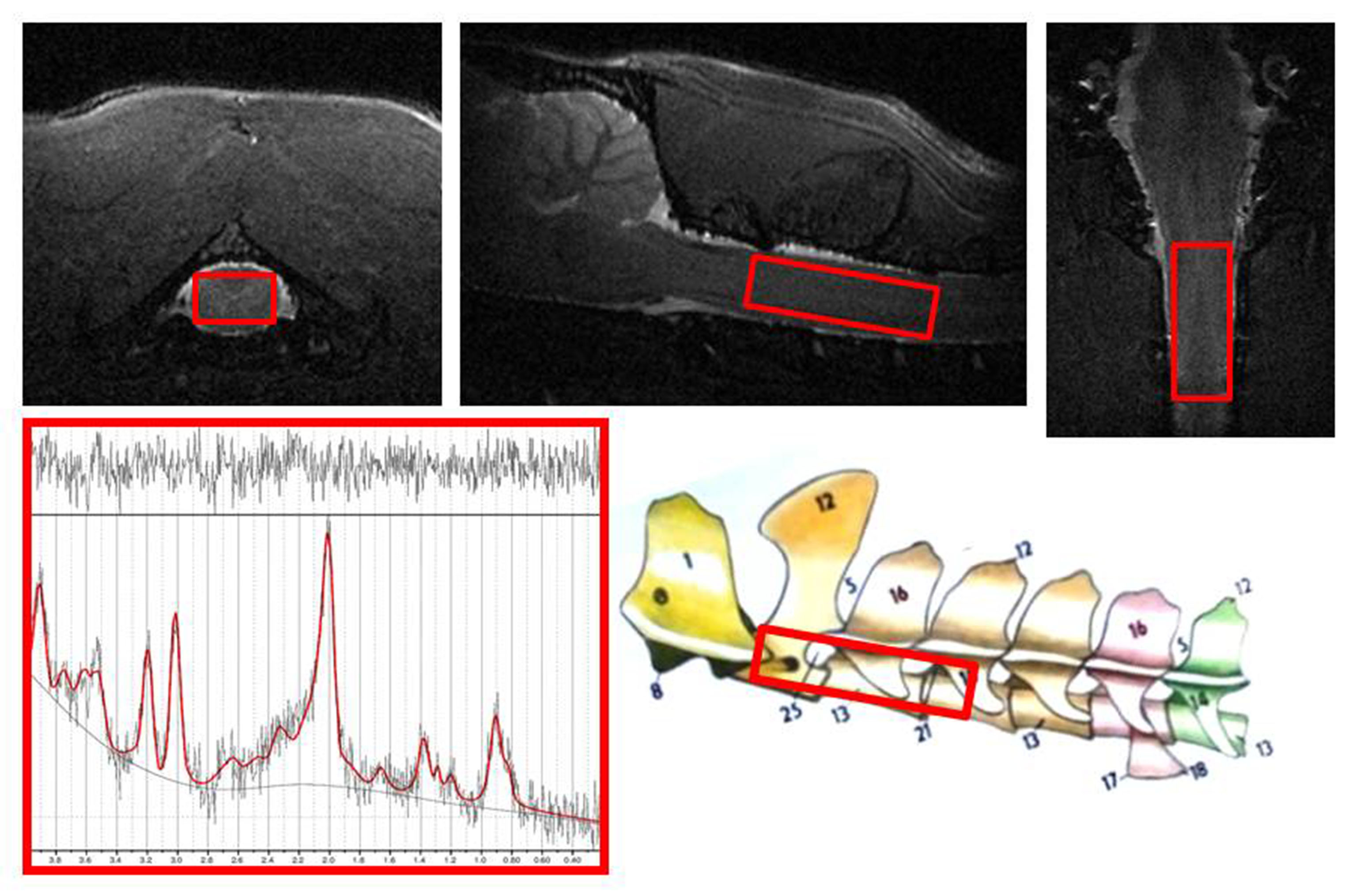
Figure shows representative in vivo 1H MR spectra
obtained by the SVS technique in the rat spinal cord (C2-C4).
The localization of the voxel (nominal voxel size: 3.5x2x7.5 mm3) is
shown on the morphological T2-weighted reference MR images in
orthogonal planes.
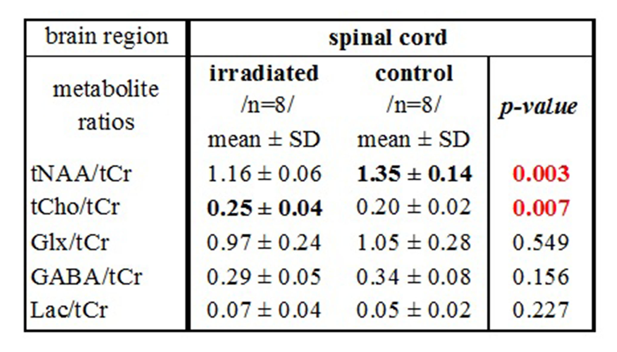
Table shows metabolite ratios in the spinal cord (C2-C4) of irradiated and control animal groups. Using independent-samples 2-tailed t-tests were obtained p-values
expressing statistical differences in metabolite ratios between groups.