2292
Association of central arterial stiffness with hippocampal blood flow and N-acetyl aspartate in hypertensive Dahl salt sensitive ratsSamuel O Ajamu1, Rachel C Fenner1, Yulia N Grigorova1, James P Karchner2, Edward G Lakatta1, Mustapha M Bouhrara2, Richard G Spencer2, Olga V Fedorova1, and Kenneth W Fishbein2
1Laboratory of Cardiovascular Science, National Institute of Aging, Baltimore, MD, United States, 2Laboratory of Clinical Investigation, National Institute of Aging, Baltimore, MD, United States
Synopsis
Central arterial stiffness (CAS) is associated with hypertension and is likely associated with stiffening of cerebral artery walls, with attendant cerebral hypoperfusion, neuronal density loss and cognitive decline. We sought to explore associations between pulse wave velocity (PWV), a marker of CAS, and hippocampal blood flow and neuronal density in hypertensive Dahl salt sensitive (Dahl-S) rats, which exhibit age-associated memory loss. We observed direct correlations between greater PWV, lower cerebral blood flow (CBF) and lower N-acetyl aspartate/total creatine ratio (NAA/tCr) in the hippocampus, supporting the role of CAS in cerebrovascular dysfunction and decline in cognitive performance with hypertension and aging.
Introduction
The global burden of dementia is projected to increase by 50% over the next decade. Further elucidation of the underlying pathophysiology will be required for the development of effective therapeutics. Hypertension and diseases of the vascular wall may be significant contributors to dementia, including Alzheimer’s disease.1,2,3 However, the mechanism of this process is poorly understood. CAS, as reflected by increased pulse wave velocity (PWV), is a state in which the compliance of a vessel such as the aorta decreases, in part due to a reduction in the elastin-to-collagen ratio in the vessel wall.4 CAS is known to be accelerated by systemic hypertension and may be accompanied by increased stiffness in the cerebral arteries. To study this, we used Dahl salt sensitive (Dahl-S) rats which develop hypertension, CAS and hippocampal memory decline as they develop a variable degree of moderate hypertension with age while on a normal salt diet.5 With this model, we sought to understand the link between arterial stiffening in the systemic circulation and deficits in cerebral perfusion and neuronal density in the hippocampus.Methods
Dahl-S rats (n=12; Charles River Laboratories) were fed with normal salt diet (0.5% NaCl) until age 22 weeks, when systolic blood pressure (SBP) and PWV were measured. SBP was measured in conscious rats via tail-cuff plethysmography (IITC Inc, Woodland Hills, CA). PWV was measured in rats sedated with 2.5% isoflurane in oxygen. Measurements were performed at the transverse aortic arch and abdominal aorta via the transit time method using a 12 MHz Doppler probe (Sonos 5500, Hewlett-Packard, Andover, MA).4 N-acetyl aspartate (NAA) concentration was measured in the right hippocampus using a 7T Bruker Biospec MRI scanner (Billerica, MA). Rats were sedated with 2% isoflurane in oxygen with vital signs monitored throughout (Respirator Monitor, SA Instruments, Stony Brook, NY). Respiration rate was maintained at 45-55 min-1 by small variations in the inhalation mixture. For data acquisition, a 2.5mm×1.5mm×2.5 mm spectroscopic voxel was placed in the right hippocampus, with acquisition performed using a PRESS sequence with CHESS water suppression and TE/TR= 15.7 ms/1s, spectral width = 6010 Hz and 128 averages. The ratio of NAA to total creatine, an indicator of neuronal density, was calculated using LCModel. Cerebral blood flow (CBF) was measured using continuous arterial spin labeling (CASL) in the coronal slice with the largest hippocampal area. Data were acquired with an EPI sequence with parameters TE/TR= 28 ms/ 5 s, 128 averages, 0.469mm×0.469mm×1 mm voxel size and labeling time of 2 s. The NESMA filter6 was applied to the resulting images and CBF was estimated in both the whole brain and hippocampus. CBF values were calculated using standard methods6 assuming values for the brain/blood partition coefficient of 𝜆=0.9ml/g, labeling efficiency of α=0.85, T1, Blood = 2300 ms8, labeling duration τ = 2 s and PLD =0.1 s.7,8 Statistical analysis: Regression analyses were performed using Microsoft Excel.Results
At 6 months of age, the Dahl-S rats displayed moderate hypertension (SBP: 164±13; mean±SD, as compared to a 3 months baseline of 142±5 mmHg; mean±SD). Linear regression revealed a positive correlation between SBP and PWV (Figure 1). Representative CBF maps where hippocampal CBF data was acquired (Figure 2). PWV was negatively correlated with both cerebral perfusion (Figure 3) and NAA/tCr (Figure 4) in the hippocampus. Hippocampal CBF positively correlated with hippocampal NAA (Figure 5).Discussion
Numerous studies have linked hypertension with CAS, as quantified by PWV. However, little is known about the relationship between the effects of increased PWV on cerebral blood flow and neuronal mass. We found that in a Dahl-S rat model of hypertension, greater aortic PWV was associated with decreased hippocampal perfusion, indicating a potential pathophysiologic mechanism for impaired neuronal function in hypertension and a possible link with cognitive impairment. The potential functional significance of this finding is indicated by the strong and statistically significant positive correlation between hippocampal CBF and NAA/tCr. NAA, which is a well-validated marker of cerebral neuronal density, is known to be directly associated with cognitive function.9,10 Given the central importance of the hippocampus in the memory manifestations of dementia, this may provide an important mechanistic link between systemic circulation and dementia and indicate PWV as a potential therapeutic target for the prevention and treatment of dementia.Conclusion
In the Dahl-S rat model of hypertension, greater PWV, a marker for CAS, was associated with decreased hippocampal perfusion and decreased neuronal mass, providing a potential mechanism for the cognitive decline with age observed in this animal model.Acknowledgements
The work was supported by the Intramural Research Program of the National Institute on Aging of the National Institutes of Health.References
References
- Iadecola C, Davisson RL. Hypertension and cerebrovascular dysfunction. Cell Metab 2008;7(6),476-84.
- Mitchell GF, Effects of central arterial aging on the structure and function of the peripheral vasculature: implications for end-organ damage. J Appl Physiol (1985), 2008; 105(5), 1652-60.
- Mitchell GF, et al. Arterial stiffness, pressure and flow pulsatility and brain structure and function: the Age, Gene/Environment Susceptibility--Reykjavik study. Brain 2011;134(Pt 11), 3398-407.
- Grigorova YN, et al. Dietary sodium restriction reduces arterial stiffness, vascular TGF-beta-dependent fibrosis and marinobufagenin in young normotensive rats. Int J Mol Sci 2018;19(10), E3168.
- Fedorova OV, Grigorova YN, Long J, McPherson R, Juhasz O, Wei W, Zernetkina V, Petrashevskaya N, Fishbein KW, Spencer RG, Rapp PR, Lakatta EG. Accelerated age-dependent cardiovascular and cognitive decline in Dahl-S rats is associated with elevated levels of an endogenous Na/K-ATPase inhibitor. Hypertension 2017;70:AP441.
- Bouhrara M, Lee DY, Rejimon AC, Bergeron CM, Spencer RG. Spatially adaptive unsupervised multispectral nonlocal filtering for improved cerebral blood flow mapping using arterial spin labeling magnetic resonance imaging, J Neurosci Methods 2018;309:121-131.
- Lu H, Leoni R, Silva AC, Stein EA, Yang Y. High-field continuous arterial spin labeling with long labeling duration: reduced confounds from blood transit time and postlabeling delay. Magn Reson Med 2010;64(6),1557-66.
- Barbier EL, Lawrence KS, Grillon E, Koretsky AP, Décorps M. A model of blood–brain barrier permeability to water: Accounting for blood inflow and longitudinal relaxation effects. Magn Reson Med 2002;47:1100-1109.
- Baslow MH. N -acetylaspartate in the vertebrate brain: metabolism and function. Neurochem Res 2003;28:941-53. 10. Dicke U, Roth G. Neuronal factors determining high intelligence. Phil Trans Royal Soc B 2016;371(1685):20150180.
- Dicke U, Roth G. Neuronal factors determining high intelligence. Phil Trans Royal Soc B 2016;371(1685):20150180.
Figures
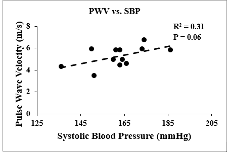
Figure 1: Central Arterial
Stiffness (CAS) measured as PWV vs. Systolic Blood Pressure (SBP). PWV, a
marker of CAS, was positively correlated with SBP, indicating that the latter
contributes to vessel wall stiffening.
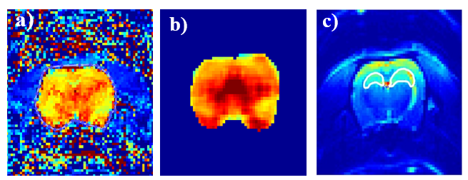
Figure 2: a) Unfiltered CBF map from
CASL. b) Corresponding CBF map from NESMA-filtered data. c) EPI image of same
slice showing ROI for hippocampus.
An ROI of the largest hippocampal axial slice created from an unfiltered
CASL image produce a whole brain CBF map. The proton density image allowed for
the proper manual segmentation of the hippocampal region in calculating
hippocampal CBF.
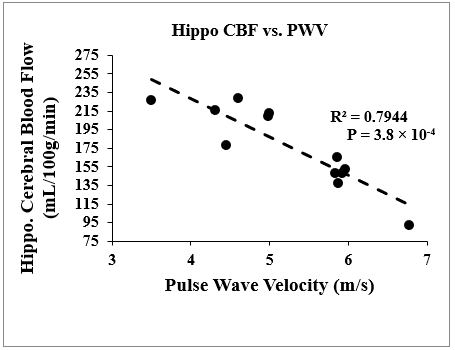
Figure 3: Hippocampal
Cerebral Blood Flow vs. PWV. PWV has a negative
correlation with hippocampal CBF, which suggests that PWV contributes to hippocampal
CBF changes.
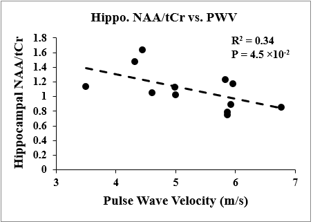
Figure 4: Hippocampal
N-Acetyl Aspartate/ Total Creatine vs. PWV. A negative correlation between
Hippocampal NAA/tCr and PWV implies that PWV has an influence on this marker
for neuronal density. This trend in conjunction with the correlation of Hippo.
CBF to PWV provides a potential link between systemic circulation and cerebral
health.
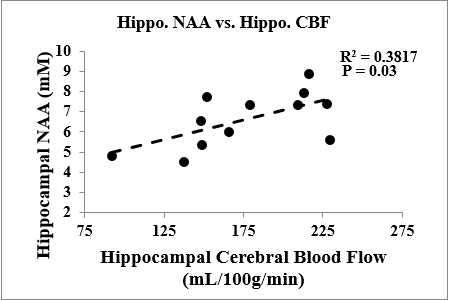
Figure 5: Hippocampal
NAA vs. Hippocampal CBF. Hippocampal NAA concentration positively correlates
with hippocampal CBF. This is expected as decreased cerebral blood
flow will directly affect neuronal density.