2291
Assessment of Thalamic Metabolic Processes in a Migraine Model: A Relaxation Enhanced Diffusion Weighted MRS Study at 21.1 T1Chemical and Biomedical Engineering, Florida State University, Tallahassee, FL, United States, 2National High Magnetic Field Laboratory, Florida State University, Tallahassee, FL, United States, 3NeuroSpin, Paris, France, 4Neurosciences, Huntington Medical Research Institutes, Pasadena, CA, United States
Synopsis
To date, there is a lack of characterization of metabolic markers in migraine studies. Though numerous studies implicate cerebral dysfunction, robust biomarkers are yet to be identified in the migraine brain. It thus follows that identification of specific metabolic changes, potentially influenced by excitatory and inhibitory neurotransmitters, may improve understanding of migraine onset and propagation while opening new avenues for therapy development.
With the goal of evaluating metabolic processes, diffusion-weighed spectroscopy is used to identify longitudinal changes in the in vivo thalamus of an acute rodent of migraine model.
Introduction
Acute and sustained biochemical changes, prominently with lactate, taurine and total creatine, have been identified previously in a 1H-MRS neocortical study [1], indicative of increased neural activity/glycolysis and supported by dynamically altered sodium homeostasis [2]. These metabolites demonstrated dynamic and longitudinal changes in concentration following intraperitoneal (IP) injection of nitroglycerin (NTG). However, other potential metabolites of interest, namely those attributed to excitability (e.g., NAA and glutamate signals in neurons, myoinositol, choline and glutamine signals in astrocytes [4,5]), did not display significant concentration changes with NTG. However, diffusion-weighted (DW-MRS) [6-8,10] may prove more sensitive for detecting cell specific alterations in endogenous metabolites with migraine onset.
In
this study, DW-MRS was employed to measure diffusion properties and
compartmental changes in an NTG rat model. Compartmentalization and shuttling
of metabolites between cells is believed, during the ictal state, to be an
important factor in migraine [10] that impacts
metabolism and function. Due to its central role in migraine, the thalamus is
the target of these in vivo
investigations, for which the high sensitivity of 21.1-T MRS will be leveraged
to monitor concentration and diffusional alteration metabolites during the progression
of migraine.
Material and Methods
Animal Model: Under sedation in the magnet, ten Sprague-Dawley male rats were administered in situ with an IP injection of either 10 mg/kg of NTG to provide conditions of a migraine analogue (n=6) or saline (n=4) to serve as controls.
MR Acquisitions: All scans were performed using the 21.1-T ultra-wide bore magnet at the US National High Magnetic Field Laboratory and a linear 1H/23Na birdcage coil. DW-MRS was acquired using a relaxation enhanced MRS (RE-MRS) sequence to target upfield metabolites from a 3x4x3-mm voxel localized in the thalamus and without water suppression. Selective bandwidth excitation was accomplished with a 5-ms sinc-10 pulse, with a 5-ms 180° SLR refocusing pulse, exciting a band between 0-4 ppm. Localization was achieved using six 5-ms adiabatic pulses by adiabatic selective refocusing (3D LASER). Due to fiber orientation in the thalamus, three diffusion weightings were applied in equally weighted directions using an optimized diffusion time of 15 ms to achieve b values of 0, 1640 and 3857 s/mm2. With TE/TR=64/2500 ms and NA=120, the total acquisition time of 15 min. A total of two pre-injection scans (for baseline data) and five post-injection scans were acquired (~2.5-h post injection).
Data Analysis: Data was acquired as a partial echo, and magnitude spectra were generated after apodization (10-Hz exponential line broadening) and Fourier transform. No other deconvolution of spectral components was performed. The apparent diffusion coefficient (ADC) values were calculated for each metabolite using a mono-exponential fit. Unweighted (b=0 s/mm2) spectra also were used to track signal intensity changes, with the intensity time course normalized to the average of the baseline (pre-injection) signal for each peak. A mixed model ANOVA with repeated measures was used to establish within and between group significances.
Results
Significant changes are reported in both the concentration and ADC for lactate and taurine, compared to both baseline and control cohorts, as early at 15-min post-NTG administration. The significances and trends observed with lactate and taurine are consistent with our prior neocortex study [1]. Glx, identified as the spectral overlap between glutamate, GABA and glutamine, shows delayed increases and significances in signal intensity for NTG-injected rats compared to control. However, Glx ADC is altered significantly at only one time-point (75-min post-NTG) from control ADC values, suggesting potential short-term alteration of compartmentalization with quick re-normalization. No significant sustained differences were observed in the concentrations or ADC of other metabolites.Discussion
Early and sustained significances in lactate and taurine indicate impaired metabolism, neuroprotective action, as well as metabolic shuttling/remodeling with temporal dependence to the onset of triggered migraine. Interestingly, with short acquisition times, Glx signals were distinguishable from its precursor glutamine [3,9]—a major advantage as these closely related neurotransmitters have very distinct functions and compartmental distributions, evidenced by their different ADCs. These distinct spectroscopic signatures are potentially sensitive to subtle microstructural changes in restricted compartments. The results also indicate that ictal glutamate levels are increased in the thalamus, with a delayed time course, supporting hyperexcitability in migraine.Conclusions
1H DW RE-MRS supports energetic impacts potentially related to mitochondrial dysfunction in the migraine brain. Impacted glycolysis produces increased lactate, which also has been linked theoretically to taurine and glutamate increases [4,5], potentially as protection against glutamate excitotoxicity. Interestingly, the observed increases indicate either increased intracellular metabolism or impaired cellular uptake, both highly energy dependent processes and representative of thalamic hyperexcitability.Acknowledgements
This work was supported by the NIH (R01-NS072497 and RO1-NS102395) and User Collaborations Grant Program (to SCG) from the National High Magnetic Field Laboratory, which is funded by the NSF (DMR-1644779) and the State of Florida.References
[1] Abad N, Rosenberg JT, Roussel T, Grice DC, Harrington MG, Grant SC. Metabolic Assessment of a Migraine Model Using Relaxation-Enhanced 1H Spectroscopy at Ultrahigh Field. Magnetic Resonance in Medicine 2017;79:1266-1275.
[2] Abad N, Rosenberg JT, Hike DC, Harrington MG, Grant SC. Dynamic sodium imaging at ultra-high field reveals progression in a preclinical migraine model. Pain 2018;159:2058-2065.
[3] Arngrim N, Schytz HW, Britze J, Amin FM, Vestergaard MB, Hougaard A, Wolfram F, de Koning PJH, Olsen KS, Secher NH, Larsson HBW, Olesen J, Ashina M. Migraine induced by hypoxia: an MRI spectroscopy and angiography study. Brain 2016;139:723-737.
[4] Danbolt NC. Glutamate uptake. Prog Neurobiol 2001;65:1-105.
[5] Erecińska M SI. Metabolism and role of glutamate in mammalian brain. Prog Neurobiol. 1990;35:245.
[6] Ligneul C, Valette J. Probing metabolite diffusion at ultra-short time scales in the mouse brain using optimized oscillating gradients and "short"-echo-time diffusion-weighted MRS. NMR Biomed 2017;30:10.1002/nbm.3671. Epub 2016 Nov 28.
[7] Ronen I, Ercan E, Webb A. Axonal and glial microstructural information obtained with diffusion-weighted magnetic resonance spectroscopy at 7T. Front Integr Neurosci 2013;7:13.
[8] Valette J, Guillermier M, Besret L, Boumezbeur F, Hantraye P, Lebon V. Optimized diffusion-weighted spectroscopy for measuring brain glutamate apparent diffusion coefficient on a whole-body MR system. NMR Biomed 2005;18:527-533.
[9] Zielman R, Teeuwisse WM, Bakels F, Van der Grond J, Webb A, van Buchem MA, Ferrari MD, Kruit MC, Terwindt GM. Biochemical changes in the brain of hemiplegic migraine patients measured with 7 tesla H-1-MRS. Cephalalgia 2014;34:959-967.
[10] Zielman R, Wijnen JP, Webb A, Onderwater GLJ, Ronen I, Ferrari MD, Kan HE, Terwindt GM, Kruit MC. Cortical glutamate in migraine. Brain 2017;140:1859-1871.
[11] Papp EA, Leergaard TB, Calabrese E, Johnson GA, Bjaalie JG. Waxholm Space atlas of the Sprague Dawley rat brain. Neuroimage 2014;97:374-386.
Figures
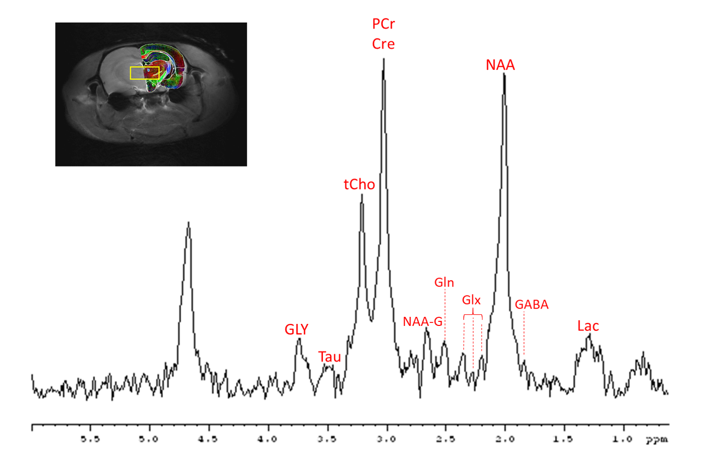
Figure 1. Representative in vivo rat brain thalamus spectra acquired at 21.1 T displaying metabolite peak assignment. Insert demonstrates the 3x4x3-mm voxel placement in the coronal anatomical direction, overlaid with a FA map [11] (three primary colors, red, green and blue were assigned to DWI(x), DWI(y) and DWI(z)).
DW-MRS spectra were acquired over 3 b-values of 30, 1640 and 3857 s/mm2, TE/TR=64/2500 ms with 120 averages was used to achieve a total scan time of 15 min.
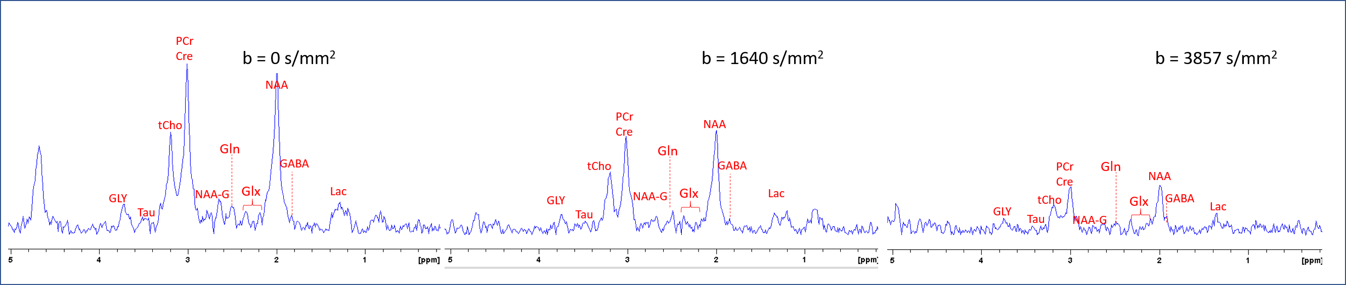
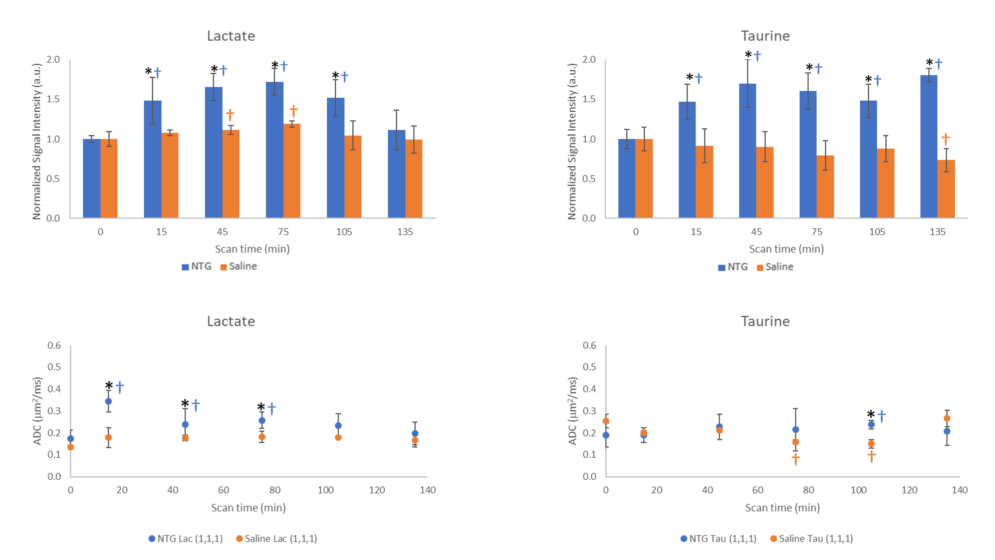
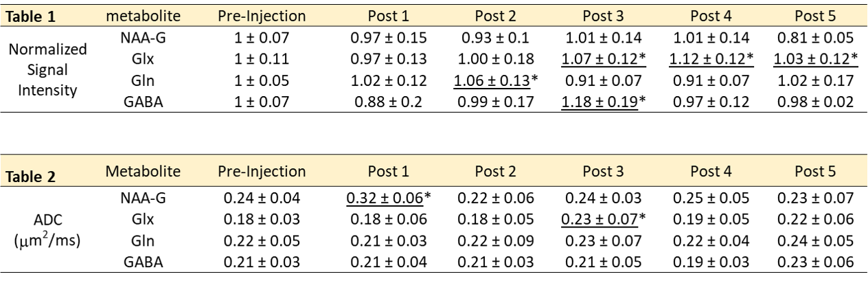
Table 1 Normalized signal intensities (mean ± SD) for only NTG cohorts
Table 2 ADC measurements (mean ± SD) for only NTG cohorts
Statistical significances are *p<0.05 (LSD) for comparisons with the saline control (data not shown).