2287
Investigating the Role of Glutamine Transporters in Breast Cancer1The Russell H. Morgan Department of Radiology and Radiological Science, The Johns Hopkins University School of Medicine, Baltimore, MD, United States, 2The Sidney Kimmel Comprehensive Cancer Center, The Johns Hopkins University School of Medicine, Baltimore, MD, United States
Synopsis
We are investigating aberrant glutamine metabolism in breast cancer to discover potential novel therapeutic targets to inhibit a critical energy source for cancer cells. We have examined glutamine and glutamate levels using 1H magnetic resonance spectroscopy (MRS) across multiple breast cancer cell types and have begun to look at the molecular mechanisms of glutamine addiction using qRT-PCR. We identified SLC38A3 to be overexpressed in nearly every breast cancer cell line examined, which may, in the future, serve as a potential target in breast cancer.
Purpose
Altered metabolism is a hallmark of cancer. Glutamine metabolism has been demonstrated, in multiple cancers, to be upregulated.1,2 Glutamine is a nonessential amino acid that provides both energy, in the form of precursors for the tricarboxylic acid (TCA) cycle, and as a source of other important metabolites including essential amino acids and purines.2 Despite the documented dependence of breast cancer on glutamine, a molecular mechanism of this dependence has not been established to date.3 We are investigating the molecular mechanism of specific genes in breast cancer cells to establish the role of specific enzymes in glutamine metabolism which may lead to novel therapeutic targets in breast cancer.Methods
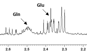
Glutamine and glutamate levels were measured using high-resolution (HR) 1H magnetic resonance spectroscopy (MRS) from cell extracts across multiple breast cell lines, including nonmalignant (MCF-12A, MCF-10A), nonmetastatic estrogen/progesterone receptor positive (MCF-7, BT-474), and triple-negative metastatic (MDA-MB-231, MDA-MB-468) cells, and quantified using previously established methods (Figure 1).4 We also measured mRNA expression levels of genes relevant to glutamine metabolism in a panel of nine breast epithelial cells by quantitative RT-PCR to identify over- or under-expression of these enzymes/transporters. For all studies, unpaired, two-sample t-tests were used, with p-value < 0.05 considered statistically significant. We found SLC38A3 to be overexpressed in almost all malignant cell lines when compared to nonmalignant cell lines. To determine the relevance of our findings as a potential therapeutic target, we examined the TCGA database for expression levels of SLC38A3 in primary tumors of breast cancer patients.Results
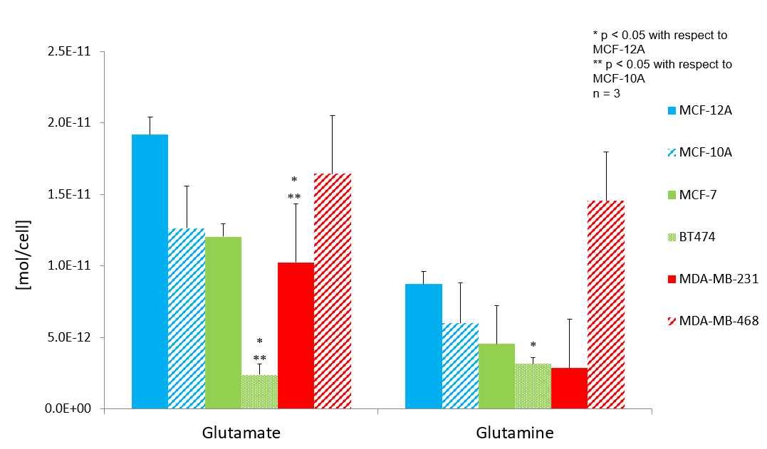
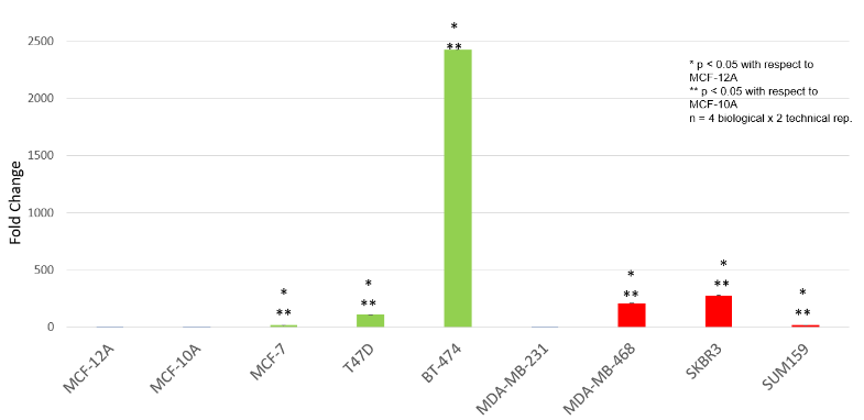
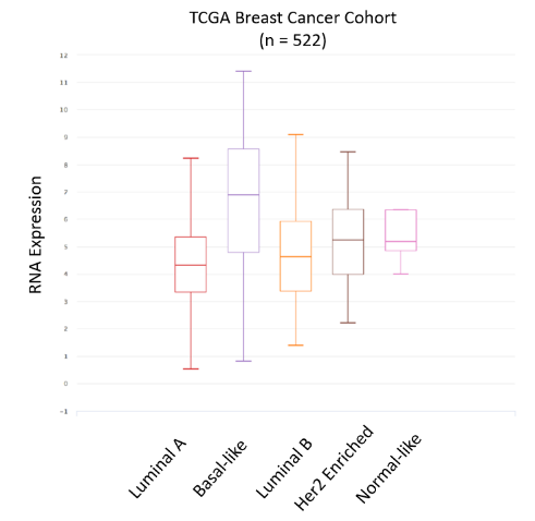
We measured (Figure 1) and quantified (Figure 2) 1H HR MR spectra from water-soluble cell extracts across six different human breast epithelial cell lines, i.e. nonmalignant (MCF-12A, MCF-10A), weakly metastatic luminal (MCF-7, BT-474), and highly metastatic triple-negative (MDA-MB-231, MDA-MB-468) cells. We observed a statistically significant decrease in glutamine and glutamate in MDA-MD-231 cells, and a statistically significant decrease in glutamate in BT-474 cells as compared to the two non-malignant cell lines. To determine the source of these differences in glutamine and glutamate levels in the identified breast cancer cells versus non-malignant cells, we utilized qRT-PCR to screen a panel of 17 glutamine/glutamate metabolism-and-transport-related genes, i.e. Glud1, Glud2, GLS 1, GLS 2, SLC1A5, SLC3A2, SLC7A5, SLC7A11, SLC6A14, SLC7A7, SLC7A8, SLC38A1, SLC38A2, SLC38A3, SLC38A5, and SLC38A7. Of these 17 genes, SLC38A3 was the only gene to be overexpressed in all breast cancer cell lines except MDA-MB-231 cells when compared to both non-malignant cell lines, i.e. MCF-12A and MCF-10A. We also examined the TCGA database and found SLC38A3 to be expressed across all breast cancer subtypes.Discussion
Our 1H HR MRS data revealed significant decreases in glutamate and/or glutamine in only two breast cancer cell lines (MDA-MB-231 and BT474) as compared to non-malignant cells, within our panel of six breast cell lines. Our qRT-PCR data identified SLC38A3 to be overexpressed in nearly all breast cancer cell lines studied within a larger panel of nine breast cell lines, relative to both nonmalignant cell lines. SLC38A3 is a glutamine transporter which has not previously been investigated in breast cancer, and is overexpressed in metastatic non-small cell lung cancer.5 This transporter transports glutamine, histidine, and asparagine out of the cell along with sodium ions.6Conclusions
We have identified SLC38A3 to be overexpressed in nearly all breast cancer cell lines studied in a panel of nine breast cell lines. We will further evaluate SLC38A3 regarding its potential therapeutic usefulness in breast cancer, as well as its effects on cellular glutamine and glutamate levels in breast cancer cells.Acknowledgements
We thank all members of the Division of Cancer Imaging Research in The Russell H. Morgan Department of Radiology and Radiological Science for their help and support.References
1. Souba, WW. Annals of Surgery, 1993. 218(6): 715-728.
2. Wise, DR., Thompson, CB. Trends Biochem. Sci., 2010. 35(8):247-433.
3. Cha, Y. J., Kim, E., Koo, J. S. Int. J. Mol. Sci. 2018, 19, 907.
4. Chan KW, Jiang L, Cheng M, Wijnen JP, Liu G, Huang P, van Zijl PC, McMahon MT, Glunde K. NMR Biomed. 2016 Jun;29(6):806-16.
5. Wang, Y., Fu, L., Cui, M., Wang, Y., Xu, Y., Ki, M., Mi, J. Cancer Letter. 2017. 393(8-15).
6. Bhutia, Y.D., Ganapathy, V. Biochimica et Biophysica Acta. 2016. 1863(2531-2539).
Figures