2280
Probing Cellular Metabolite Diffusion Characteristics for Intact Human Prostate Tissue with HRMAS Diffusion Ordered MRSSamuel Hernandez1,2, marlon tilgner3, Peter Caravan3, and Leo L Cheng1
1Radiology and Pathlogy, MGH/Harvard Medical School, Charlestown, MA, United States, 2Dartmouth College, Hanover, NH, United States, 3MGH/Harvard Medical School, Charlestown, MA, United States
Synopsis
Prostate Cancer (PCa) is the second leading cause of cancer death among men, however, in clinic at present, there is still a lack of sensitive biomarkers that can assist accurate diagnoses for PCa patients. Our laboratory has been engaged in the discovery of PCa metabolomic markers in the past decade using intact tissue high-resolution magic angle spinning magnetic resonance spectroscopy (HRMAS MRS). In this study, we further our endeavor by investigations of prostate tissue with diffusion ordered MRS to include the physical properties of cellular metabolites through evaluations of their diffusion characteristics.in the biomarker discovery.
Introduction
Prostate Cancer (PCa) is the second leading cause of cancer death among men. Currently, an elevation of blood prostate specific antigen (PSA) level often results in surgical biopsies to determine if PCa is present. In the presence of PCa, biopsy cores are used to determine Gleason Scores (GS) through histopathology to assess PCa severity. However, the GS thus obtained from a random prostate biopsy often cannot accurately reflect PCa aggressiveness. Additional biomarkers are needed to assist precision medicine for individual PCa patients. The ongoing studies in our laboratory attempt to realize the potential utility of metabolite changes in the prostate due to PCa for more accurate diagnosis and patient prognosis. We have focused on quantifying prostate metabolites in intact tissue using high-resolution magic angle spinning magnetic resonance spectroscopy (HRMAS MRS) and performing metabolomic analyses to correlate tissue metabolic and metabolomic changes arising from PCa. Here we extend this work to evaluate metabolite diffusion in prostate and searched their correlations with PCa. Diffusion MRS can resolve different metabolites in a biological sample based on their sizes and shapes. Diffusion may be altered in different cellular compartments and this can be resolved using multiexponential analysis. Changes in metabolite diffusion may provide an additional dimension of metabolite profiling in PCa. We developed rotor-synchronized HRMAS Diffusion Ordered Spectroscopy (DOSY) for analysis of prostate tissue metabolite diffusions with radio-frequency pulses under field gradients.Methods
Rotor-synchronized DOSY experiments were measured on a Bruker AVANCE III HD MR spectrometer at 4°C and under the HRMAS condition of 3600 Hz. Samples analyzed were: 10 mM solution of a prostate specific metabolite mixture, spermine and citrate; 20 mM agarose gel of spermine and citrate; intact prostate tissues from five PCa patients. DOSY measures metabolite signal intensities (I) as a function exponentially dependent on the strength of the pulse gradient (G): I ~ exp (c*G2), or ln(I) ~ c*G2, where c is an experimental constant that depends on the gyromagnetic ratio, the diffusion delays, as well as the diffusion coefficient (D). Therefore, by measuring metabolite intensities under different strengths of pulse gradients, D values of metabolites can be determined from the slopes of ln(I) ~ G2.Results
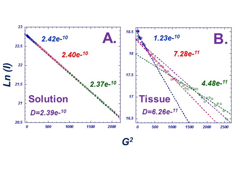
Fig 1A presents spermine diffusion characteristics measured in the metabolite mixture solution, while Fig 1B shows the diffusion behavior of spermine in a prostate tissue sample. The variation of spermine diffusion coefficients can be estimated by using different experimental regions in the diffusion curve (the beginning, mid, and ending 20 spectra). In solution, the same regional analyses of the ln(I) vs G2 curve give the the same diffusion coefficient. However in tissue there is a clear deviation from linearity suggesting the existence of tissue spermine in multi-compartment cellular environments. Similar diffusion differences can also be seen between spermine in gel, which closely followed that in solution, and in tissue. In addition to spermine (Spm), the multi-compartment phenomenon could also be observed to various degrees for other tissue metabolites including citrate (Cit), creatine (Cre), lactate (Lac) and alanine (Ala), as shown in Fig 2. Metabolites in different sample diffuse differently likely due to their different micro-environments. Quantitative evaluations of diffusion coefficients measured for different metabolites in different tissues show strong intra-samples correlations among different metabolites, as shown in Fig 3 where the fast diffusion components as represented by the beginning 20 measured points (blue point in Fig 1, or values in Fig 2A) present significant linear correlations between citrate and spermine, and citrate and creatine. However, the diffusion rates are different for different metabolites with Cit:Cre=1:1, but Cit:Spm=4:3.Discussion
Here, we present our initial diffusion (DOSY) studies of cellular metabolites in intact human prostate tissues and compared them with metabolite mixture solution and gel standard samples measured under the same experimental conditions. While our standard samples confirmed the ability of DOSY in quantifying unified metabolite diffusion coefficients in homogenous chemical and physical environments, the variations in diffusion coefficients seen for each metabolite in each tissue samples indicated the heterogenous environments within which they reside. These changes in diffusion based on compartmentalization present the potential for consideration as PCa markers. Quantification of other cellular metabolites; evaluation of their correlations with prostate pathologies; and modeling compartmentalization analysis are currently underway in our laboratory.Conclusion
Our study has demonstrated the ability of DOSY in quantifying cellular metabolite diffusion coefficients from intact tissue with the assistance of the HRMAS method. Investigations of metabolite diffusion characteristics present the potential for more comprehensive understanding of disease metabolomic markers according to their cellular micro-environments.Acknowledgements
We gratefully acknowledge the support of the Massachusetts General Hospital Athinoula A. Martinos Center for Biomedical Imaging.References
No reference found.Figures
A
Comparison Diffusion Coefficients between Spermine in Solution and Tissue. A)
10 mM spermine and citrate mixture solution prepared with D2O, and B) a human prostate tissue sample of
~10mg. Solution DOSY measurements of spermine showed a linear trend under a
magnetic field gradient, and strongly confirm an exclusive diffusion
coefficient for spermine in a homogeneous medium. However, DOSY measurements of
spermine in tissue showed nonlinear behavior suggesting its existence in different
biological compartments with varying diffusion coefficients that can be
estimated by using the beginning (blue),
middle (red), and ending (green)
spectra (20 data points for each).
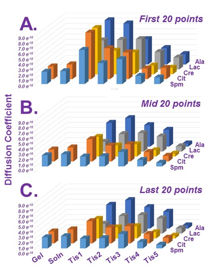
Diffusion
Coefficients. Diffusion coefficients for different
metabolites in solution, a gel, and in different human prostate tissue samples
analyzed as in Fig 1, where they are
divided into three individual segments of twenty points (beginning 20, A, middle 20, B, and ending 20, C) to
visualize variations in diffusion coefficients. The analysis showed different
diffusion coefficients in tissue samples while those in solution and gel showed
no change over their fractioned sets.
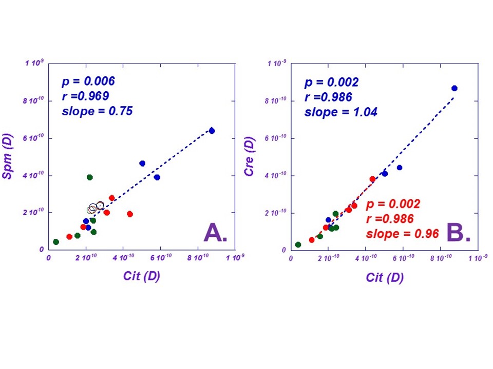
Comparisons
of Diffusion Coefficient Linearity between Citrate and Spermine, and Citrate
and Creatine. Diffusion coefficients in Fig 2 (beginning 20, blue solid circle, middle 20, red, and
ending 20, green)
are plotted to show relationships between different metabolites in tissue: A) Citrate vs. Spermine (open circles:
solution and gel), and B) Citrate
vs. Creating. The linearities between Cit and Spm, and Cit and Cre suggest the parallel
diffusion coefficient changes related to different cellular compartments
conditions in different tissue samples, while metabolite diffusion rates are
different for different metabolites as shown by different slope values
presented in the Figures.