2276
Role of combining NMR metabolomics data of intestinal mucosal biopsies, blood plasma and urine for determining biomarker/s for Celiac Disease (CeD)1Department of NMR & MRI Facility, All India Institute of Medical Sciences (AIIMS), New Delhi, India, 2Department of Pathology, All India Institute of medical sciences, New Delhi, India, 3Department of Gastroenterology and human Nutrition, All India Institute of Medical Sciences, New Delhi, India
Synopsis
NMR based metabonomics of small intestinal mucosal biopsies, blood plasma and urine samples from same set of patients demonstrated the underlying biochemical abnormalities and biomarker/s for celiac disease (CeD). Intestinal mucosa of CeD patients had higher levels of proline and allantoin while lower glycine, histidine and GPC compared to controls. Metabolome of blood plasma of CeD patients showed significantly higher concentration of proline, arginine and β-hydroxybutyrate while urine had higher proline, allantoin and β-hydroxybutyrate compared to healthy controls. These findings indicated metabolic abnormalities associated with villous atrophy seen in CeD and suggested proline, arginine and allantoin may serve as biomarker/s.
Introduction
Celiac disease (CeD) is an autoimmune enteropathy caused by ingestion of gluten in genetically predisposed individuals. Serological tests such as IgA anti-tissue transglutaminase (tTG) antibodies and anti endomysial antibodies (EMA) followed by endoscopic evaluation and histopathology of small intestinal biopsy are used for the diagnosis of CeD. Serological tests such as IgA anti-tTG antibodies have low sensitivity in regard to histological abnormalities.1 Therefore, histopathological evaluation of small intestinal biopsies is still regarded as the gold standard for the diagnosis of CeD. Thus, there is need of non-invasive biomarker/s which may compliment traditional serologic tests for the diagnosis of CeD. The present study investigates the metabolic profile of small intestinal mucosal biopsies, blood plasma and urine using in-vitro NMR spectroscopy to determine the biomarker/s for villous atrophy.
Patients and Methods
Sixty treatment naive CeD patients (n=60; mean age 28.2±11.4 yrs) were recruited and small intestinal mucosal biopsies, blood and urine were obtained from each patients. Thirty patients with functional dyspepsia (n=30; mean age 32.0 ± 9.6 yrs) where intestinal mucosa appeared endoscopically/histologically normal were recruited as disease controls (DC). Healthy controls (HC; n=30; mean age 28.2 ± 4.5 yrs) were also recruited for the comparison of metabolic profile of blood plasma and urine. Institute ethics committee approved the study and an informed consent was taken from each participant. During the endoscopic examination, small intestinal mucosal biopsies were collected for NMR spectroscopy. Blood and urine samples from all CeD patients and HC were collected in morning pre-prandial. Water soluble metabolites were extracted from intestinal mucosal samples using perchloric acid extraction method and lyophilized powder obtained was dissolved in deuterium oxide. Sodium trimethyl silyl- (2,2,3,3-H4) propionate (TSP) was added as a standard for chemical shift reference and for quantification of metabolites present in intestinal biopsies and urine. While formate was used for the quantification of metabolites present in blood. NMR experiments were carried out at 700 MHz NMR spectrometer (Agilent, U.S.A.). Mann Whitney U test was used for comparison of metabolites and a p-value <0.05 was considered significant. Partial least square discriminant analysis (PLS-DA) was also carried using Unscrambler 10.2 (CAMO, Oslo, Norway).Results
Figure 1 represents venn diagram showing overlapping of metabolites in intestinal mucosal biopsies, blood plasma and urine in CeD. Elevated concentration of proline (Pro) and allantoin (Alln) while lower level of glycine (Gly), histidine (His) and glycerophosphocholine (GPC) was observed in the small intestinal mucosa of CeD patients with CeD in comparison to DC. While the metabolic profile of blood plasma of CeD was characterized with significantly higher level of Pro, arginine (Arg), glycine (Gly), alanine (Ala) and β-hydroxybutyrate (β-OHB) compared to HC. Metabolome of urine of CeD patients showed higher level of β-OHB, Pro, Alln, aminohippurate while lower level of N-methylnicotinamide.Discusson
To the best of our knowledge, this is the first study that investigated the metabolic profile of small intestinal mucosal biopsies, blood plasma and urine in same cohort of patients. A significantly higher concentration of Pro was observed in all the three biological specimens; intestinal mucosal biopsies, blood plasma and urine of CeD patients compared to controls. Small intestinal mucosa requires certain amino acids such as glutamine, Pro, Arg for the synthesis of polymaines which is essential for proliferation, differentiation and repair of intestinal epithelial cells.2 Additionally, elevated Arg levels were observed in blood plasma of CeD patients. Arginine also serves as a precursor for the synthesis of nitric oxide, which has implications in the mucosal protection.3 Thus, elevated Pro and Arg levels indicated compromised repair and healing mechanism of small intestinal mucosa and may serve as biomarkers for villous atrophy for CeD.
Allantoin is a degradation product of purine metabolism and is produced from the oxidation of uric acid by reactive oxygen species in humans.4 Thus, elevated level of Alln in small intestinal mucosa and urine of CeD patients suggested increased oxidative stress which would have contributed for intestinal inflammation and may be used as a biomarker for CeD.
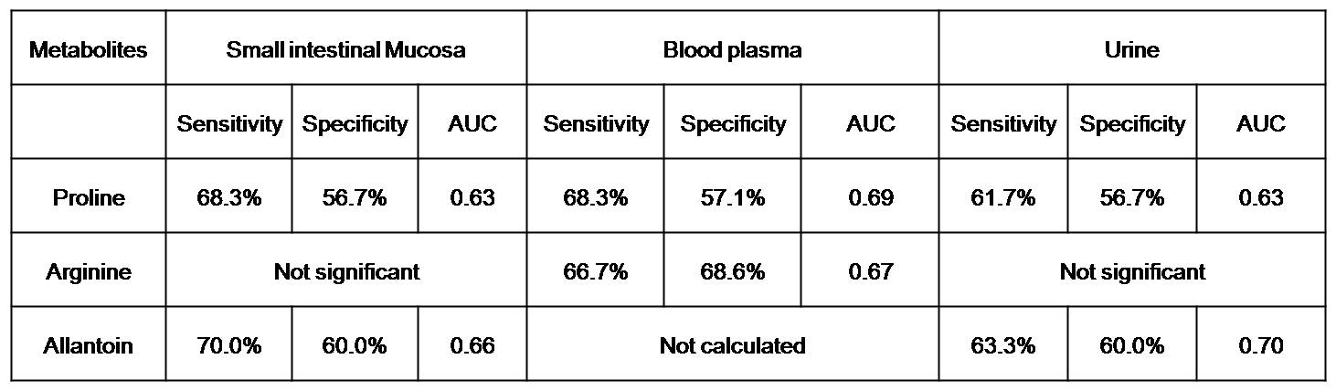
The sensitivity, specificity and area under the curve were determined using ROC analyses. The result showed that Pro, Arg and Alln may have diagnostic utility in differentiating CeD patients from controls (Table 1). Furthermore, PLS-DA score plot revealed separate clusters for CeD patients and controls based on the metabolic profile of intestinal mucosal biopsies, blood plasma and urine (Figure 2).
Conclusion
Present study highlighted the potential utility of NMR based metabonomics for determining biomarker/s for CeD. Metabolites such as Pro, Arg and Alln may serve as biomarker/s for assessing villous atrophy in CeD.Acknowledgements
US thanks the Department of Biotechnology, Government of India for financial support (BT/BioCARe/01/233/2010-11) while NRJ thanks SERB, Government of India for J.C. Bose fellowship.References
1) Rostami K, Kerckhaert J, Tiemessen R, et al. Sensitivity of antiendomysium and antigliadin antibodies in untreated celiac disease: disappointing in clinical practice. Am J Gastroenterol. 1999;94(4):888-894.
2) Wu G, Bazer FW, Burghardt RC, et al. Proline and hydroxyproline metabolism: implications for animal and human nutrition. Amino Acids. 2011;40(4):1053-1063.
3) Luiking YC, Ten Have GA, Wolfe RR, et al. Arginine de novo and nitric oxide production in disease states. Am J Physiol Endocrinol Metab. 2012;303(10):E1177-1189.
4) Dryland PA, Love DR, Walker MF, et al. Allantoin as A Biomarker of Inflammation in an Inflammatory Bowel Disease Mouse Model: NMR Analysis of Urine. TOBCJ. 2008;1:1-6.
Figures