2275
Potential Celiac disease (CeD) patients may serve as pre-clinical model to understand pathogenesis of Celiac Disease: A NMR metabonomics approach1Department of NMR & MRI Facility, All India Institute of Medical Sciences (AIIMS), New Delhi, India, 2Department of Gastroenterology and human Nutrition, All India Institute of Medical Sciences, New Delhi, India, 3Department of Pathology, All India Institute of medical sciences, New Delhi, India
Synopsis
Potential celiac disease (CeD) patients have positive CeD associated antibodies (anti-tissue tansglutaminase antibodies) and HLA-DQ2 and/or HLA-DQ8 genotype but no intestinal inflammation. NMR based metabonomic study of blood plasma of potential CeD patients demonstrated a distinct metabolic fingerprint characterized by raised histidine and proline in comparison to healthy controls. The changes in histidine suggested compromised cytoprotective mechanism while elevated arginine level indicated altered functioning of intestinal cells in potential CeD. These altered metabolic activities could be the initial event that precede the pathogenesis of CeD and may contribute to intestinal inflammation which results in villous atrophy.
Introduction: Potential CeD patients are characterized by CeD specific serological markers (anti-tTG antibody and anti-endomysial antibody) and positive HLA-DQ2 and/or HLA-DQ8 genotype, however their small intestine is histopathologically normal. It has been reported that only one-third of potential CeD patients develop villous atrophy after 3 years of follow-up study.1 Thus, it may be hypothesized that potential CeD patients may serve a pre-clinical model to understand the early pathophysiological changes occurring in CeD. The present study investigates the metabolic profile of blood plasma of potential CeD patients using NMR based targeted metabonomics to understand the biochemical and pathophysiological processes of CeD development.
Patients and Methods: Sixty four patients with CeD (n = 64; mean age 28.5 ± 11.4 yrs), seven potential CeD patients (n=7; mean age 27.7 ± 12.5 yrs) and fifty healthy controls (n = 50; mean age 28.8 ± 4.6 yrs) were recruited. An informed consent was taken and the institute ethics committee approved the study. All patients were treated according to the standard treatment regimen. Diagnosis of CeD was made on the guidelines of European Society of Pediatric Gastroenterology Hepatology and Nutrition. Blood samples were collected in morning pre-prandial and plasma was separated and stored at -80 °C until NMR analysis. One-dimensional Carr-Purcell-Meiboom-Gill Sequence and two dimensional total correlation spectroscopy experiments were performed at 700 MHz (Agilent, U.S.A.) and the concentration of metabolites was determined. Mann Whitney (SPSS 20.0) test was used for comparison of metabolite levels in different groups and p-value <0.05 was considered significant.
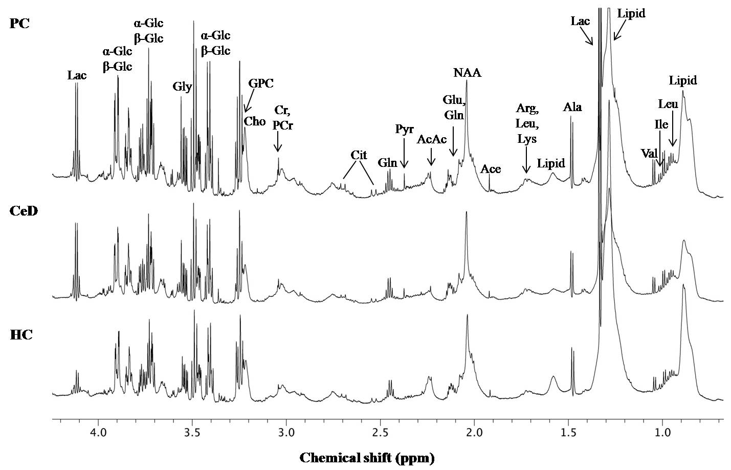
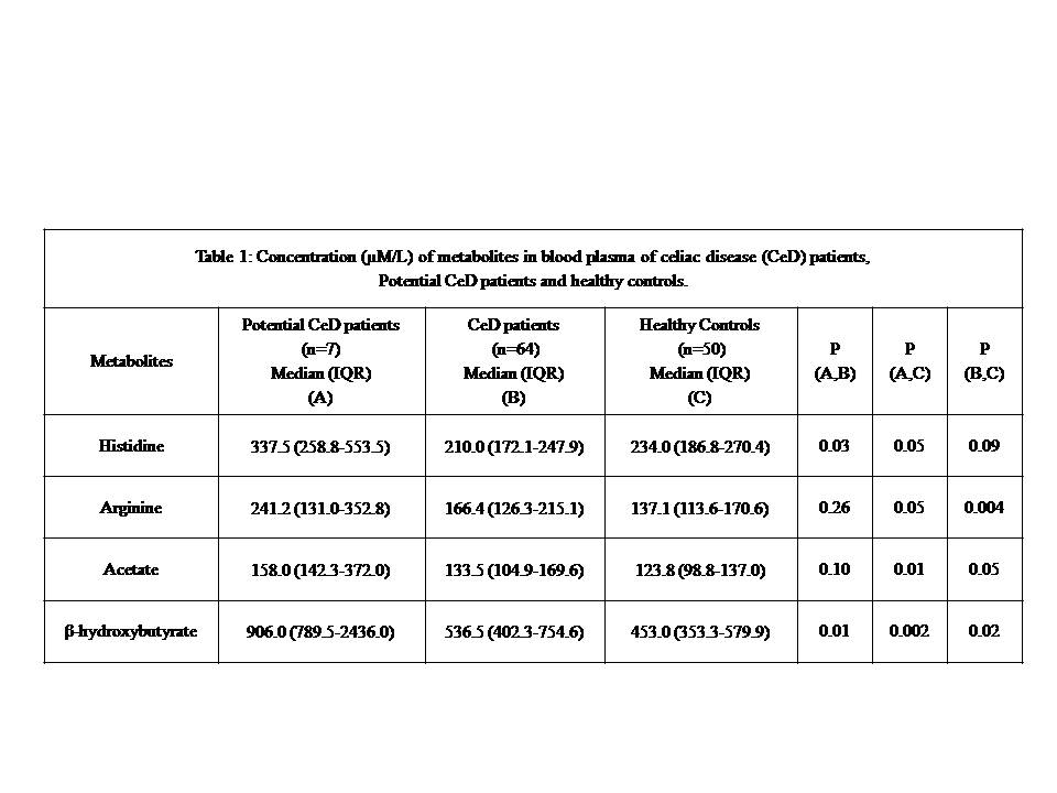
Results: Figure 1 shows the representative proton NMR spectra of blood plasma of a patient with CeD, potential CeD and HC. In all 40 metabolites were assigned using 1D and 2D NMR. The concentration of metabolites that showed statistical difference in the blood plasma of potential CeD patients, patients with CeD and HC are presented in Table1. The concentration of histidine was significantly elevated in blood plasma of potential CeD, while arginine (Arg), acetate (Ace) and β-hydroxybutyrate (β-OHB) were higher in both potential CeD and CeD patients compared to HC. Potential CeD patients also showed higher level of His and β-OHB as compared to CeD patients.
Discussion: This is the first study that demonstrated the metabolic pattern of blood plasma of patients with CeD in comparison to CeD patients and HC using NMR based targeted metabonomics. A higher concentration of His was observed in blood plasma of potential CeD patients as compared to both CeD patients and HC. Elevated His level in blood plasma of potential CeD patients may be attributed to the impaired activity of transporters which are responsible for its absorption in small intestine. Histidine has been shown to have antioxidative and anti-inflammatory effects.2 Hence, impaired uptake of His in small intestinal mucosa of potential CeD patients may lead to the compromised cytoprotective mechanism and contribute to the intestinal inflammation which results in villous atrophy.
Further, our data showed significantly increased concentration of Arg in the blood plasma of both potential CeD patients and CeD patients than HC. Arginine serves as a precursor for the synthesis of many important biological compounds such as nitric oxide, ornithine and polyamines. These metabolites have implications in providing protection against oxidative stress and in maintaining mucosal integrity.3,4 Elevated Arg concentration in blood plasma of potential CeD patients might be attributed to the impaired metabolism of Arg by the intestinal cells which may eventually lead to the development of villous atrophy in potential CeD patients over time.
A significantly higher concentration of Ace in both potential CeD and CeD patients compared to HC suggested increased catabolism of lipids in order to meet energy requirements in potential CeD patients as well as in CeD patients. Furthermore, both potential CeD and CeD patients had elevated level of β-OHB in comparison to HC. The level of β-OHB in potential CeD were also higher compared to CeD patients. These findings suggested that the potential CeD patients excessively metabolize ketone bodies to meet the high energy requirement of the body for protecting against gluten induced immune response.
Conclusion: Present study demonstrated a distinct metabolic fingerprint of blood plasma in potential CeD patients. The results suggested that patients with potential CeD patients have modulated metabolic pathways that helped in delaying or inhibiting the immune response and thereby maintain the integrity of intestinal mucosa.
Acknowledgements
US thanks the Department of Biotechnology, Government of India for financial support (BT/BioCARe/01/233/2010-11) while NRJ thanks SERB, Government of India for J.C. Bose fellowship.References
1. Tosco A, Salvati VM, Auricchio R, et al. Natural history of potential celiac disease in children. Clin Gastroenterol Hepatol. 2011;9(4):320-325.
2. Yan SL, Wu ST, Yin MC, et al. Protective effects from carnosine and histidine on acetaminophen- induced liver injury. J Food Sci. 2009;74(8):H259-265.
3. Luiking YC, Ten Have GA, Wolfe RR, et al. Arginine de novo and nitric oxide production in disease states. Am J Physiol Endocrinol Metab. 2012;303(10):E1177-1189.
4. Fritz JH. Arginine Cools the Inflamed Gut. Infect Immun. 2013; 81(10): 3500-3502.
Figures